-
Články
- Vzdělávání
- Časopisy
Top články
Nové číslo
- Témata
- Kongresy
- Videa
- Podcasty
Nové podcasty
Reklama- Kariéra
Doporučené pozice
Reklama- Praxe
microRNAs Regulate Cell-to-Cell Variability of Endogenous Target Gene Expression in Developing Mouse Thymocytes
microRNAs are integral to many developmental processes and may 'canalise' development by reducing cell-to-cell variation in gene expression. This idea is supported by computational studies that have modeled the impact of microRNAs on the expression of their targets and the construction of artificial incoherent feedforward loops using synthetic biology tools. Here we show that this interesting principle of microRNA regulation actually occurs in a mammalian developmental system. We examine cell-to-cell variation of protein expression in developing mouse thymocytes by quantitative flow cytometry and find that the absence of microRNAs results in increased cell-to-cell variation in the expression of the microRNA target Cd69. Mechanistically, T cell receptor signaling induces both Cd69 and miR-17 and miR-20a, two microRNAs that target Cd69. Co-regulation of microRNAs and their target mRNA dampens the expression of Cd69 and forms an incoherent feedforward loop that reduces cell-to-cell variation on CD69 expression. In addition, miR-181, which also targets Cd69 and is a known modulator of T cell receptor signaling, also affects cell-to-cell variation of CD69 expression. The ability of microRNAs to control the uniformity of gene expression across mammalian cell populations may be important for normal development and for disease.
Published in the journal: . PLoS Genet 11(2): e32767. doi:10.1371/journal.pgen.1005020
Category: Research Article
doi: https://doi.org/10.1371/journal.pgen.1005020Summary
microRNAs are integral to many developmental processes and may 'canalise' development by reducing cell-to-cell variation in gene expression. This idea is supported by computational studies that have modeled the impact of microRNAs on the expression of their targets and the construction of artificial incoherent feedforward loops using synthetic biology tools. Here we show that this interesting principle of microRNA regulation actually occurs in a mammalian developmental system. We examine cell-to-cell variation of protein expression in developing mouse thymocytes by quantitative flow cytometry and find that the absence of microRNAs results in increased cell-to-cell variation in the expression of the microRNA target Cd69. Mechanistically, T cell receptor signaling induces both Cd69 and miR-17 and miR-20a, two microRNAs that target Cd69. Co-regulation of microRNAs and their target mRNA dampens the expression of Cd69 and forms an incoherent feedforward loop that reduces cell-to-cell variation on CD69 expression. In addition, miR-181, which also targets Cd69 and is a known modulator of T cell receptor signaling, also affects cell-to-cell variation of CD69 expression. The ability of microRNAs to control the uniformity of gene expression across mammalian cell populations may be important for normal development and for disease.
Introduction
The complexity of developmental processes in metazoans relies on mechanisms that confer a degree of robustness against environmental and genetic variation [1]. microRNAs are small non-coding RNAs that negatively regulate gene expression at the post-transcriptional level by reducing mRNA stability and/or translation. Their role in dampening gene expression makes microRNAs potential building blocks for gene regulatory circuits that can stabilize gene regulatory networks [2–5].
Gene expression is subject to intrinsic stochasticity associated with mRNA transcription and translation, as well as extrinsic noise such as fluctuations in upstream regulators. Gene expression noise is not restricted to protein coding genes: the expression of primary microRNA transcripts, their processing into pre-microRNAs, nuclear export, processing into mature microRNAs, association with RISC components, etc., presumably all have stochastic components. The participation of microRNAs in the regulation of protein-coding genes could therefore add noise contained in both microRNA and protein-coding systems. Feed-forward loops (FFLs) are recurrent network motifs that can reduce gene expression noise by buffering fluctuations in upstream regulators [6]. Placing the expression of a microRNA and its target mRNA under the control of common upstream regulators can link the production of mRNAs to the production of microRNAs that target the mRNAs. Theoretical considerations [2] and computational simulations [7, 8] suggest that this circuit topology, which resembles an incoherent FFL, allows microRNAs to buffer protein expression against fluctuations in the activity of upstream regulators [9]. In silico models predict that FFL regulation enables microRNAs to reduce not only the level of target gene expression, but also cell-to-cell variability [7, 8]. Data from synthetic circuits indicate that co-expression of microRNAs and target mRNAs can reduce temporal fluctuations and in some cases cell-to-cell variability in reporter gene expression [7, 10].
Emerging experimental evidence supports a role for microRNAs in biological robustness [2]. microRNAs affect several phenotypic traits in Drosophila, for example by stabilizing the regulation of the enhancer of split transcription factor to guide sensory organ development under conditions of environmental flux [11]. Loss of microRNAs can increase the variation of primordial germ cell numbers [12, 13] and sensory bristles [14], and quantitative phenotypic traits in the Drosophila cuticle [15]. These data demonstrate that microRNAs can buffer variation in phenotypic traits, but it is not clear whether this is achieved by reduced variation in the expression of microRNA target genes or the operation of thresholds for phenotypic outcomes [16]. Zebrafish miR-26b and ctdsp2 mRNA are encoded by the same primary transcript, and ctdsp2 mRNA is a target of miR-26b [17]. The processing of miR-26b is developmentally controlled during neuronal differentiation, effectively initiating a microRNA-mediated incoherent FFL but the consequences for cell-to-cell variation in the expression of ctdsp2 have yet to be established [17]. microRNAs can dampen temporal oscillations in gene expression in C. elegans [18] and reduce fluctuations in the average expression of reporter constructs in mammalian cells [19]. Measurements at the population level, but not in individual cells, showed that methyl CpG-binding protein 2 (MeCP2) acts through BDNF to induce the neuronal miRNA miR-132, which feeds back to repress MeCP2 [20]. However, simple negative feedback loops like this may increase noise as determined experimentally and computationally [7]. The miR-17-92 family forms a complex network with Cyclin D1 in neuronal progenitors, and the variability of Cyclin D1 expression was increased by heterozygosity in Dicer [21]. The relationship between microRNAs and variability of target gene expression is complicated in this system, since miR-17-92 is required for the differentiation of mouse cortical neuronal progenitors [22], and reduced microRNA expression affects the frequency of proliferating neuronal progenitors as well as the expression of Ccnd1 within them [21, 22]. That loss of microRNAs can also result in reduced variability in the expression of pluripotency markers was recently demonstrated for mouse ES cells [23].
Here we address the impact of the microRNA biogenesis pathway on cell-to-cell variability of endogenous gene expression in mouse thymocytes (developing T cells). This system offers a number of key advantages. T cell development proceeds in a series of discrete developmental steps that are defined by the expression of cell surface markers [24]. This allows for (i) the precise definition and isolation of cell populations at specific developmental stages for the molecular characterization of microRNA and target mRNA expression, (ii) the use of developmentally regulated Cre transgenes for the synchronous deletion of conditional alleles of the RNase III enzyme Dicer, (iii) the verification of reduced microRNA expression at defined developmental stages, and (vi) like-for-like comparisons between control and Dicer-deficient cells at the same developmental stage. Thymocytes readily form cell suspensions that are ideally suited for analysis and sorting by flow cytometry, and high-quality reagents are available to enable quantitative flow cytometry at the single cell level [25]. Using this approach we demonstrate that microRNAs can reduce cell-to-cell variation of target gene expression in mammalian cells. The activation-induced microRNA target CD69 was regulated by microRNAs in two different ways. miR-181a affected variation by modulating the responsiveness of thymocytes to activation signals, acting at least in part upstream on the target mRNA Cd69. Members of the miR-17-92 cluster were co-regulated with the target mRNA Cd69, resembling an activation-induced incoherent FFL.
Results
microRNA-dependent regulation of gene expression in developing thymocytes
We previously characterised an experimental system where a developmentally regulated Lck-Cre transgene deletes a conditional Dicer allele in developing mouse thymocytes [26]. As a result, the expression of Dicer-dependent microRNAs was reduced by ∼90% at the CD4 CD8 double positive (DP) stage of development (Fig. 1A) [26]. miReduce analysis [27] of 3'UTR motifs associated with post-transcriptional de-repression in Lck-Cre DP thymocytes (see GSE57511) showed enrichment for microRNAs miR-181, miR-17 and miR-142 (Fig. 1B).
Fig. 1. microRNA-dependent regulation of gene expression in developing thymocytes. 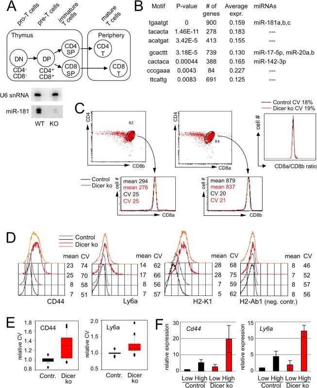
A) Schematic of T cell differentiation. Abbreviations are DN: double negative for CD4 and CD8; DP: double positive for CD4 and CD8; Periphery: peripheral lymphoid organs. The expression of mature microRNAs, including miR-181a, is reduced by ∼ 90% at the DP stage of development as demonstrated by northern blotting with U6 snRNA as a loading control, data from [26]. B) 3'UTR motifs and microRNAs associated with positive fold-change of transcript levels in Dicer-deficient DP thymocytes as determined by miReduce [27]. 'Average expr.' denotes the fold-change in the expression of mRNAs with the indicated 3'UTR motifs between Dicer-deficient and control DP thymocytes. C) Control and Dicer-deficient DP thymocytes as defined by staining for CD4 versus CD8a (left) and CD4 versus CD8b (middle). Both the mean expression and the CV of CD8a and CD8b are comparable between control and Dicer-deficient DP thymocytes. The ratio of CD8a/CD8b expression was calculated for individual control (left) and Dicer-deficient (middle) DP thymocytes. The CV of these ratios represents experimental noise (right) [25]. D) CD44, Ly6a and H2-Kb are examples of proteins encoded by transcripts that are deregulated in Dicer-deficient DP thymocytes. The panels show representative flow cytometry histograms of CD44, Ly6a and H2-K1 expression by Dicerlox/lox (black/grey) and DicerΔ/Δ (red/orange) DP thymocytes gated on low levels of T cell receptor (TCR) expression. The MHC class II antigen H-2Ab1 is not expressed by mouse T cells and served as a negative control. Numbers indicate the mean expression level and the coefficient of variation (CV). Representative of 3–5 biological replicates. E) Analysis of 10–30 biological replicates showed an increase in the CV of CD44 protein expression of 30% and an increase in the CV of Ly6a protein expression of 15% in Dicer-deficient versus control DP thymocytes. F) DP thymocytes were sorted by flow cytometry according to the level of CD44 and Ly6a expression by individual cells. Analysis of the sorted populations by quantitative RT-PCR indicates that protein expression as assessed by flow cytometry predicts mRNA expression. We evaluated flow cytometry as an approach to determine protein expression by individual cells. To estimate technical noise we examined CD8a and CD8b, which are expressed as obligate heterodimers in DP thymocytes. The mean expression and the coefficient of variation (CV) of CD8a and CD8b were very similar for control and Dicer-deficient DP thymocytes (Fig. 1C, left and centre), as was the ratio of CD8a/CD8b expression for individual control and Dicer-deficient DP thymocytes (Fig. 1D, right). The CV of these ratios defines the upper bound of technical noise [25]. Based on published criteria for quantitative flow cytometry [25] we identified antibodies directed against putative microRNA targets including CD44, a predicted target of miR-21 (www.targetscan.org) and established target of miR-34 [28] as well as the predicted miR-181 targets Ly6a and H2-K1 (www.targetscan.org; Fig. 1D). As expected based on elevated mRNA expression (see GSE57511), Dicer-deficient thymocytes showed higher average expression of CD44, Ly6a and H2-K1 than control cells. Interestingly, the cell-to-cell variation of CD44, Ly6a and H2-K1 expression was also increased in Dicer-deficient thymocytes (Fig. 1D, E) while there was no change in the negative staining control, MHC class II (H2-Ab1; Fig. 1D).
We used the CV as a stringent measure of variation because in contrast to the standard deviation (SD), the CV is expected to decrease as the mean expression increases (CV = standard deviation/mean). An increase in the CV at the same time as an increase in mean protein expression therefore unambiguously indicates an increase in cell-to-cell variation (Fig. 1D, E). As the CV is expected to decrease with the level of expression, the finding of an increased CV in Dicer-deficient cells that expressed higher protein levels prompted several control experiments. First, we asked whether the increased CV was explained by residual microRNA-retaining cells. Experimental mixing and computational modeling experiments indicated that this was highly unlikely (S1 Fig.). Second, the apparent impact on the CV could be due to technical limitations in the detection of low levels of protein expression: if microRNAs reduce expression below the sensitivity or our instrumentation we would detect expression—and associated noise—only in Dicer-deficient cells but not in wild type cells. To address this concern we asked whether the level of protein expression detected in wild type and Dicer-deficient cells was biologically meaningful. We sorted control and Dicer-deficient thymocytes according to the level of CD44 and Ly6a protein detected by flow cytometry and carried out quantitative reverse transcriptase (RT)-PCR for Cd44 and Ly6a transcripts (Fig. 1F). The data correlated CD44 and Ly6a protein expression with the abundance of Cd44 and Ly6a mRNA in both control and Dicer-deficient DP thymocytes. Although Cd44, Ly6a and H2-K1 mRNAs are not confirmed direct microRNA targets in thymocytes, these data demonstrate that our instrumentation discriminates meaningful levels of protein expression.
Dicer-deficient DP thymocytes show increased variation in inducible CD69 expression
To unambiguously determine the impact of microRNAs on cell-to-cell variability of target gene expression on a direct microRNA target in thymocytes we focused on CD69, which is inducibly expressed in response to T-cell activation [29]. CD69 controls cell migration and sphingosine 1-phosphate signaling [30], and the Cd69 mRNA is a well-characterised target of miR-181 and other microRNAs [31–33]. In response to activation signals through the T cell receptor (TCR), DP thymocytes initiated the expression of CD69 (Fig. 2A), and graded activation signals induced a proportional increase of Cd69 mRNA and CD69 protein (S2 Fig.). As expected for an established microRNA target, average CD69 levels were higher in Dicer-deficient than in control DP thymocytes (S2B Fig.). In addition, Dicer-deficient DP thymocytes showed an increase in the CV of CD69 expression (Fig. 2B), and this increase was seen over a range of activation conditions (Fig. 2C). The broader distribution of CD69 expression among Dicer-deficient DP cells was due in part to a greater fraction of CD69hi cells (Fig. 2D, characterized by the co-expression of CD25 Fig. 2A). In addition, Dicer-deficient DP thymocytes showed an increased CV of CD69 expression within the CD69hi CD25+ subset (Fig. 2E, F). Hence, Dicer-deficient DP thymocytes showed increased cell-to-cell variability in the expression of the microRNA target CD69. This was true whether the CV was assessed for the entire DP thymocyte population, or separately for the CD69+ population or the CD69high CD25+ subset (Fig. 2G).
Fig. 2. Increased CV of inducible CD69 expression in Dicer-deficient thymocytes. 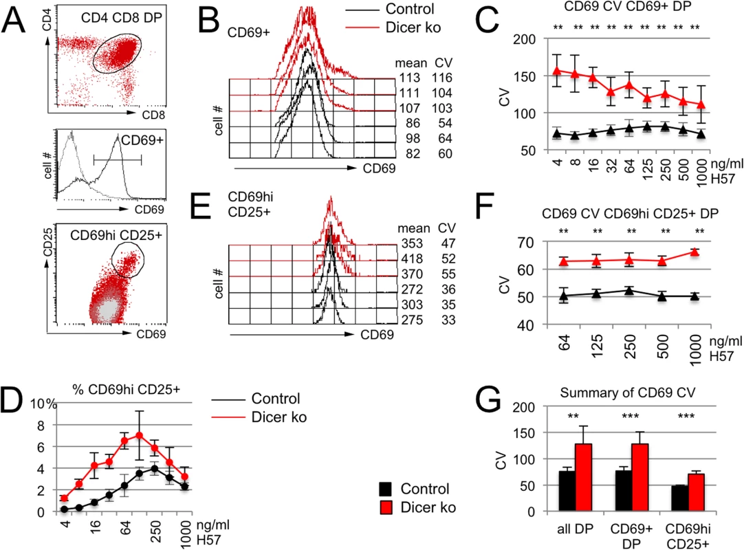
A) Activation of CD4+ CD8+ DP thymocytes (oval, top) results in CD69 expression (horizontal line defines CD69+ cells, middle) and definition of CD69hi cells (oval, bottom). B) Histogram overlays of CD69 expression by CD69+ DP thymocytes activated for 18 hours with 125ng H57/ml. Mean and CV of CD69 expression are indicated. C) The CV of CD69 expression is higher in Dicer-deficient than in control CD69+ DP thymocytes over a range of activation conditions (n = 4–7 per data point, ** P<0.001). See S1 Table for additional data. D) The frequency of CD69hi CD25+ DP thymocytes is higher in Dicer-deficient than in control DP thymocytes. E) Histogram overlays of CD69 expression by CD69hi CD25+ thymocytes activated for 18 hours with 125ng H57/ml. Mean and CV of CD69 expression are indicated. See S1 Table for additional data. F) The CV of CD69 expression is higher in Dicer-deficient than in control CD69hi CD25+ DP thymocytes (n = 4 per data point, ** P<0.001). See S1 Table for additional data. G) Summary of CD69 CV data for Dicer-deficient DP thymocytes, CD69+ DP thymocytes and CD69hi CD25+ DP thymocytes. Taken together, our results indicate that microRNAs can shape not only the level but also the cell-to-cell variability of protein expression in developing thymocytes. To investigate the underlying mechanisms we next identified endogenous microRNAs that target Cd69 in DP thymocytes.
A dual fluorescence reporter system identifies endogenous microRNAs that target the Cd69 3'UTR in DP thymocytes
The Cd69 3'UTR contains predicted sites for miR-181, miR-130 and miR-17/20 (http://www.targetscan.org) and there is firm experimental evidence for Cd69 regulation by miR-181a, miR-130 and the miR-17-92 cluster (which encodes the microRNAs miR-17, -18, -19a, -19b, -20a, and -92 [34] in T lymphocytes [31–33].
To evaluate the impact of endogenous microRNAs on the expression of proteins linked to the 3'UTR of Cd69 we developed a dual fluorescence reporter construct. The construct encodes the two fluorescent reporter proteins, eGFP and mCherry, under the control of separate retroviral long terminal repeat (LTR) and mouse Pgk promoters, as well as a cloning site 3’ of the eGFP transcript (Fig. 3A). In a manner similar to luciferase reporter constructs, 3’ UTRs of interest can be cloned into this site to measure their impact on the expression of GFP relative to mCherry. In contrast to heterologous reporter assays, however, this system allows to delineate the biological activity of endogenously expressed microRNAs after retroviral gene transfer of the reporter construct into primary cells. We characterised this dual fluorescence reporter system in mature CD4+ T cells that were isolated from lymph nodes and activated in vitro to render them receptive to retroviral gene transfer (S3A Fig.). To determine the impact of Dicer on the expression of eGFP linked to the 3'UTR of Cd69 we transduced wild type and CD4Cre Dicerlox/lox (Dicer-deficient) [35] CD4+ T cells with the reporter construct containing the entire Cd69 3'UTR (Fig. 3A, Cd69 3'UTR). Fig. 3B shows a representative dot plot of mCherry and eGFP-Cd69 3'UTR expression in control (black) versus Dicer-deficient CD4+ T cells (red). Compared to the empty control vector, wild type CD4+ T cells expressed eGFP-Cd69 3'UTR at a lower level and Dicer-deficient CD4+ T cells expressed eGFP-Cd69 3'UTR at a higher level (Fig. 3B, C), indicating that as well as repressive miRNA-binding sites, the CD69 3’ UTR may contain sequences that enhance expression. Mutation of the miR-181 site in the Cd69 3'UTR did not measurably affect the expression of eGFP in mature CD4+ T cells (Fig. 3C), which express only low levels of the developmentally regulated miR-181 [31]. However, deletion of the miR-130 and particularly the miR-17/20 site resulted in increased eGFP expression in wild type CD4+ T cells (Fig. 3C).
Fig. 3. A dual fluorescence reporter system identifies endogenous microRNAs that target the Cd69 3'UTR in DP thymocytes. 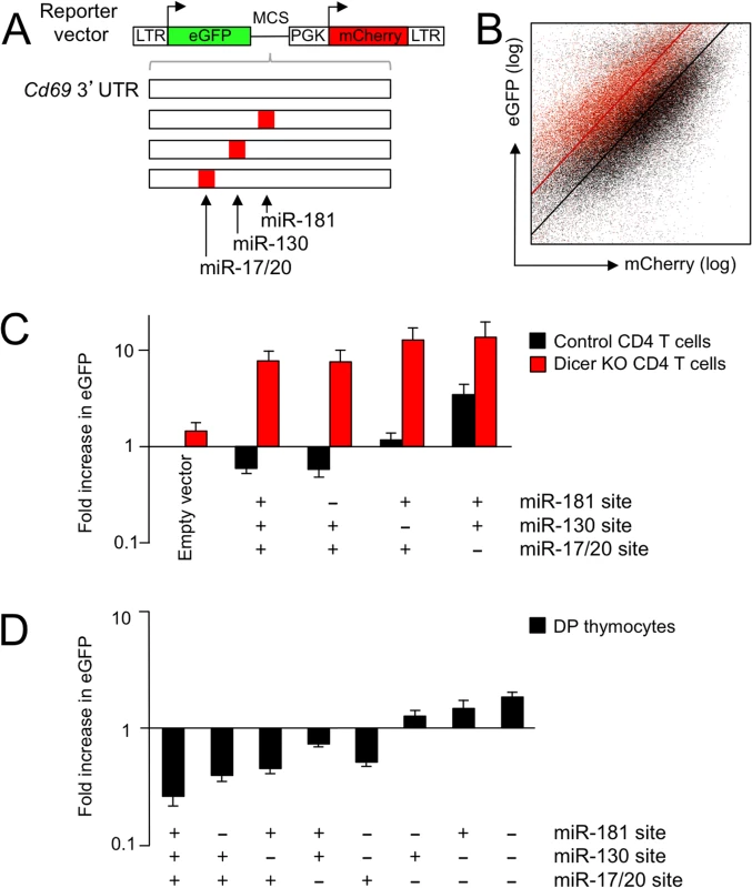
A) Dual fluorescence reporter based on retroviral vectors encoding eGFP followed by a multiple cloning site and mCherry for normalisation. Introduction of a 3'UTR containing relevant microRNA sites is predicted to downregulate eGFP expression relative to the mCherry control. The 842 nt 3’ UTR of Cd69 contains predicted binding sites for miR-181, miR-130 and miR-17-20 starting at positions 255, 354 and 391, respectively, which were mutated alone and in combination. B) Representative log-log dot plot of mCherry and eGFP-Cd69 3'UTR expression by control (black) and Dicer-deficient mature CD4+ T cells isolated from lymph nodes (red). Fitted lines were used to calculate de-repression of eGFP. See S3A Fig. for additional data. C) Expression of eGFP obtained with empty vector and the indicated 3'UTR constructs in control (black) and Dicer-deficient mature CD4+ T cells isolated from lymph nodes (red, n = 4–14 per data point, mean ± SE). D) Expression of eGFP obtained with the indicated 3'UTR constructs in control DP thymocytes (n = 4–14 per data point, mean ± SE). See S3B Fig. for additional data. Next, thymocytes were transduced with Cd69 3'UTR reporter constructs and maintained for 24 hours in reaggregation thymic organ cultures until the expression of fluorescent reporters by CD4+ CD8+ DP thymocytes was recorded by flow cytometry. In contrast to mature CD4+ T cells, the miR-181 site affected eGFP-Cd69 3'UTR expression in CD4+ CD8+ DP thymocytes, which express maximal levels of the developmentally regulated miR-181 [31]. The predicted sites for miR-130 and miR-17/20 within the Cd69 3' UTR also affected eGFP reporter gene expression in DP thymocytes.
Taken together, these results show that eGFP-Cd69 3'UTR expression was Dicer-dependent in mature CD4+ T cells (Dicer-deficient thymocytes could not be used successfully for retroviral gene transduction and subsequent reaggregate organ cultures) and that the impact of predicted microRNA binding sites reflected the developmental regulation of microRNAs [31]. We focused our subsequent analysis on miR-181a and the miR-17-92 cluster.
microRNA-181a controls cell-to-cell variability in CD69 expression
To explore the influence of miR-181 on the CV of CD69 expression we analysed DP thymocytes deficient in mir-181ab1, which accounts for most of the miR-181a and -b copies in DP thymocytes [36]. Following activation, miR-181-deficient DP thymocytes showed increased mean CD69 expression (control = 245 ± 17, mean miR-181 ko = 278 ± 10, n = 26, P<10–10, 2-tailed T-test). Interestingly, CD69 expression in miR-181-deficient DP thymocytes also showed an increased CV (Fig. 4A) over a range of activation conditions (Fig. 4B). The increased CV was due mainly to a higher fraction of CD69hi cells among miR-181-deficient DP thymocytes (Fig. 4C). The CV of CD69 expression within the CD69hi subset was only mildly affected (Fig. 4D).
Fig. 4. miR-181a controls cell-to-cell variation in CD69 expression. 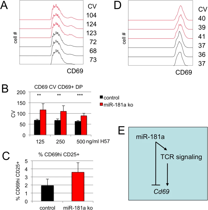
A) Histogram overlays of CD69 expression by control (black) and miR-181a-deficient (red) DP thymocytes activated for 18 hours with 125ng H57/ml. Histograms are gated on CD69+ cells. B) The CV of CD69 expression is higher in miR-181a-deficient than in control DP thymocytes (n = 7–8, ** p<0.005, *** p<0.0005). C) The frequency of CD69 hi CD25+ DP cells is higher in miR-181a-deficient than in control DP thymocytes (n = 12, P<10–5). D) The CV of CD69 expression in miR-181a-deficient and control CD69 hi CD25+ DP thymocytes is slightly higher than in control thymocytes. E) Model for the action of miR-181 upstream of TCR signaling and on Cd69 mRNA. These results show that miR-181 is an important determinant of cell-to-cell variability in CD69 expression in activated DP thymocytes, and is required to restrict the fraction of CD69hi DP cells. This is consistent with a role for miR-181 as a modulator of TCR signaling [36–38] (Fig. 4E).
miR-17 and miR-20a form an incoherent positive feedforward loop with the target mRNA Cd69
miRNA expression responds to T-cell activation signals [34, 35, 39–45]. Many microRNAs are downregulated upon T-cell activation [40–43], but the expression of the miR-17-92 cluster is upregulated in activated mouse and human T cells [45]. Since the miR-17-92 cluster encodes microRNAs that target the Cd69 3'UTR, including miR-17 and miR-20a (Fig. 3), we investigated how the expression of miR-17 and miR-20a was affected by the activation of DP thymocytes.
We applied graded stimuli (0, 0.1, 1 and 10μg H57/ml) to induce a graded increase in Cd69 mRNA expression (Fig. 5A). Interestingly, this graded increase in Cd69 mRNA was accompanied by a proportional upregulation of miR-17 and miR-20a expression (Fig. 5A).
Fig. 5. miR-17 and miR-20 form an incoherent positive feedback loop with the target mRNA Cd69. 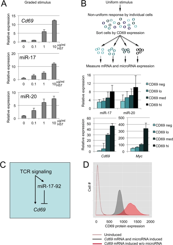
A) The strength of activation signals (0.1, 1, 10 μg/ml H57) determines the expression of Cd69, miR-17 and miR-20a (normalised to snRNA-135 and -202, n = 2–3, mean ± SE). B) Perceived signal strength varies among individual DP thymocytes and determines the expression of Cd69, miR-17 and miR-20a (microRNA expression is normalised to snoRNA-135 and snoRNA-202). At a fixed extracellular signal of 1u/ml H57, the fold-change in miR-17 and miR-20 relative to CD69 negative DP and normalised to snoRNA-135 was proportional to the expression of CD69 (n = 3, mean ± SD). C) The microRNA target mRNA Cd69 and microRNAs of the miR-17-92 cluster are co-regulated in response to activation signals and form an incoherent feed-forward loop downstream of the TCR. D) Modeling CD69 protein expression with and without microRNA feed-forward regulation. The state of the system is described by five major variables: the number of mRNAs transcribed from the TF gene, the number of TF molecules, the number of miRNAs, the number of mRNAs and the number of target proteins. Of these variables we can estimate the number of mRNA copies and the number of microRNA copies. As detailed in the legend to S4 Fig., the number of Cd69 mRNA copies was estimated as 0 in resting and 6 in activated cells, miR-17 and miR-20 were estimated as 6–12 copies per cell the resting state and 30–60 copies per cell after activation. Simulations of transcriptional networks were carried out using the Gillespie exact stochastic simulation algorithm, programmed and analysed using R based on a microRNA feed-forward model [8] to simulate CD69 protein expression in resting T cells (unfilled histogram), in a scenario where activation increases Cd69 mRNA but the expression of miR-17 and miR-20a remained the same as in resting T cells (activated without microRNA FFL, filled red histogram), and in a scenario where activation increases both Cd69 mRNA and miR-17 and miR-20a expression (activated with microRNA FFL, filled grey histogram). The plot represents 10,000 simulations. The model predicts that thymocyte activation with co-regulation of Cd69 mRNA and miR-17/miR-20a reduces the mean (887 versus 1300) and the CV (10.2 versus 14.6) of CD69 expression compared to the regulation of Cd69 mRNA alone (P<10–4). See S4 Fig. for details of the underlying circuitry, the parameters used, and a model based on microRNA effects on mRNA degradation [49]. Next, we asked how miR-17 and miR-20a expression was related to the range of responses by individual cells to a uniform extracellular signal. When stimulated with a fixed concentration of TCR antibody, DP thymocytes expressed a range of CD69 protein, from undetectable to high (see Fig. 2A). We applied a uniform stimulus (1μg H57/ml) and sorted DP thymocytes that expressed no detectable CD69 protein (CD69 neg), low levels of CD69 (CD69 lo), intermediate levels of CD69 (CD69 med) or high levels of CD69 (CD69 hi). Increasing expression of CD69 protein correlated with increasing Cd69 mRNA levels, and with incremental expression of miR-17 and miR-20a (Fig. 5B).
Hence, activation signals of increasing strength induce a proportional upregulation of the microRNAs miR-17 and miR-20a and the target mRNA Cd69. Furthermore, cells exposed to a uniform stimulus show a range of responses, and the induction of microRNAs and mRNA target is coordinated with the expression of the protein encoded by the target mRNA in individual cells. These findings suggest that miR-17, miR-20a and Cd69 are co-regulated. Mechanistically, the transcription factor Myc provides a link between thymocyte activation and the coordinated regulation of Cd69 and the miR-17-92 cluster. Myc expression is upregulated by signals that drive lymphocyte activation and mediates downstream transcriptional responses [46] (Fig. 5B). Myc and Cd69 are induced by shared signaling pathways downstream of the TCR [47], and Myc directly activates transcription of the miR-17-92 cluster [48]. These data indicate that the microRNA target mRNA Cd69 and microRNAs of the miR-17-92 cluster form an incoherent feed-forward loop in response to TCR signaling (Fig. 5C).
The coordinated regulation of miR-17, miR-20a and Cd69 in response to TCR signaling provides a potential mechanism for restricting cell-to-cell variability of microRNA target gene expression. To explore this idea further we implemented computational models of noise regulation by microRNAs. In one model, a microRNA and target mRNA are induced together and the microRNAs inhibits the translation of the mRNA as part of an incoherent feedforward loop [8] (S4A Fig.). In an alternative model, a co-regulated pair of microRNA and mRNA interact to induce mRNA degradation [49] (S4B Fig.). Both models predict that microRNA feedforward regulation reduces the mean and the CV of target expression. To implement a more specific model of CD69 regulation we estimated the mRNA copy numbers for Cd69 and the microRNA copy numbers for miR-17 and miR-20a in resting and activated T cells (see legend Fig. 5D). This model predicts that thymocyte activation results in mean CD69 expression of 887 with a CV of 10.2% when Cd69 mRNA and miR-17/miR-20a are induced together (activated with microRNA FFL, filled grey histogram in Fig. 5D). In contrast, induction of Cd69 mRNA without upregulation of miR-17/miR-20a results in a higher mean (1300) and increased CV (14.6%, activated without microRNA FFL, filled red histogram in Fig. 5D, P<10–4). This result is consistent with our experimental data where the mean and CV of activation-induced CD69 expression were significantly elevated in Dicer-deficient thymocytes: in the absence of a functional microRNA biogenesis pathway, the activation-driven increase in Cd69 mRNA was not balanced by increased miR-17 and miR-20a expression.
Discussion
microRNAs are essential for mammalian development [50] due to their diverse range of regulatory roles in gene expression. They facilitate developmental transitions by the reciprocal regulation of microRNAs and their targets in cell types derived from a common progenitor [23] and by participating in regulatory circuits with switch-like functions [5], buffer against environmental and genetic variation [2–5], limit intrinsic transcriptional noise (by allowing mRNA 'overproduction' and post-transcriptional removal of excess transcripts) [2, 51] and reduce extrinsic noise as part of FFLs [2, 7, 8, 10], as demonstrated in the current study for a mammalian developmental system.
The inducible expression of the established microRNA target Cd69 [31–33] allowed us to explore molecular mechanisms by which microRNAs affect the cell-to-cell variability of target gene expression in thymocytes. miR-181 is a known modulator of TCR signal transduction [36–38] and our data show that the deletion of mir-181ab1 affected the CV of CD69 expression mainly by altering the proportion of thymocytes that expressed CD69 at high levels. The expression of miR-181a is downregulated as thymocytes mature [31] and in this way may account for developmentally regulated changes in the responsiveness of thymocytes to TCR signaling. Our data are consistent with this model and further suggest that developmental regulation of miR-181a reduces cell-to-cell variability of thymocyte responses to TCR signaling. A different mechanism applies to the regulation of CD69 by miR-17 and miR-20a, two microRNAs of the miR-17-92 cluster. Our data show that the expression of these microRNAs is induced together with Cd69 mRNA in response to TCR signals, and that the expression of CD69 protein, Cd69 mRNA and miR-17/miR20a is proportional in thymocytes. This co-regulation of microRNAs and target mRNA has the potential for feed-forward regulation. While the specific circuitry that places Cd69 and miR-17-92 under the shared control of TCR signaling remains to be elucidated, Myc and Cd69 are induced by shared signaling pathways downstream of the TCR [47], and Myc directly activates transcription of the miR-17-92 cluster [48].
Computational and experimental data suggest that FFLs can confer microRNA-mediated robustness of target gene expression by reducing noise that originates upstream of the transcription of the target mRNA itself [8, 10]. Modeling the impact of microRNA feedforward regulation either by translational inhibition or mRNA degradation predicted a reduction in the mean and CV of target expression [8, 49]. This was confirmed by modeling the experimentally estimated copy numbers of Cd69 mRNA and miR-17 and miR-20a in resting and activated T cells. Of note, while all models captured the ability of microRNAs to reduce both the average expression and the CV of microRNA targets, they nevertheless overestimated the actual impact of microRNA-mediated feed-forward regulation. Neither model fully predicted the complexity of the data, specifically the experimentally observed skewing of expression at the top end of the expression spectrum. This indicates that the current models do not fully capture the integration of microRNAs into biological circuits and their impact on gene expression.
TCR signaling drives developmental decisions in thymocytes according to a specific set of rules: too little signal results in a failure to differentiate ('neglect'), too much signal results in activation-induced cell death ('negative selection') [24]. Intermediate signals induce thymocyte differentiation ('positive selection') towards CD4-expressing T helper and CD8-expressing cytotoxic T cells. The nature and strength of signals also directs differentiation towards specialized T cell subsets such as regulatory T cells (Treg cells) and natural killer T cells (NK-T). The functionality of CD4, CD8, Treg and NK-T cells depends on their TCR specificity and it is therefore critical that signal strength and lineage choice are appropriately matched [24, 52]. microRNAs are intimately involved in T cell lineage choices [26, 35, 36–38, 53–57]. The ability to mount predictable responses to extracellular signal is therefore as important for T cell development as it is for other developmental decisions and we speculate that the exploration of microRNA-mediated regulation of cell-to-cell variation in gene expression in other cell types will prove relevant for understanding normal development and disease.
Materials and Methods
Mouse work was done according the UK Animals (Scientific Procedures) Act under the authority of project licences issued by the Home Office, UK. LckCre Dicer [26], CD4Cre Dicer [35] and mir-181ab1-deficient mice [36] have been described. Fixation and intracellular staining of thymocytes were done as described [39], Antibodies used were RM4-5 (anti-CD4), 53-6.7 (anti-CD8a), 53-5.8 (anti-CD8b), PC61 (anti-CD25), H1.2F3 (anti-CD69), IM7 (anti-CD44), E13-161.7 (anti-Ly6a), AF6-88.5 (anti-H2-K1), and 11-5.2 (anti-H2-Ak; Becton Dickinson) and cells were analysed and sorted on FACS Calibur, LSR II and FACS Aria instruments (Becton Dickinson, Oxford, UK).
Mature CD4+ T cells were activated with anti-CD3 and anti-CD28 for 24 hours, thymocytes were activated with the indicated concentrations of plate-bound T cell receptor beta antibody H57-597 and 2ug/ml of anti CD28 (37.51) for 18 hours.
Dual Fluorescence reporter constructs were based on pMSCVpuro plasmids (Clontech) and contained cDNAs for the fluorescent reporter proteins eGFP under the control of the retroviral LTR and mCherry under the control a separate Pgk promoter, as well as a cloning site in the 3’ UTR of eGFP for the introduction of 3’ UTRs. 3’ UTR fragments were cloned from lymphocyte cDNA and microRNA site mutations introduced by PCR. Retrovirus was produced and activated mature CD4+ T cells or newborn thymocytes were transduced by spin infection as described [58]. Cells were reaggregated with dissociated stromal cells from deoxyguanosine-treated embryonic thymi as described [59], recovered 24 hours later and reporter fluorescence was assessed by flow cytometry. To model the relationship between GFP and mCherry we used orthogonal linear regression, with the relative level of eGFP to mCherry calculated as the slope of the fitted line. These ratios of eGFP expression to cherry expression are normalised to the eGFP/mCherry ratio of the empty vector, to quantify the change in eGFP expression in experimental vectors compared to the empty vector. By comparing eGFP expression from control and Dicer-deficient cells the level of miRNA-dependent repression can also be observed.
RNA was extracted from three biological replicates of Dicerlox/lox and DicerΔ/Δ DP thymocytes, and processed for Affymetrix Mouse Genome 430 2.1 array hybridisation as described [58]. Gene expression array data have been deposited at Geo under accession number GSE57511. Array data were analysed using dChip (http://www.dchip.org). Microarray probe sets were mapped to Refseq transcripts [60]. microRNA sequences were from miRBase [61]. 3' UTR nucleotide motifs were identified using miReduce [27].
Total RNA was isolated using RNAbee (Tel-Test, Friendswood, TX) and reverse transcribed. PCR reactions included 2x SYBR PCR Master Mix (Qiagen), 300nM primers and 2 μl of cDNA as a template in 50μl reaction volume. Cycle conditions were 94°C for 8 min, 40 cycles of 94°C for 30 sec, 55°C for 30 sec, 72°C for 1 min, followed by plate read. All primers amplified specific cDNAs with at least 95% efficiency. Data were normalized to the geometrical average of two housekeeping genes, using the CT method as outlined in the Applied Biosystems protocol for reverse transcriptase-PCR. Primer sequences were (5' to 3'):
Ywhaz fw CGTTGTAGGAGCCCGTAGGTCAT rev TCTGGTTGCGAAGCATTGGG
Ube fw AGGAGGCTGATGAAGGAGCTTGA rev TGGTTTGAATGGATACTCTGCTGGA
Computational modeling of microRNA effects on target gene expression was done as described [8, 49].
Supporting Information
Zdroje
1. Waddington C. H. (1959). Canalization of development and genetic assimilation of acquired characters. Nature 183 : 1634–1638 13666845
2. Hornstein E., Shomron N. (2006). Canalization of development by microRNAs. Nat. Genet. 38: S20–S24. 16736020
3. Tsang J, Zhu J, van Oudenaarden A. 2007. MicroRNA-mediated feedback and feedforward loops are recurrent network motifs in mammals. Mol Cell 26 : 753–767. 17560377
4. Herranz H., Cohen S. M. (2010). MicroRNAs and gene regulatory networks: managing the impact of noise in biological systems. Genes Dev. 24 : 1339–1344. doi: 10.1101/gad.1937010 20595229
5. Ebert M. S., Sharp P. A. (2012) Roles for microRNAs in conferring robustness to biological processes. Cell 149 : 515–524. doi: 10.1016/j.cell.2012.04.005 22541426
6. Mangan S, Alon U. (2003) Structure and function of the feed-forward loop network motif. Proc Natl Acad Sci USA. 100 : 11980–11985 14530388
7. Bleris L., Xie Z, Glass D, Adadey A, Sontag E, et al. (2011) Synthetic incoherent feedforward circuits show adaptation to the amount of their genetic template. Mol. Syst. Biol. 7 : 519. doi: 10.1038/msb.2011.49 21811230
8. Osella M, Bosia C, Corá D, Caselle M. (2011) The Role of incoherent microRNA-mediated feedforward loops in noise buffering. PLoS Comput Biol. 7: e1001101. doi: 10.1371/journal.pcbi.1001101 21423718
9. Swain PS, Elowitz MB, Siggia ED (2002). Intrinsic and extrinsic contributions to stochasticity in gene expression. Proc Natl Acad Sci U S A. 99 : 12795–12800. 12237400
10. Siciliano V, Garzilli I, Fracassi C, Criscuolo S, Ventre S, et al. (2013) miRNAs confer phenotypic robustness to gene networks by suppressing biological noise. Nat Commun. 4 : 2364. doi: 10.1038/ncomms3364 24077216
11. Li X., Cassidy J. J., Reinke C. A., Fischboeck S. Carthew R. W. (2009) A microRNA imparts robustness against environmental fluctuation during development. Cell 137 : 273–282. doi: 10.1016/j.cell.2009.01.058 19379693
12. Yohn C. B., Pusateri L., Barbosa V., and Lehmann R. (2003) Malignant brain tumor and three novel genes are required for Drosophila germ-cell formation. Genetics 165 : 1889–1900. 14704174
13. Kugler JM, Chen YW, Weng R, Cohen SM. (2013) Maternal loss of miRNAs leads to increased variance in primordial germ cell numbers in Drosophila melanogaster. G3 (Bethesda). 3 : 1573–1576. doi: 10.1534/g3.113.007591 23893743
14. Li Y, Wang F, Lee JA, Gao FB. (2006) MicroRNA-9a ensures the precise specification of sensory organ precursors in Drosophila. Genes Dev. 20 : 2793–805. 17015424
15. Arif S., Murat S., Almudi I., Nunes M. D., Bortolamiol-Becet D., et al. (2013) Evolution of mir-92a underlies natural morphological variation in Drosophila melanogaster. Curr Biol. 23 : 523–528. doi: 10.1016/j.cub.2013.02.018 23453955
16. Cohen SM, Brennecke J, Stark A. (2006) Denoising feedback loops by thresholding—a new role for microRNAs. Genes Dev. 20 : 2769–2772. 17043305
17. Dill H., Linder B., Fehr A., and Fischer U. (2012). Intronic miR-26b controls neuronal differentiation by repressing its host transcript, ctdsp2. Genes Dev. 26 : 25–30. doi: 10.1101/gad.177774.111 22215807
18. Dh Kim, Grün D, van Oudenaarden A. (2013). Dampening of expression oscillations by synchronous regulation of a microRNA and its target. Nat Genet. 45 : 1337–1344 doi: 10.1038/ng.2763 24036951
19. Nakamoto M, Jin P, O'Donnell WT, Warren ST (2005) Physiological identification of human transcripts translationally regulated by a specific microRNA. Hum Mol Genet 14 : 3813–3821. 16239240
20. Klein M.E., Lioy D.T., Ma L., Impey S., Mandel G, et al. (2007). Homeostatic regulation of MeCP2 expression by a CREB-induced microRNA. Nat. Neurosci. 10 : 1513–1514 17994015
21. Ghosh T, Aprea J, Nardelli J, Engel H, Selinger C, et al. (2014). MicroRNAs establish robustness and adaptability of a critical gene network to regulate progenitor fate decisions during cortical neurogenesis. Cell Reports 7 : 1779–1788. doi: 10.1016/j.celrep.2014.05.029 24931612
22. Bian S, Hong J, Li Q, Schebelle L., Pollock A, et al. (2013). MicroRNA cluster miR-17-92 regulates neural stem cell expansion and transition to intermediate progenitors in the developing mouse neocortex. Cell Reports, 3, 1398–1406. doi: 10.1016/j.celrep.2013.03.037 23623502
23. Kumar RM, Cahan P, Shalek AK, Satija R, DaleyKeyser AJ, et al. (2014). Deconstructing transcriptional heterogeneity in pluripotent stem cells. Nature 516 : 56–61. doi: 10.1038/nature13920 25471879
24. Kisielow P, von Boehmer H. (1995). Development and selection of T cells: facts and puzzles. Adv Immunol. 58 : 87–209. 7741032
25. Feinerman O, Veiga J, Dorfman JR, Germain RN, Altan-Bonnet G. (2008) Variability and robustness in T cell activation from regulated heterogeneity in protein levels. Science 321 : 1081–1084. doi: 10.1126/science.1158013 18719282
26. Cobb BS, Nesterova TB, Thompson E, Hertweck A, O'Connor E, et al. (2005). T cell lineage choice and differentiation in the absence of the RNAse III enzyme dicer. J. Exp. Med. 201 : 1367–1373 15867090
27. Sood P, Krek A, Zavolan M, Macino G, Rajewsky N. Cell-type-specific signatures of microRNAs on target mRNA expression. Proc Natl Acad Sci U S A. 2006. 103 : 2746–2751. 16477010
28. Liu C, Kelnar K, Liu B, Chen X, Calhoun-Davis T, et al. (2011) The microRNA miR-34a inhibits prostate cancer stem cells and metastasis by directly repressing CD44. Nat Med. 17 : 211–215. doi: 10.1038/nm.2284 21240262
29. Sancho D, Gomez M, Sanchez-Madrid F (2005) CD69 is an immunoregulatory molecule induced following activation. Trends in Immunology 26 : 136–140 15745855
30. Shiow LR, Rosen DB, Brdickova N, Xu Y, An J, et al. (2006) CD69 acts downstream of interferon-alpha/beta to inhibit S1P1 and lymphocyte egress from lymphoid organs. Nature 440 : 540–544 16525420
31. Neilson JR, Zheng GX, Burge CB, Sharp PA. (2007) Dynamic regulation of miRNA expression in ordered stages of cellular development. Genes Dev. 21 : 578–589. 17344418
32. Zhang N, Bevan MJ. (2010) Dicer controls CD8+ T-cell activation, migration, and survival. Proc Natl Acad Sci U S A. 107 : 21629–21634. doi: 10.1073/pnas.1016299107 21098294
33. de Kouchkovsky D, Esensten JH, Rosenthal WL, Morar MM, Bluestone JA, et al. (2013) microRNA-17-92 regulates IL-10 production by regulatory T cells and control of experimental autoimmune encephalomyelitis. J Immunol. 191 : 1594–605. doi: 10.4049/jimmunol.1203567 23858035
34. Tanzer A, Stadler PF. (2004) Molecular evolution of a microRNA cluster. J Mol Biol. 339 : 327–335. 15136036
35. Cobb BS, Hertweck A, Smith J, O'Connor E, Graf D, et al. (2006). A role for Dicer in immune regulation. J. Exp. Med. 203 : 2519–2527. 17060477
36. Fragoso R, Mao T, Wang S, Schaffert S, Gong X, et al. (2012). Modulating the strength and threshold of NOTCH oncogenic signals by mir-181a-1/b-1. PLoS Genet. 8: e1002855. doi: 10.1371/journal.pgen.1002855 22916024
37. Li QJ, Chau J, Ebert PJ, Sylvester G, Min H, et al. (2007) miR-181a is an intrinsic modulator of T cell sensitivity and selection. Cell 129 : 147–161 17382377
38. Ebert PJ, Jiang S, Xie J, Li QJ, Davis MM. (2009) An endogenous positively selecting peptide enhances mature T cell responses and becomes an autoantigen in the absence of microRNA miR-181a. Nat. Immunol. 10 : 1162–1169 doi: 10.1038/ni.1797 19801983
39. Haasch D, Chen YW, Reilly RM, Chiou XG, Koterski S, et al. (2002) T cell activation induces a noncoding RNA transcript sensitive to inhibition by immunosuppressant drugs and encoded by the proto-oncogene, BIC. Cell Immunol. 217 : 78–86. 12426003
40. Monticelli S, Ansel KM, Xiao C, Socci ND, Krichevsky AM, et al. (2005). MicroRNA profiling of the murine hematopoietic system. Genome Biol. 6: R71. 16086853
41. Barski A, Jothi R, Cuddapah S, Cui K, Roh TY, et al. (2009). Chromatin poises miRNA - and protein-coding genes for expression. Genome Res. 19 : 1742–1751. doi: 10.1101/gr.090951.109 19713549
42. Sandberg R, Neilson JR, Sarma A, Sharp PA, Burge CB. (2008) Proliferating cells express mRNAs with shortened 3' untranslated regions and fewer microRNA target sites. Science. 320 : 1643–1647. doi: 10.1126/science.1155390 18566288
43. Jiang S, Li C, Olive V, Lykken E, Feng F, et al. (2011) Molecular dissection of the miR-17-92 cluster's critical dual roles in promoting Th1 responses and preventing inducible Treg differentiation. Blood 118 : 5487–5497. doi: 10.1182/blood-2011-05-355644 21972292
44. Wu H, Neilson JR, Kumar P, Manocha M, Shankar P, et al. (2007). miRNA profiling of naïve, effector and memory CD8 T cells. PLoS One. 2: e1020. 17925868
45. Bronevetsky Y, Villarino AV, Eisley CJ, Barbeau R, Barczak AJ, et al. (2013) T cell activation induces proteasomal degradation of Argonaute and rapid remodeling of the microRNA repertoire. J Exp Med. 210 : 417–432. doi: 10.1084/jem.20111717 23382546
46. Kelly K, Siebenlist U. (1988). Mitogenic activation of normal T cells leads to increased initiation of transcription in the c-myc locus. J Biol Chem. 263 : 4828–4831. 3127392
47. Patrussi L, Savino MT, Pellegrini M, Paccani SR, Migliaccio E, et al. (2005) Cooperation and selectivity of the two Grb2 binding sites of p52Shc in T-cell antigen receptor signaling to Ras family GTPases and Myc-dependent survival. Oncogene 24 : 2218–2228 15688026
48. O'Donnell K.A., Wentzel E.A., Zeller K.I., Dang C.V., Mendell J.T. (2005) c-Myc-regulated microRNAs modulate E2F1 expression. Nature 435 : 839–843. 15944709
49. Mukherji S, Ebert MS, Zheng GX, Tsang JS, Sharp PA, et al. (2011). MicroRNAs can generate thresholds in target gene expression. Nat Genet. 43 : 854–859. doi: 10.1038/ng.905 21857679
50. Bernstein E, Kim SY, Carmell MA, Murchison EP, Alcorn H, et al. (2003) Dicer is essential for mouse development. Nat Genet. 35 : 215–217. 14528307
51. Raser J.M., and O’Shea E.K. (2005). Noise in gene expression: origins, consequences, and control. Science 309 : 2010–2013. 16179466
52. Gascoigne NR, Palmer E. (2011). Signaling in thymic selection. Curr Opin Immunol. 23 : 207–212 doi: 10.1016/j.coi.2010.12.017 21242076
53. Muljo SA, Ansel KM, Kanellopoulou C, Livingston DM, Rao A, et al. (2005) Aberrant T cell differentiation in the absence of Dicer. J Exp Med. 202 : 261–269. 16009718
54. Lu LF, Boldin MP, Chaudhry A, Lin LL, Taganov KD, et al. (2010) Function of miR-146a in controlling Treg cell-mediated regulation of Th1 responses. Cell 142 : 914–929. doi: 10.1016/j.cell.2010.08.012 20850013
55. Henao-Mejia J, Williams A, Goff LA, Staron M, Licona-Limón P, et al. (2013). The microRNA miR-181 is a critical cellular metabolic rheostat essential for NKT cell ontogenesis and lymphocyte development and homeostasis. Immunity 38 : 984–997. doi: 10.1016/j.immuni.2013.02.021 23623381
56. Ziętara N, Łyszkiewicz M, Witzlau K, Naumann R, Hurwitz R, et al. (2013) Critical role for miR-181a/b-1 in agonist selection of invariant natural killer T cells. Proc Natl Acad Sci USA 110 : 7407–7412. doi: 10.1073/pnas.1221984110 23589855
57. Rupp LJ, Brady BL, Carpenter AC, De Obaldia ME, Bhandoola A, et al. (2014) The microRNA Biogenesis Machinery Modulates Lineage Commitment during αβ T Cell Development. J Immunol. 193 : 4032–4042. doi: 10.4049/jimmunol.1401359 25217159
58. Sauer S, Bruno L, Hertweck A, Finlay D, Leleu M, et al. (2008) T cell receptor signaling controls Foxp3 expression via PI3K, Akt, and mTOR. Proc Natl Acad Sci USA 105 : 7797–7802. doi: 10.1073/pnas.0800928105 18509048
59. Merkenschlager M, Graf D, Lovatt M, Bommhardt U, Zamoyska R, et al. (1997) How many thymocytes audition for selection? J Exp Med. 186 : 1149–1158. 9314563
60. Pruitt KD, Brown GR, Hiatt SM, Thibaud-Nissen F, Astashyn A, et al. (2014). RefSeq: an update on mammalian reference sequences. Nucleic Acids Res. 42: D756–63. doi: 10.1093/nar/gkt1114 24259432
61. Kozomara A, Griffiths-Jones S. (2014) miRBase: annotating high confidence microRNAs using deep sequencing data. Nucleic Acids Res. 42: D68–73. doi: 10.1093/nar/gkt1181 24275495
62. Wöbke TK, von Knethen A, Steinhilber D, Sorg BL (2013). CD69 is a TGF-β/1α,25-dihydroxyvitamin D3 target gene in monocytes. PLoS One 8: e64635. doi: 10.1371/journal.pone.0064635 23696902
63. Battich N, Stoeger T, Pelkmans L (2013). Image-based transcriptomics in thousands of single human cells at single-molecule resolution. Nat Methods 10 : 1127–1133. doi: 10.1038/nmeth.2657 24097269
64. Kirigin FF, Lindstedt K, Sellars M, Ciofani M, Low SL, et al. (2012) Dynamic microRNA gene transcription and processing during T cell development. J Immunol. 188 : 3257–3267. doi: 10.4049/jimmunol.1103175 22379031
Štítky
Genetika Reprodukční medicína
Článek 2014 Reviewer Thank YouČlánek Closing the Gap between Knowledge and Clinical Application: Challenges for Genomic TranslationČlánek Discovery of CTCF-Sensitive Cis-Spliced Fusion RNAs between Adjacent Genes in Human Prostate CellsČlánek K-homology Nuclear Ribonucleoproteins Regulate Floral Organ Identity and Determinacy in ArabidopsisČlánek A Nitric Oxide Regulated Small RNA Controls Expression of Genes Involved in Redox Homeostasis inČlánek Contribution of the Two Genes Encoding Histone Variant H3.3 to Viability and Fertility in MiceČlánek The Genetic Architecture of the Genome-Wide Transcriptional Response to ER Stress in the Mouse
Článek vyšel v časopisePLOS Genetics
Nejčtenější tento týden
2015 Číslo 2- Akutní intermitentní porfyrie
- Farmakogenetické testování pomáhá předcházet nežádoucím efektům léčiv
- Růst a vývoj dětí narozených pomocí IVF
- Hypogonadotropní hypogonadismus u žen a vliv na výsledky reprodukce po IVF
- Molekulární vyšetření pro stanovení prognózy pacientů s chronickou lymfocytární leukémií
-
Všechny články tohoto čísla
- 2014 Reviewer Thank You
- Systematic Cell-Based Phenotyping of Missense Alleles Empowers Rare Variant Association Studies: A Case for and Myocardial Infarction
- African Glucose-6-Phosphate Dehydrogenase Alleles Associated with Protection from Severe Malaria in Heterozygous Females in Tanzania
- Genomics of Divergence along a Continuum of Parapatric Population Differentiation
- microRNAs Regulate Cell-to-Cell Variability of Endogenous Target Gene Expression in Developing Mouse Thymocytes
- A Rolling Circle Replication Mechanism Produces Multimeric Lariats of Mitochondrial DNA in
- Closing the Gap between Knowledge and Clinical Application: Challenges for Genomic Translation
- Partially Redundant Enhancers Cooperatively Maintain Mammalian Expression Above a Critical Functional Threshold
- Discovery of Transcription Factors and Regulatory Regions Driving Tumor Development by ATAC-seq and FAIRE-seq Open Chromatin Profiling
- Mutations in Result in Ocular Coloboma, Microcornea and Cataracts
- A Genome-Wide Hybrid Incompatibility Landscape between and
- Recurrent Evolution of Melanism in South American Felids
- Discovery of CTCF-Sensitive Cis-Spliced Fusion RNAs between Adjacent Genes in Human Prostate Cells
- Tissue Expression Pattern of PMK-2 p38 MAPK Is Established by the miR-58 Family in
- Essential Role for Endogenous siRNAs during Meiosis in Mouse Oocytes
- Matrix Metalloproteinase 2 Is Required for Ovulation and Corpus Luteum Formation in
- Evolutionary Signatures amongst Disease Genes Permit Novel Methods for Gene Prioritization and Construction of Informative Gene-Based Networks
- RR-1 Cuticular Protein TcCPR4 Is Required for Formation of Pore Canals in Rigid Cuticle
- GC-Content Evolution in Bacterial Genomes: The Biased Gene Conversion Hypothesis Expands
- Proteotoxic Stress Induces Phosphorylation of p62/SQSTM1 by ULK1 to Regulate Selective Autophagic Clearance of Protein Aggregates
- K-homology Nuclear Ribonucleoproteins Regulate Floral Organ Identity and Determinacy in Arabidopsis
- A Nitric Oxide Regulated Small RNA Controls Expression of Genes Involved in Redox Homeostasis in
- HYPER RECOMBINATION1 of the THO/TREX Complex Plays a Role in Controlling Transcription of the Gene in Arabidopsis
- Mitochondrial and Cytoplasmic ROS Have Opposing Effects on Lifespan
- Structured Observations Reveal Slow HIV-1 CTL Escape
- An Integrative Multi-scale Analysis of the Dynamic DNA Methylation Landscape in Aging
- Combining Natural Sequence Variation with High Throughput Mutational Data to Reveal Protein Interaction Sites
- Transhydrogenase Promotes the Robustness and Evolvability of Deficient in NADPH Production
- Regulators of Autophagosome Formation in Muscles
- Genomic Selection and Association Mapping in Rice (): Effect of Trait Genetic Architecture, Training Population Composition, Marker Number and Statistical Model on Accuracy of Rice Genomic Selection in Elite, Tropical Rice Breeding Lines
- Eye Selector Logic for a Coordinated Cell Cycle Exit
- Inflammation-Induced Cell Proliferation Potentiates DNA Damage-Induced Mutations
- The DNA Polymerase δ Has a Role in the Deposition of Transcriptionally Active Epigenetic Marks, Development and Flowering
- Contribution of the Two Genes Encoding Histone Variant H3.3 to Viability and Fertility in Mice
- Membrane Recognition and Dynamics of the RNA Degradosome
- P-TEFb, the Super Elongation Complex and Mediator Regulate a Subset of Non-paused Genes during Early Embryo Development
- is a Long Non-coding RNA in JNK Signaling in Epithelial Shape Changes during Drosophila Dorsal Closure
- A Pleiotropy-Informed Bayesian False Discovery Rate Adapted to a Shared Control Design Finds New Disease Associations From GWAS Summary Statistics
- Genome-wide Association Study Identifies Shared Risk Loci Common to Two Malignancies in Golden Retrievers
- and Hyperdrive Mechanisms (in Mouse Meiosis)
- Elevated In Vivo Levels of a Single Transcription Factor Directly Convert Satellite Glia into Oligodendrocyte-like Cells
- Systemic Delivery of MicroRNA-101 Potently Inhibits Hepatocellular Carcinoma by Repressing Multiple Targets
- Pooled Sequencing of 531 Genes in Inflammatory Bowel Disease Identifies an Associated Rare Variant in and Implicates Other Immune Related Genes
- Abscission Is Regulated by the ESCRT-III Protein Shrub in Germline Stem Cells
- Temperature Stress Mediates Decanalization and Dominance of Gene Expression in
- Transcriptome Wide Annotation of Eukaryotic RNase III Reactivity and Degradation Signals
- The Exosome Component Rrp6 Is Required for RNA Polymerase II Termination at Specific Targets of the Nrd1-Nab3 Pathway
- Sex-specific -regulatory Variation on the X Chromosome
- Regulation of Toll-like Receptor Signaling by the SF3a mRNA Splicing Complex
- Modeling of the Human Alveolar Rhabdomyosarcoma Chromosome Translocation in Mouse Myoblasts Using CRISPR-Cas9 Nuclease
- Asymmetry of the Budding Yeast Tem1 GTPase at Spindle Poles Is Required for Spindle Positioning But Not for Mitotic Exit
- TIM Binds Importin α1, and Acts as an Adapter to Transport PER to the Nucleus
- Antagonistic Roles for KNOX1 and KNOX2 Genes in Patterning the Land Plant Body Plan Following an Ancient Gene Duplication
- The Genetic Architecture of the Genome-Wide Transcriptional Response to ER Stress in the Mouse
- Fatty Acid Synthase Cooperates with Glyoxalase 1 to Protect against Sugar Toxicity
- Region-Specific Activation of mRNA Translation by Inhibition of Bruno-Mediated Repression
- An Essential Role of the Arginine Vasotocin System in Mate-Guarding Behaviors in Triadic Relationships of Medaka Fish ()
- Interaction between the tRNA-Binding and C-Terminal Domains of Yeast Gcn2 Regulates Kinase Activity In Vivo
- Hyper-Acetylation of Histone H3K56 Limits Break-Induced Replication by Inhibiting Extensive Repair Synthesis
- Prodomain Removal Enables Neto to Stabilize Glutamate Receptors at the Neuromuscular Junction
- Recent Selective Sweeps in North American Show Signatures of Soft Sweeps
- Identification and Functional Analysis of Healing Regulators in
- A Multi-Megabase Copy Number Gain Causes Maternal Transmission Ratio Distortion on Mouse Chromosome 2
- Drosophila Casein Kinase I Alpha Regulates Homolog Pairing and Genome Organization by Modulating Condensin II Subunit Cap-H2 Levels
- The Hippo Pathway Regulates Homeostatic Growth of Stem Cell Niche Precursors in the Ovary
- PLOS Genetics
- Archiv čísel
- Aktuální číslo
- Informace o časopisu
Nejčtenější v tomto čísle- Genomic Selection and Association Mapping in Rice (): Effect of Trait Genetic Architecture, Training Population Composition, Marker Number and Statistical Model on Accuracy of Rice Genomic Selection in Elite, Tropical Rice Breeding Lines
- Discovery of Transcription Factors and Regulatory Regions Driving Tumor Development by ATAC-seq and FAIRE-seq Open Chromatin Profiling
- Evolutionary Signatures amongst Disease Genes Permit Novel Methods for Gene Prioritization and Construction of Informative Gene-Based Networks
- Proteotoxic Stress Induces Phosphorylation of p62/SQSTM1 by ULK1 to Regulate Selective Autophagic Clearance of Protein Aggregates
Kurzy
Zvyšte si kvalifikaci online z pohodlí domova
Autoři: prof. MUDr. Vladimír Palička, CSc., Dr.h.c., doc. MUDr. Václav Vyskočil, Ph.D., MUDr. Petr Kasalický, CSc., MUDr. Jan Rosa, Ing. Pavel Havlík, Ing. Jan Adam, Hana Hejnová, DiS., Jana Křenková
Autoři: MUDr. Irena Krčmová, CSc.
Autoři: MDDr. Eleonóra Ivančová, PhD., MHA
Autoři: prof. MUDr. Eva Kubala Havrdová, DrSc.
Všechny kurzyPřihlášení#ADS_BOTTOM_SCRIPTS#Zapomenuté hesloZadejte e-mailovou adresu, se kterou jste vytvářel(a) účet, budou Vám na ni zaslány informace k nastavení nového hesla.
- Vzdělávání


