-
Články
- Vzdělávání
- Časopisy
Top články
Nové číslo
- Témata
- Kongresy
- Videa
- Podcasty
Nové podcasty
Reklama- Kariéra
Doporučené pozice
Reklama- Praxe
The Genetic Architecture of the Genome-Wide Transcriptional Response to ER Stress in the Mouse
Genetic variation among individuals can greatly impact the severity of disease outcomes. To understand the effects of different genetic backgrounds on disease presentation, we focused on ER stress, an important cellular stressor that impacts many human diseases. We examined how genetic variation affects ER stress response, at the RNA level, in eight laboratory mouse strains and their hybrid progeny. We find that each mouse strain responds in a unique way to ER stress, and we characterized the patterns of genetic variation that underlie the differences in ER stress response between the strains. We find that the strains show major differences in their inflammatory response to ER stress, a critical component to disease. The results of this study are important for understanding potential ways in which genetic variation in ER stress response could impact disease, and lays the groundwork for future studies in human patients.
Published in the journal: . PLoS Genet 11(2): e32767. doi:10.1371/journal.pgen.1004924
Category: Research Article
doi: https://doi.org/10.1371/journal.pgen.1004924Summary
Genetic variation among individuals can greatly impact the severity of disease outcomes. To understand the effects of different genetic backgrounds on disease presentation, we focused on ER stress, an important cellular stressor that impacts many human diseases. We examined how genetic variation affects ER stress response, at the RNA level, in eight laboratory mouse strains and their hybrid progeny. We find that each mouse strain responds in a unique way to ER stress, and we characterized the patterns of genetic variation that underlie the differences in ER stress response between the strains. We find that the strains show major differences in their inflammatory response to ER stress, a critical component to disease. The results of this study are important for understanding potential ways in which genetic variation in ER stress response could impact disease, and lays the groundwork for future studies in human patients.
Introduction
The endoplasmic reticulum (ER) is a large cellular organelle that is involved in protein processing, lipid metabolism, and calcium storage. ER stress occurs when misfolded proteins accumulate in the lumen of the ER [1]. A cell responds to ER stress with the unfolded protein response (UPR), which consists of three main signaling branches, IRE1, ATF6 and PERK [1]. The UPR returns the ER to homeostasis by inducing the expression of chaperones and other proteins involved in refolding or degrading misfolded proteins. If ER stress cannot be resolved, the cell will initiate a program of apoptosis, leading to cell death [1].
ER stress is a critical component of many diseases. In some cases, altered ER stress responses can be a primary cause of disease [2]. However, more commonly the response to ER stress is an important modifier of disease severity [2]. Numerous studies have demonstrated that altering ER stress responses in the mouse, either pharmacologically or genetically, has profound effects on disease outcome. This has been demonstrated in different diseases, such as type 2 diabetes, certain forms of cancer, and neurodegenerative diseases. For example, in various mouse models of diabetes, genetic manipulation of CHOP, a pro-apoptotic factor mediated by the PERK pathway, prevents ER stress-induced β cell death, resulting in more normal glucose homeostasis [3, 4]. In a form of familial amyotrophic lateral sclerosis, a neurodegenerative motor neuron disease caused by mutations in SOD1, ER stress pathways are activated, but different genetic alterations to the PERK pathway can ameliorate [5] or accelerate [6] the disease. These experimental genetic examples are a proof-of-principle that genetic variation in ER stress response may modify the outcome of certain diseases.
While the ER stress response pathway is well studied, it is only recently that we are beginning to understand how genetic variation impacts an individual’s response to ER stress. ER stress-responsive gene expression is variable among immortalized human B cells and this variation is likely heritable [7, 8]. We have recently shown that ER stress responsive gene expression is also highly variable across wild-derived, inbred Drosophila melanogaster strains, and that susceptibility to ER stress in these strains is associated with SNPs in canonical ER stress genes, such as Xbp1, as well as many novel candidate ER stress genes [9]. Although one need not be surprised that there is inter-individual variability in ER stress response, these studies lay the foundation for understanding how genetic variation in novel and known components of ER stress response modulate the overall response of the cell to an overload of misfolded proteins.
Identifying the variable elements of the ER stress response is a critical first step in understanding how ER stress responses impact disease. Studying variation in ER stress response also provides the opportunity to nominate and eventually functionally validate new genes that influence the ER stress response, genes that may be missed by studying only one inbred laboratory strain [9]. Studies of human ER stress variation are limited to immortalized cell lines [7, 8] and cannot be extended to in vivo studies. The mouse, however, is uniquely suited for understanding the genetic variation in ER stress response, both in cultured cell lines as well as in vivo, and allows for direct extension to and testing in models of human disease.
Tools for studying genome-wide genetic variation in the mouse are now becoming widely available. For example, the mouse Collaborative Cross (CC) is a large panel of new recombinant-inbred (RI) strains that are derived from eight existing laboratory and wild-derived strains [10]. Together these eight founder strains capture ~90% of the known genetic variation, including SNPs and structural variants, in all available mouse strains [10] and the amount of genetic variation present mirrors that of the human population [11, 12, 13]. Through a carefully designed breeding scheme, each RI strain equally captures a randomized portion of the genomes of each of the eight founder strains [14]. After scoring genotypes of these RI strains, rapid progress in the study of systems genetics and complex traits can be made by phenotypic analysis [14, 15].
To study the extent and genetic architecture of ER stress transcriptional response variation in the mouse, we took advantage of the genetic diversity in the eight founder strains employed in the CC. To identify the genes that are most variable in their transcript-level response to ER stress, we used tunicamycin to induce ER stress in mouse embryonic fibroblasts (MEFs) derived from the CC founder strains. We found that hundreds of genes vary among strains in their ER stress-induced transcript-level responses. Strikingly, the most variable response genes are enriched in inflammation-related transcription factor binding sites and in functions related to inflammatory response and are not major, recognized canonical ER stress genes. To uncover the genetic architecture underlying these strain differences in ER stress-induced gene expression, we measured gene expression in ER stress treated MEFs from a subset of F1 crosses of the eight CC founder strains. We show that variation in ER stress response is controlled by a unique genetic architecture that is not detectable under healthy conditions. The bulk of strain differences in ER stress-induced gene expression derive from differences in cis-regulatory control, rather than differences in trans-regulatory control. We also find significant effects of ER stress on changes in allele-specific responses on key ER stress response genes. Together these data suggest that there is a decipherable genetic network controlling differences in a basic cellular response like ER stress. Our results also may have important implications for mouse genetic background selection, identifying disease modifiers, and understanding the plasticity of the ER stress response.
Results and Discussion
ER stress-induced expression across genetically diverse mouse strains
To evaluate the extent of genetic variation in ER stress-induced gene expression, MEFs derived from the eight founder strains of the CC [10] were exposed to tunicamycin (TM) (or DMSO control). TM inhibits glycosylation, causing the accumulation of misfolded proteins in the lumen of the ER, triggering a robust ER stress response (from here forward, ER stress refers to TM-induced ER stress). TM is a commonly used pharmacological agent used to experimentally induce ER stress in MEFs [16] and produces a strong transcriptional response to ER stress that models the ER stress that can occur during disease. MEFs were exposed to 2ug/ml of TM for four hours. This concentration and exposure time is sufficient to induce a strong ER stress transcriptional response, while secondary effects of TM are still absent. Following TM exposure, cells were harvested and RNA was subjected to RNAseq. For each strain, ER stress-induced gene expression in TM-treated cells was compared to expression from DMSO-treated control cells. We defined an ER stress-induced gene as one whose RNA levels are upregulated after TM exposure by at least 1.5 fold (FDR 1%) in at least one of the eight strains. By this criterion, there were a total of 2,182 ER stress-induced genes in these eight mouse strains (S1 Table; Fig. 1A). Among the ER stress-induced genes, 214 (10%) are upregulated in all eight strains and are designated ‘common ER stress-induced genes’. The set of common ER stress-induced genes shows enrichment for ER stress function (e.g. GO:0006986: response to unfolded protein, q = 1.45×10⁻⁴ and GO:0034976: response to endoplasmic reticulum stress, q = 0.048; Table 1). Some of these common induced genes include canonical ER stress genes such as Xbp1, CHOP (Ddit3), Bip (Hspa5), Atf4, and Hyou1 (Fig. 1B). Additionally, common ER stress-induced genes are enriched for genes with the NFYA and C/EBPα transcription factor binding sites (Table 2). NFYA interacts with the ER stress transcription factor ATF6, to bind to the ER stress responsive elements, ERSE and ERSEII [17, 18]. C/EBPα interacts with CHOP, a PERK/ATF4 induced transcription factor, under ER stress. Strikingly, we did not observe enrichment for functions related to apoptosis and cell death, indicating that apoptosis signaling has not been initiated in the MEFs. The enrichment of canonical ER stress genes in the set of common ER stress-induced genes is a ‘proof-of-principle’ that TM sufficiently induces the ER stress response in all eight strains and that observations made from these studies reflect strong ER stress responses.
Fig. 1. ER stress-induced genes. 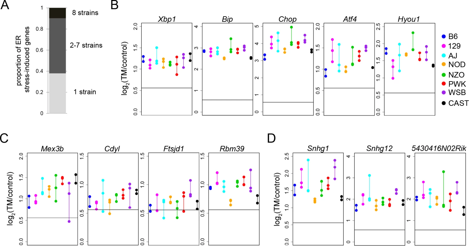
(A) Proportion of ER stress-induced genes that are shared among strains and genes that are uniquely upregulated in only one strain. Change in gene expression is displayed for common ER stress-induced genes with known ER stress functions (B), genes with functions that regulate gene expression (C), and noncoding RNAs (D). For each strain (different colors), all biological replicates are displayed as points connected by a vertical line (B-D). Horizontal line represents 1.5 fold change (B-D). TM: tunicamycin. Tab. 2. Transcription factor binding site analysis. 
Among the common ER stress-induced genes are some genes with no previously known function in the ER stress response. These putative ER stress genes fall into diverse categories. In some cases, genes not previously implicated in ER stress response have functions in processes that are important to the ER stress response. These include genes involved in Golgi trafficking (i.e. Rab39b), oxidoreductase activity (Oxnad1), and energy metabolism (Cpox). This co-occurrence of novel putative ER stress genes with canonical ER stress genes, across all eight strains, suggests that these response genes might be important in ER stress and warrant future study.
In addition to the enrichment of ER stress-related functions, common ER stress-induced genes are also enriched for functions involved in transcription (GO:0006350: transcription − q = 0.0038 and GO:0045449: regulation of transcription − q = 0.0319; Table 1). Forty-seven of the 214 common ER stress-induced genes are involved in the ‘regulation of transcription’ (GO:0045449). Some of these genes encode known ER stress transcription factors such as Xbp1 and Atf4. However, other transcription factors, like Arid5a, have no previously known role in ER stress. Even more striking, at least 15 of these transcription factors are unstudied zinc-finger proteins with no known function (e.g. Zfp191 and Zfp202). This enrichment for transcription factors with unknown function reinforces the idea that that the ER stress response involves a complex network of gene regulation, as condition-dependent elevation of transcription factor expression nearly always stimulates expression of downstream target genes as well [19].
A large component of the ER stress response involves changes in gene expression through different mechanisms including transcriptional regulation, RNA degradation, and sequestration of RNAs to stress granules [19]. In addition to the enrichment in transcription-related functions, we also identified genes that regulate gene expression through other mechanisms. Some of these genes function in chromatin remodeling (Cdyl), RNA binding (Mex3b), and RNA metabolism (Ftsjd1 and Rbm39) (Fig. 1C). Non-coding RNAs (ncRNAs) are contributors to another mechanism of regulating gene expression [20]. We also identified a number of ncRNAs that are upregulated across all eight strains by ER stress. These include the lincRNAs Snhg1 and Snhg12 and other ncRNAs like 2410006H16Rik, 5430416N02Rik, and 9430008C03Rik (Fig. 1D). Little is known of the exact role of ncRNAs in regulating the ER stress response. However, recent studies have suggested that ncRNAs might be important in fine-tuning the ER stress response. For example, the ncRNA Gadd7 regulates reactive oxygen species induced ER stress through a feed-forward loop [21] and certain miRNAs are now known to be regulated by ER stress [22, 23, 24]. For the most part, however, the mechanisms of these actions are unknown. The fact that these ncRNAs are all upregulated over two-fold in response to ER stress across eight diverse mouse strains, suggests that they may play an important role in ER stress response.
ER stress-induced expression is variable among mouse strains
The eight genetically diverse founder strains of the CC provide the opportunity to identify the genes that display variation in ER stress-induced expression. Due to the strong conservation of the ER stress response, studying these genes in the mouse may provide clues as to which portions of the response network can be subject to variation in the human population. In fact, most ER-stress induced genes are not upregulated in all eight strains. Of the 2,182 ER stress-induced genes, 829 (38%) are uniquely upregulated by ER stress in only one of the eight strains. 1139 genes (52%) showed shared up-regulation in two to seven strains (S1 Table; Fig. 1A).
To identify the genes with the most variable ER stress-induced expression among strains, gene expression results were tested using linear models to assess differences among the eight strains. 14.5% of ER stress-induced genes (317/2182 genes) show a significant strain effect on their induced expression (FDR 1%, S2 Table). Strikingly, the ten genes with the most significant strain effect all have very clear roles in inflammation (in order of statistical significance of strain effect: Cxcl2, Apobec1, Plk2, Agpat9, Ccl20, Pdgfb, Lif, Cxcl1, Mmp9, and Clec4e; Fig. 2A). GO analysis of the ER stress-induced genes with a strain effect further demonstrated a strong enrichment for inflammation functions. The three most significant GO categories are cytokine activity (GO:0005125: q = 1.49×10⁻⁶), chemokine activity (GO:0008009: q = 2.09×10⁻⁵), and chemokine receptor binding (GO:0042379: q = 1.73×10⁻⁵) (Table 1). We did not find enrichment of genes specifically involved in apoptosis signaling. Genes showing the most variable ER-stress-induced expression are also enriched for the inflammation related transcription factor binding sites, RelA and NF-kB1 (Table 2), but are not enriched for transcription factor binding sites related to ER stress signaling. The RelA and NF-kB1 transcription factors are members of the NF-κB family of transcription factors and dimerize in various combinations to control gene expression in response to stimuli such as inflammation [25]. In fact, we find that transcript levels of four members of the NF-κB family of transcription factors show significant variation among strains: Nfkb1 (q = 0.009; encoding NF-kB1), Nfkb2 (q = 0.003; encoding NF-kB2), Rela (q = 0.03; encoding RelA), and Relb (q = 0.002; encoding RelB) (S2 Table).
Fig. 2. ER stress-induced genes with a strain effect are enriched for inflammation functions. 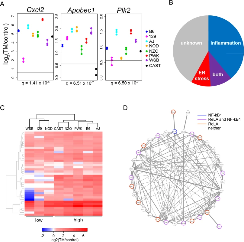
(A) The top three genes with the strongest strain effect. Horizontal line represents 1.5 fold. For each strain (different colors), all biological replicates are displayed as points connected by a vertical line. Horizontal line represents 1.5 fold; q = FDR correction of the strain effect. (B) Literature search identifies the proportion of genes with inflammation functions, ER stress functions, inflammation and ER stress functions (both), or unknown ER stress functions in the 100 genes with the strongest strain effect (FDR <0.01%). (C) Cluster analysis indicates that strains are either in a low (WSB, 129, and NOD) or high (CAST, NZO, PWK, B6, and A) inflammation group. Cluster analysis was performed with the 39 inflammation genes among the 100 genes with the strongest strain effect. For gene identities see S1 Fig. (D) Correlation network showing that inflammation genes are highly correlated in their response to ER stress. Edge thickness represents the correlation coefficient between the two nodes. Only significant pairwise correlations are displayed (Spearman’s; P < 0.05). Genes that contain ReLA and NF-kB1 transcription factor binding sites are color coded. All pairwise correlations can be found in S4 Table. TM: tunicamycin. A literature search indicates that 39 of the top 100 genes with the most significant strain effect (FDR <0.01%), have very clear roles in inflammation, 9 have known roles in ER stress response (i.e. Atf3 and Derl3), 10 have roles in both inflammation and ER stress, and 42 have no known function in either inflammation or ER stress (S3 Table; Fig. 2B). Cluster analysis with the 39 inflammation genes indicates that the eight strains fall into either a low (WSB, 129, and NOD) or high (CAST, NZO, PWK, B6, and A) inflammation response group (S1 Fig.; Fig. 2C). To understand how these inflammation genes might function within a network, we performed a correlation analysis. These 39 inflammation genes function in a highly correlated network. Half contain either a RelA or a NF-kB binding site, or both (S4 Table; Fig. 2D).
ER stress response and inflammation are interconnected in complex ways. ER stress can induce and can be a consequence of inflammatory responses. ER stress induces NF-kB signaling by various routes. Ire1 activation during ER stress results in the degradation of IkBa, an inhibitor of NF-kB signaling, resulting in increased NF-kB signaling [26]. PERK activation during ER stress results in global translation attenuation, which in turn results in decrease of IkBa production and increased NF-kB signaling [26]. Recent evidence suggests that Atf6 can also influence NF-kB signaling, but the exact role of Atf6 in this process is still unclear [27]. Because we do not find variation in the expression levels of key ER stress genes like Ire1, PERK or Atf6, it is likely that the variation we observe originates from genes encoding effectors of NF-kB signaling or NF-kB transcription factors themselves. The enrichment in NF-kB binding sites and the strong correlations among the inflammation genes further support this. More extensive work will be needed to identify which are the primary variable response genes and which are secondary. Nevertheless, inflammatory response to ER stress appears to be an important difference between strains and might be an attribute of stress response that is also variable in humans. The intersection between inflammation and ER stress might be a fruitful avenue for identifying important modifiers of human disease.
Inference of cis-regulatory variation in ER stress response
The fact that there is variation in ER stress-induced transcript levels among eight genetically diverse mouse strains suggests that there is genetic variation underlying these differences. In order to understand how genetic variation impacts the ER stress response, we need to know how genetic variation influences gene expression differences among strains under non-stressed conditions and how it changes once ER stress is applied. Transcriptional variation may arise from polymorphisms in one (or a few) transcription factors (trans-), or each response gene could harbor polymorphisms affecting its own expression (cis-).
The use of F1 crosses in this study was specifically designed to allow us to quantify the magnitude of cis-regulatory variation and to partition the inter-strain variance in expression into its cis - and trans - components [28, 29, 30, 31]. For genes that show an expression difference between two strains, identification of cis - and trans-regulatory differences requires comparing the relative allelic expression in the F1 to the relative expression of the parental strains [28, 29, 30, 31]. In the F1, the two parental alleles are exposed to the same trans - factors in the same levels and combinations. Thus, the ratio of allelic expression is a direct measure of cis-regulatory differences between parental strains—if the allelic ratio matches the ratio of the parental expression levels, the expression difference is attributed to cis-regulation. If the allelic ratio differs from the ratio of parental expression levels, then the difference is attributable to trans-regulation [29]. We focused on five F1 crosses. In all five crosses, B6 was the maternal strain and the paternal strain in each cross was one of five CC founder strains (129, NOD, NZO, WSB, and CAST; see materials and methods for F1 cross abbreviations). B6 was maintained as the maternal strain in all F1 crosses to avoid parent-of–origin and imprinting effects, but future studies with reciprocal crosses are warranted, as nothing is known about how parent-of-origin and imprinting influence ER stress responses. MEFs from each F1 cross were exposed to control and ER stress conditions, and transcript levels were measured by RNA-seq.
cis-/trans - analysis can only be performed for a gene that has sufficiently high expression and that harbors SNPs within the transcribed region that discriminate between parental alleles. In this study, we only considered informative genes with at least two such SNPs. The number of informative genes for analysis ranged from 2246 to 7,954 (xNOD: 2246, x129 : 2843, xNZO: 2910, xWSB: 3285, xCAST: 7954; S5 Table). For the rest of this analysis, unless otherwise noted, focus will mainly be on the xCAST F1 cross because this combination has the largest number of informative genes.
ER stress does not change the general pattern of regulatory divergence
The regulatory control of ER stress-induced expression is complex. The three arms of the UPR all induce major transcription factors (e.g. Xbp1, Atf6, and Atf4) and those transcription factors can activate other transcription factors [1, 19]. A polymorphism that affects the function of one of these would result in many trans-regulatory differences in their targets genes. On the other hand, some target genes contain ER stress responsive elements (ERSREs) that act as binding sites for these transcription factors [18, 32, 33, 34, 35]. A polymorphism in an ERSRE or other regulatory element could result in a cis-regulatory difference among strains for a particular target gene. To identify which of these possibilities contributes to variation in ER stress induced transcriptional variation, we measured the regulatory divergence under control and TM conditions and compared the results.
Overall, we found that TM has little effect on the proportion and magnitude of cis-regulatory differences among strains. Under both control and TM conditions, we find a similar number of regulatory differences (cis - and trans-) between B6 and CAST—244 genes under control conditions and 264 genes under TM conditions. In fact, under both conditions, 82% of genes with regulatory differences between B6 and CAST can be attributed to a cis-regulatory difference (control: 201/244 genes; TM: 216/264 genes; S7 Table; Fig. 3A). The magnitude of the median cis-regulatory differences, under control conditions, was significantly larger than the magnitude of the median trans-regulatory differences (control median magnitude: cis: 0.52, trans: 0.11; p < 3.746×10⁻⁸) and this pattern was unchanged by the application of TM (TM median magnitude—cis: 0.50, trans: 0.20; p < 0.03) (Fig. 3B). The median amount of divergence among strains explained by cis-regulatory differences (% cis) is also unaffected by TM conditions (C: 0.49, TM: 0.50; Fig. 3C). It appears that ER stress does not alter the overall proportion of cis-/trans - regulation between B6 and CAST. A portion of the regulatory variation is also attributable to a combination of cis - and trans - variation, but these are not further considered in this study (S6 Table and S7). We found similar results for the other four F1 crosses (S2 Fig.; S6 Table, S8–S11).
Fig. 3. ER stress does not alter the regulatory landscape between B6 and CAST. 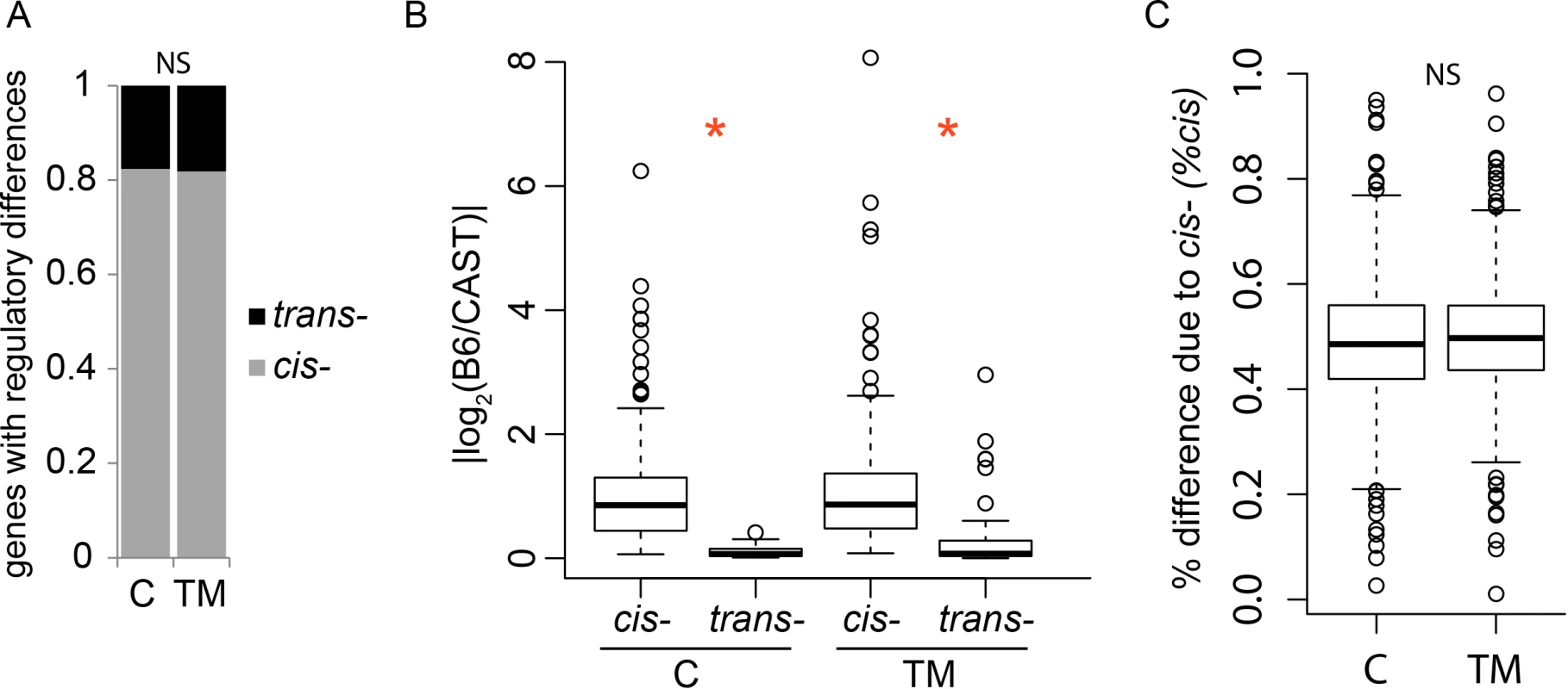
(A) Distribution of genes that display a cis- or trans-regulatory difference between B6 and CAST under control and TM conditions. There is no significant change in the proportion of cis-regulatory differences between conditions. (B) The magnitude of the regulatory difference between B6 and CAST. Under both conditions, the cis-regulatory differences are larger than trans-regulatory differences (*, control: p < 3.746×10⁻⁸; TM: p < 0.03). (C) The amount of divergence between B6 and CAST attributable to cis-differences is not affected by TM. NS: not significant. C: control. TM: tunicamycin. For boxplots in (B) and (C) the boxes represent the interquartile range, the whiskers represent 1.5X interquartile range, and open circles are outliers. This is the first study in genetically-diverse inbred mouse strains to examine the regulatory variation under ER stress. We find that most of the regulatory variation under control conditions is attributable to cis-regulatory variation parallels results from previous studies of regulatory variation in the CC [14] and other crosses [30]. Because of the hierarchical nature of the transcriptional response to ER stress, one might predict that ER stress regulatory variation would arise from a small number of trans-acting polymorphisms in the major transcription factors. Instead, just as we observed under control conditions, we find that cis-regulatory variation dominates under TM conditions. The fact that so few regulatory differences under TM conditions appear to arise from trans-acting polymorphisms, indicates polymorphisms in major transcription factors do not influence ER stress-induced transcriptional differences. This is supported by our observation that canonical ER stress transcription factors do not differ in their expression among strains (see above). Instead, genetic polymorphisms in promoters, enhancers, or other elements in individual target genes drive ER stress-induced transcriptional variation.
ER stress uncovers unique regulatory variation among strains
We next examined whether there was overlap in the genes that showed regulatory differences between B6 and CAST, under both control and TM conditions. We find that there are a number of genes that show the same regulatory difference between B6 and CAST under both control and TM conditions. Of all the genes that show a cis-regulatory difference between B6 and CAST (control: 201 and TM: 216, see above), 136 genes show a cis-regulatory difference under both conditions (control: 67% or 136/201; TM: 63% or 136/216; S7 Table; Fig. 4A). Of the genes that show a trans-regulatory difference between B6 and CAST (control: 43 and TM: 48, see above), 18 genes show a trans-regulatory difference under both conditions (control: 42% or 18/43; TM: 38% or 18/48; S7 Table; Fig. 4A). The magnitudes of the cis - or trans-regulatory differences found under both conditions are highly correlated between the two conditions (r2 = 0.9; Fig. 4B). Together this indicates that this set of cis - and trans-regulatory differences that are found under both conditions represent a group of genes whose regulatory differences between B6 and CAST are not influenced by ER stress.
Fig. 4. ER stress alters the genes that display regulatory differences between B6 and CAST. 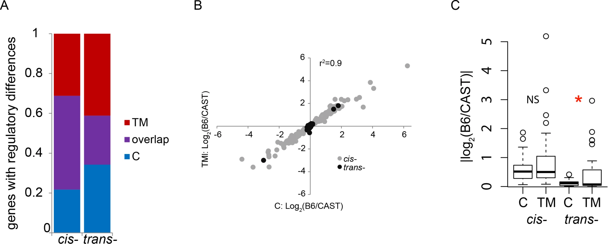
(A) Distribution of genes showing cis- or trans-regulatory differences found only under TM conditions, found only under control conditions, or found in both conditions. (B) Genes that show the same cis- or trans-regulatory difference under both conditions (‘purple’ genes in A) are strongly correlated in their magnitude. ER stress does not affect regulation of these genes. (C) Magnitude of the cis- or trans-regulatory differences unique to control or TM conditions. Magnitude of cis-regulatory differences unique to either condition is unaffected by ER stress. trans-regulatory differences unique to TM conditions show a small but significantly larger magnitude than trans-regulatory differences unique to control conditions (*, p < 0.05). NS: not significant; C: control; TM: tunicamycin. It is likely that many genes harbor genetic variation affecting ER stress response, but the functional consequences of this variation are undetectable during healthy, non-stressed states. Strain differences in ER stress response might arise from the genes that show ER stress-specific regulatory variation. In particular, genes that show regulatory variation under TM conditions, and not control conditions, might reveal which ER stress pathways are most critical to strain differences in this response. Of the genes that show regulatory variation, 23% (65/281 genes) with a cis-regulatory difference and 34% (25/73 genes) with a trans-regulatory difference are unique to the control condition (S7 Table; Fig. 4A). These regulatory differences were only detected under control conditions and were not present under ER stress conditions. 28% (80/281 genes) with cis-regulatory differences and 41% (30/73 genes) with trans-regulatory differences are unique to TM conditions (S7 Table; Fig. 4A). These regulatory differences are only detectable when ER stress is induced, and absent under control conditions. Genes that show a cis-regulatory difference unique to either control or TM conditions do not differ in their median magnitude of expression (C: 0.52, TM: 0.50; Fig. 4C). However, there is a small but significant decrease in the median magnitude of the genes with trans-regulatory differences under TM compared to control conditions (C: 0.11, TM: 0.07; p = 0.021; Fig. 4C). The majority of genes that show regulatory differences between the two strains depend on the presence or absence of ER stress.
Genes that show evidence of differential regulation among strains may point to pathways that contribute to strain differences in ER stress response. In some cases, genes with known roles in ER stress show differential regulation among strains, and these might be useful in identifying how strains differ in canonical ER stress signaling. For example, Gadd45a shows a strong cis-regulatory difference between B6 and CAST only under ER stress conditions and not under control conditions—the B6 allele expresses 2.4 fold higher than the CAST allele. This regulatory difference is only detectable under TM conditions because Gadd45a shows very low expression in control conditions, but is strongly induced by ER stress. There are seven SNPs within the Gadd45a gene that differ between B6 and CAST, one or a combination of these polymorphisms may be responsible for this cis-regulatory difference. Gadd45a is induced in an ATF4-dependent manner and modulates apoptosis signaling [36]. Under stress signaling, PERK phosphorylates eIF2α which in turn results in the selective translation of ATF4 and induction of Gadd45a transcription [36]. Understanding how Gadd45a is differentially regulated will lend insight into how different genetic backgrounds might utilize Gadd45a function and the broader ATF4 signaling pathways to influence ER stress response. On the other hand, genes that show differential regulation and have no known function in ER stress might reveal novel ways in which different mouse strains might respond to ER stress. One such gene, Cactin also shows a cis-regulatory difference between B6 and CAST only under ER stress. The B6 Cactin allele expresses ~2.2 fold higher than CAST under ER stress, while under control conditions, the alleles are equally expressed. Cactin is associated with the spliceosome in Arabidopsis [37], but it is unknown if Cactin forms part of the mammalian spliceosome. Understanding how Cactin is differentially regulated among strains could lend insight into strain differences in splicing under ER stress conditions. In general, splicing in response to ER stress is poorly understood and provides another layer of complexity among mouse strains. Genes that show ER stress specific regulatory differences among strains may identify pathways that can be nominated as potential targets of variation in human studies.
Shared regulatory differences
The strains utilized in the CC are genetically diverse and were chosen to maximize genetic differences among strains. Because the F1 crosses that we examined all have B6 as a common parental strain, we have the opportunity to compare how each of the five strains differs in their transcriptional regulation from B6. This analysis might reveal how the genetic architecture underlying ER stress response variation differs among strains. We hypothesized that if two (or more) F1 crosses (which share B6 as the maternal parental strain) showed evidence of the same regulatory difference in a particular gene, then the two (or more) non-B6 parental strains might differ from B6 in the same way, especially if the magnitude of the regulatory difference is equal. If the magnitude of the regulatory variation is different (a strain effect), it might indicate that different polymorphisms are driving the cis-regulatory differences among the F1 crosses.
We compared the genes with cis - or trans-regulatory variation in each of the five F1 crosses. Because this analysis can only be performed on informative genes of each F1 cross, we only considered genes with regulatory differences that are informative in at least two F1 crosses. Among the five F1 crosses, we find 265 genes and 322 genes with cis-regulatory differences under control and TM conditions, respectively. Under both conditions, the minority of cis-regulatory differences are shared among F1 crosses (S12 Table). Under control conditions, 25% (66/265) of genes with cis-regulatory differences are shared among two or more F1 combinations. Under TM conditions, 14.5% (47/322) of genes with cis-regulatory differences are shared among F1 combinations (Fig. 5A). TM treatment decreases the proportion of cis-regulatory differences shared among F1 combinations (From 25% to 14.5%; (χ2: p < 4×10⁻⁷; Fig. 5A). Even when cis-regulatory sharing is subdivided by number of informative strains (2, 3, 4, or 5 informative strains), TM treatment still significantly reduces the number of cis-regulatory differences shared among F1 combinations (χ2: p < 0.05; Fig. 5B). We find a similar pattern for trans-regulatory differences, where TM treatment reduces sharing between F1 combinations (S3 Fig.; S13 Table). The reduction by ER stress in the amount of shared regulatory differences among strains suggests that each strain differs from B6 in unique ways in its ER stress-induced transcriptional regulation.
Fig. 5. ER stress reduces the amount of shared cis-regulatory differences. 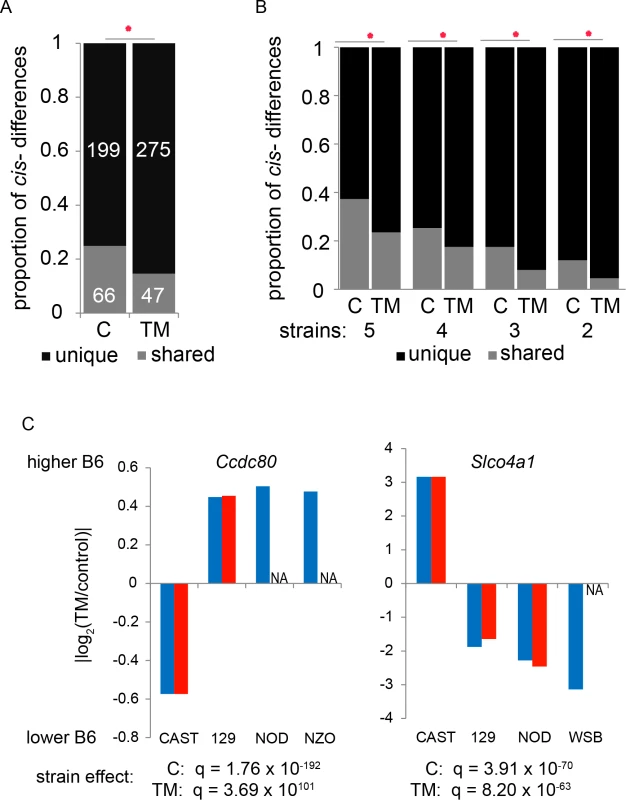
(A) The proportion of cis-regulatory differences that are unique to a particular F1 combination or shared among F1 combinations. The proportion of shared cis-regulatory differences is significantly reduced by ER stress (*, χ2: p < 4.27×10⁻⁹). (B) Broken down by genes that are informative in different numbers of strains, the proportion of genes that show cis-regulatory differences that are unique or shared is still significantly reduced by ER stress (*, χ2: p < 0.05). (C) Examples of genes that have shared cis-regulatory differences. These two examples also display a significant strain effect in the magnitude of the cis-regulatory difference under control and TM conditions. Under ER stress, Ccdc80 and Slco4a1 both show reduction in the number of strains that share a cis-regulatory difference. NA: no detectable cis-regulatory difference. The data above indicate that some genes show a signature of shared cis-regulatory variation among strains. However, the magnitude of most of this shared cis-regulatory variation differs among the F1 crosses (a strain effect). Forty genes (40/66; 61%; χ2, FDR 5%) under control conditions and 32 genes (32/47; 68%, χ2, FDR 5%) under ER stress conditions with shared cis-regulatory differences show a strain effect on the magnitude of the regulatory difference (S12 Table; examples in Fig. 5C). TM treatment significantly increases the proportion of shared cis-regulatory differences that show a strain effect (from 61% to 68%; χ2; p = 0.01). trans-regulatory differences also show more strain effects under TM treatment (S13 Table). Because most shared-regulatory variation differs in magnitude and that this strain effect increases with ER stress further supports the idea that induction of ER stress uncovers more genetic variation among strains.
Understanding the role of the genes that show strain differences in their cis-regulation may help to identity how strains respond to ER stress. Ccdc80 is one example that shows both an ER stress-induced reduction in cis-regulatory sharing and a strain effect on the magnitude of those shared cis-regulatory differences. As mentioned above, under control conditions, Ccdc80 shows cis-regulatory differences in four F1 crosses: xCAST, x129, xNOD, and xNZO. The magnitudes of these four cis-regulatory differences are significantly heterogeneous among strains (χ2, q < 10-12; Fig. 5C). The B6 allele in the xCAST cross is expressed at lower levels than the CAST allele, but the B6 allele is the more highly expressed allele in the other three F1 crosses. Under TM conditions Ccdc80 only shows cis-regulatory differences in the xCAST and x129 F1 combinations and again, there is a strong effect of strain on the shared cis-regulatory differences (χ2, q < 10-12; Fig. 5C). In the case of Ccdc80, ER stress eliminates the cis-regulatory variation between B6 and NOD and between B6 and NZO. Ccdc80 encodes a coiled-coiled domain protein which is localized to the Golgi and has been shown to relocate to the ER under certain conditions [38]. There are >100 SNPs that differ between B6 and the five strains within the Ccdc80 gene any of these or a combination could contribute to these differences. Understanding how this type of genetic diversity contributes to ER stress response will begin to elucidate the ER stress network in each strain.
ER stress-induced allele-specific expression
Because TM has a large influence on the cis-regulatory variation between B6 and the other strains, we evaluated the amount of allele-specific expression (ASE) that is influenced by TM. Because we are interested in the regulatory differences under TM conditions, we focused on those genes that showed a significant change in ASE upon TM treatment. That is, when ER stress is induced, there was a significant change in allelic ratio of expressed RNAs in the F1 cells between control and TM conditions. In all five F1 crosses, less than four percent of informative genes show a relative change in ASE upon TM treatment (S14 Table; Table 3). Often TM treatment resulted in increased expression of one allele and decreased expression of the other allele, without changing the overall expression of the gene. Less than 12% of genes in any of the F1 crosses show both a TM-induced ASE change and show TM upregulated transcript levels (Table 3).
Tab. 3. ER stress-induced allele-specific expression. 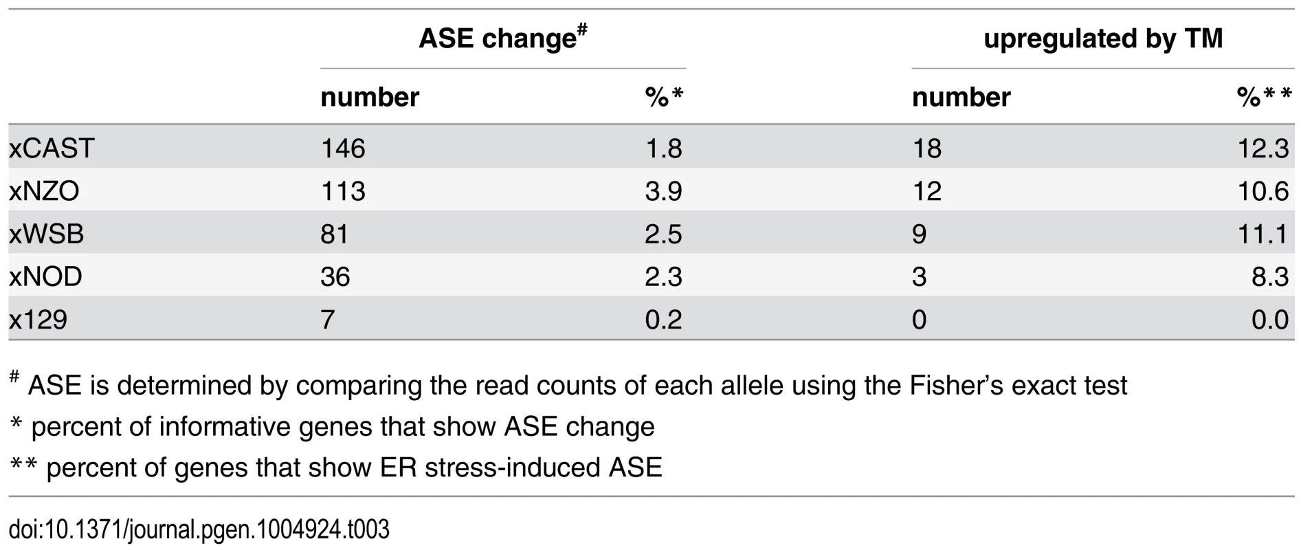
# ASE is determined by comparing the read counts of each allele using the Fisher’s exact test In the xCAST F1 cross, a total of 146 informative genes (1.8%) showed a change in ASE induced by TM. Sixty one genes (42%) under TM conditions show a decrease in the proportion of the B6 allele while 85 genes (58%) showed an increase in the proportion of the B6 allele (Fig. 6A). Genes that showed a change in ASE and also show increased expression under TM are of particular interest because this indicates that the two alleles in the F1 are differentially affected by TM. Of the genes that show a change in ASE, 18/146 genes (12%) also show increased transcript levels under TM conditions (Table 3). Nine of the genes have known functions in ER stress responses, including Bip (Hspa5), Grp75 (Hspa9), Trib3, and Sesn2. Sesn2, which is involved in a variety of stress responses [39], showed the most significant change in ASE (Fisher’s exact; q < 4.30 × 10-55), where the B6 allele responds to a lower extent than the CAST allele. Under control conditions, the Sesn2 B6 allele is expressed at 40%, but under TM conditions, B6 allelic expression is reduced to 22% (Fig. 6B). Total Sesn2 transcript responds to TM conditions with a 3.3 fold increase. However, the B6 allele shows only a 1.7 fold increase while the CAST allele shows a 4.3 fold increase. This difference in allelic response to TM drives the change in ASE. Snhg5 encodes a noncoding RNA and showed the second most significant change in ASE (Fisher’s exact; q < 5.63 × 10⁻¹⁷), where the CAST allelic response is stronger than that of B6. In this case, under control conditions, the B6 allele is expressed at 38%, but B6 allelic expression increases to 56% under TM conditions (Fig. 6B). While total Snhg5 transcript increases by 3 fold, the B6 allele increases by 4.4 fold, but the CAST allele increases by only 2.1 fold. Again, the change in ASE is driven by a strong difference in the allelic response to TM.
Fig. 6. ER stress-induced allele-specific expression (ASE). 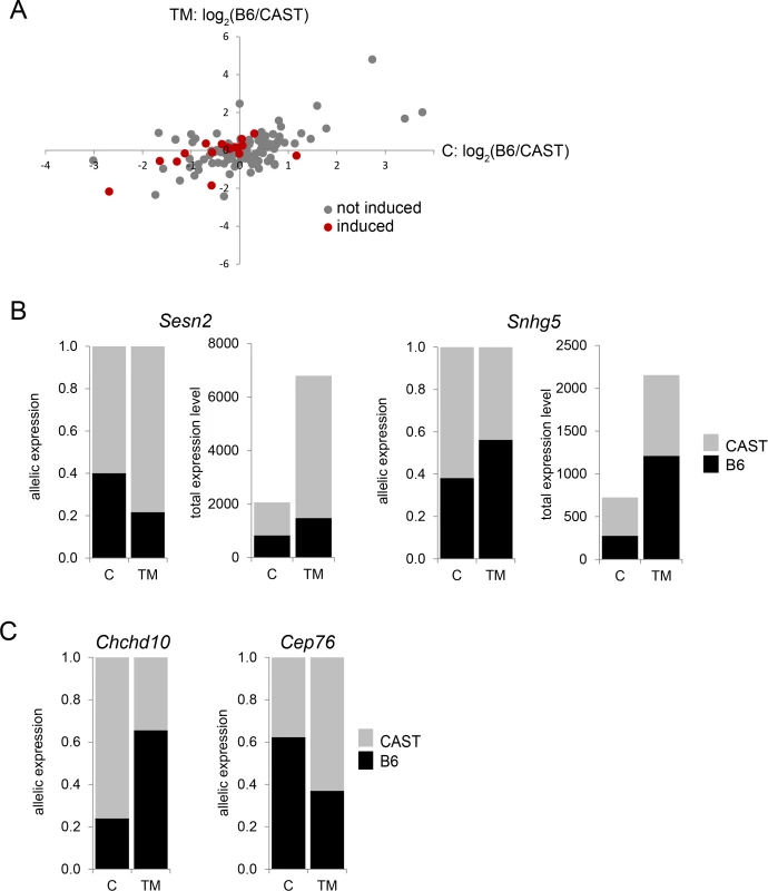
(A) Genes that show ER stress-induced change in ASE, under control and TM conditions. Genes in red are genes that also show ER stress-induced increase in transcript levels. (B) Sesn2 (Fisher’s exact; q < 4.30 × 10-55) and Snhg5 (Fisher’s exact; q < 5.63 × 10⁻¹⁷) are examples of genes that show ER stress-induced change in ASE and ER stress-induced increase in transcript levels. (C) Chchd10 (Fisher’s exact; q < 9.0 × 10⁻⁴) and Cep76 (Fisher’s exact; q < 6.23 × 10⁻⁵) are examples of genes that show ER stress-induced change in ASE, but whose transcript is not upregulated by ER stress. The majority of genes (128 genes or 88%) that show a TM-induced ASE show no TM-induced change in total transcript level. Thirteen of the 20 genes with the most significant change in ASE do not respond to TM in total transcript abundance. For example, while total transcript level does not change, Chchd10 shows a very large shift in ASE induced by TM (Fisher’s exact; q < 9.0 × 10⁻⁴; Fig. 6C). The Chchd10 B6 allele is expressed at 24% under control conditions, but under TM conditions, the B6 allele is expressed at 66%. Similarly, Cep76 shows a B6 allelic shift from 62% to 37% induced by TM (Fisher’s exact; q < 6.23 × 10⁻⁵; Fig. 6C). A change in ASE without a change in total transcript number could represent a difference in auto-regulation between the two alleles.
Conclusions
Understanding how genetic variation impacts ER stress transcriptional response will result in a richer understanding of this basic cellular response and how its variation might influence disease severity. Because the ER stress response is under intense investigation at the molecular level, studies of its variation can be anchored within the well-studied, canonical steps and genes of this essential cellular response. Identifying how the ER stress response pathway is modulated by genetic variation serves two important purposes: 1) Identifying which canonical ER stress genes can or cannot vary in their response to ER stress and 2) Identifying novel ER stress genes that may be missed by studying only a single genetic background. By studying ER stress response in light of genetic variation, we learn how the response is buffered in different individuals and genetic backgrounds, eventually leading to potential personalized therapies that can be targeted to the specific response profile of an individual. Genetic resources in model organisms allow for the dissection of the genetic architecture behind the variable response to ER stress. Here, we took advantage of emerging resources from the mouse Collaborative Cross to study the genetic architecture of the ER stress response. This important new resource allowed us to identify variable elements of the ER stress response pathway and to uncover the genetic architecture underlying such variation.
We used TM to induce ER stress in the MEFs. While TM is commonly used to experimentally induce ER stress in vivo and in vitro, other drugs like thapsigargin and dithiothreitol are also used. These drugs have very different mechanisms of action, all resulting in ER stress as an endpoint. It is likely that using these other drugs would produce slightly different results. The results we report are specific to the concentration of TM and exposure time that we describe. These experimental conditions are in common use and the effects of TM are recognized as an acceptable experimental model of physiological ER stress. Future experiments might be warranted where the concentration and timing of exposure is altered. While these caveats limit our study to some extent, we believe, given the experimental design, the principles can be broadly applied to ER stress in general.
Our results reveal that the genetic variation underlying ER stress transcript-level responses in the mouse does not involve the major known regulators of the ER stress response. We found that on the whole, most canonical ER stress genes are not variable in TM-induced expression levels among mouse strains. Instead the strains differ most in their inflammatory response to ER stress. The finding that major regulators at the top of the signaling pathway are not variable indicates that variation arises from downstream signaling elements. If major ER stress transcription factors like Xbp1, Atf4, Atf6, or CHOP, harbored polymorphisms that affected their function, then we would expect extensive ER stress-induced trans-regulatory differences among strains. However, we observed the exact opposite. We found that cis-regulatory variation dominated the ER stress regulatory landscape, indicating that response genes themselves harbor polymorphisms that affect their response. The fact that major elements of this pathway are not variable across these “healthy” mouse strains makes sense. Previous studies show that even mice that carry heterozygous null mutations in various major ER stress genes display disease phenotypes [40]. The major ER stress response genes may not tolerate damaging polymorphisms that affect function and thus would not be a target for variation.
It is becoming increasingly clear that the effect of a particular regulatory polymorphism is context dependent [41, 42, 43]. These contexts can include, but are not limited to, environmental insults and tissue and cell types. There is likely ‘hidden’ variation that is only revealed under certain contexts. Our study demonstrates the importance of studying the role of genetic variation in different cellular contexts, like ER stress. In this study we show that induction of ER stress by TM uncovers an entire layer of cis - and trans-regulatory variation among mouse strains that is not apparent under healthy, steady state conditions. ER stress also eliminates the effect of certain regulatory variation found under healthy conditions. We also found that, for a subset of genes, the two alleles of a particular gene can display differential ER stress-induced transcriptional regulation. Regulatory variation may impact gene expression in many context-specific ways. A particular variant may affect a transcription factor binding site or might alter chromatin accessibility in a way that only affects the gene expression changes induced by ER stress. Several well defined transcription factor binding sites bind canonical ER stress transcription factors under stress conditions [18, 32, 33, 34, 35]. Because most of the regulatory variation we identified is cis-, these ER stress transcription factor binding sites might be where genetic variation impacts ER stress-induced transcription. SNPs or larger variants, like Copy Number Variants (CNVs), might be driving these differences we observe between strains. These polymorphisms might affect expression through various mechanisms, including epigenetic changes. We only utilized the founder strains and their F1 progeny and thus, we cannot identify the specific polymorphisms underlying these regulatory differences in this study. However, the CC recombinant inbred strains and other resources like the Diversity Outbred mice [44] are well suited for future studies aimed at mapping, identifying, and functionally studying the polymorphisms in these or other regulatory sites that might alter ER stress responses. It should also be noted that the focus of this study was on ER stress-induced gene expression variation and does not address possible differences in protein levels. Future studies aimed at identifying specific causative polymorphisms should also integrate protein abundance to understand the role of regulatory transcriptional variation on the proteome.
ER stress has been implicated in numerous diseases, including both Mendelian diseases and complex, polygenic disorders [2]. Studying genetic variation in ER stress transcriptional response in the mouse provides the opportunity to identify links between ER stress and disease that can be easily tested in the future. To identify ER stress genes that may contribute to disease, we searched for human orthologs of genes involved in Mendelian disease (OMIM) and genes involved in complex diseases (GWAS catalog). We found that 42% (89/214) of the common ER stress-responsive genes (upregulated in all eight strains) have been implicated in either Mendelian or complex diseases. Some examples include neurological diseases (gene: Sil1, disease: Marinesco-Sjogren Syndrome; Zfp238, mental retardation), diabetes (Zfp57, neonatal diabetes mellitus), and cancers (Ddit3/CHOP, myxoid liposarcoma). 39% (125/317) of ER stress-induced genes that show differences among strains have been implicated in disease. Given the enrichment of inflammation-related functions, it is not surprising that many of the diseases associated with genes that show strain differences involve inflammation (i.e. Arhgef3, rheumatoid arthritis; Ifnar1, Crohn’s disease). Other examples of diseases associated with genes with strain differences include anemia (Slc25a38, pyridoxine-refractory sideroblastic anemia) and neurological diseases (Pdgfb, basal ganglia calcification; Bean1, spinocerebellar ataxia 31). Understanding how these disease genes function within the context of genetic variability and ER stress will likely reveal new insight into the stress response and reveal possible roles for ER stress genes in disease pathogenesis.
This study highlights the importance of using genetic variation to study basic cellular traits like ER stress. The resources from the CC allowed us to identify novel and variably responsive ER stress genes and to uncover the complex genetic architecture that drives inter-individual transcriptional variation under ER stress. Because the ER stress pathway is well conserved, this study complements previous studies of variation in humans [7, 8] and Drosophila [9]. For example, the ER stress-induced expression of the mouse gene Dhrs11 in this study and the orthologous Drosophila gene, CG10962, in a previous study [9], are both strongly influenced by genetic background. Furthermore, in Drosophila, a SNP in CG10962 is strongly associated with survival under ER stress conditions [9]. This study lays the groundwork for future studies that will be aimed at identifying the genetic polymorphisms underlying the observed variation in ER stress transcriptional response and the functional consequences of such variation. The CC RI lines are perfectly suited for this type of study [14, 45, 46, 47] and will likely prove to be an excellent resource for understanding the genetic basis for variation in basic cellular traits. Understanding how genetic variation affects the ER stress transcriptional response will increase the understanding of this conserved pathway and may provide mechanistic links to disease and disease susceptibility.
Materials and Methods
Mouse embryonic fibroblasts
Mouse embryonic fibroblasts (MEFs) were generated from founder and F1 strains used in the Collaborative Cross (MEFs were kindly provided by Dr. David Threadgill, Texas A&M University). To eliminate individual, stochastic variation, all MEFs were generated from multiple female individuals from the same litter. Thus, each is a mix of genetically identical cells derived from multiple individual mice. F1 MEFs were derived from F1 crosses where the B6 is the maternal parent. Founder and F1 strain identities are abbreviated throughout the text (see Table 4).
Tab. 4. Abbreviations for founder and F1 strain identities. 
For technical reasons, we excluded the C57BL/6J X A/J and C57BL/6J X PWK F1 crosses. Excluding these two crosses does not alter the balance of this study. All MEFs were thawed at passage three or four and cells were split 1 : 3 every 3 days (fewer than three passages) before treatment. MEFs were maintained in DMEM containing 4.5 g/L glucose (Invitrogen), 10% fetal bovine serum, L-glutamine, and pen/strep (Invitrogen).
Tunicamycin treatment and RNA isolation
Tunicamycin (TM) treatment of MEFs was performed using a common approach [48, 49, 50, 51, 52]. These conditions are typical of studies of ER stress in MEFs and are sufficient to elicit a strong ER stress response, without inducing apoptosis. Briefly, MEFs were plated at a concentration of 2 × 10⁵ cells/well in six-well plates. Cells were plated at least 18 h before treatment. TM stocks were prepared at 2 mg/ml in DMSO. To induce ER stress, MEFs were treated with a final concentration of 2 ug/ml of TM. Control cells were treated with 0.1% DMSO. MEFs were treated for 4 h. All genotypes and conditions were treated in parallel and in triplicate. At the end of the treatment, medium was aspirated out and Trizol (Invitrogen) was added for immediate isolation of total RNA by standard procedures.
Illumina mRNA sequencing and mRNA-seq alignment
mRNA sequencing was performed on total RNA from 78 MEF samples (13 genotypes X 2 treatments X 3 replicates). Single-end 100 bp mRNA-seq libraries were made with 1.5 ug of total RNA using the Illumina TruSeq RNA Sample Preparation kit (Illumina Inc., CA), per manufacturer’s guidelines. The 78 samples were multiplexed and sequenced on a total of eight lanes using the Illumina HiSeq 2500 instrument. Image analysis and base calling were performed with the provided Illumina software. RNA-seq reads were aligned to the strain-specific reference mouse genome assembly using TopHat v1.4.1 [53] with three mismatches allowed. Total expression level for each transcript, measured in FPKM (Fragments Per Kilobase-pair of exon Model), was calculated based on all mapped reads [54].
Quantification of total expression and change in expression
For both founder and F1 strains, read counts were normalized across all samples using the default normalization method (TMM) in the edgeR package in R [55, 56, 57]. For each strain and condition, principal components analysis was used to identify outlying samples. Within a strain, we required that control samples be clustered together and TM treated samples be clustered together. If a sample was not clustered with the appropriate condition, it was removed from analysis. After removal of outlying samples, there remained at least two replicates for each strain and condition combination. Remaining samples were re-normalized. TM-induced gene expression changes were assessed by comparing control vs TM treated samples using linear models with the edgeR package in R [55].
Strain effect on expression in founder strains
For each strain, a single median expression value was calculated for each gene in the control samples. For each TM replicate, TM-induced change in expression was calculated as the log2 fold change of the TM expression value and the median control expression value (log2(TM/control)), resulting in two or three values per strain per gene. To test for a strain effect in TM-induced expression, analysis of variance (ANOVA) was used to apply a simple linear model to fold change and strain, similar to the analysis previously described [58, 59]. The TM-induce expression change (yi) for the ith observation of the ith strain was:
Benjamini-Hochberg correction was applied to P values to identify the set of tests with a 1% False Discovery Rate (FDR). Cluster analysis was performed with gplots in R (V.2.11.0; http://CRAN.R-project.org/package=gplots).Allele calls in F1 and quantification of ASE
Quality assessment of the Illumina RNA-seq reads was performed using FastQC software (http://www.bioinformatics.babraham.ac.uk/projects/fastqc/) for all samples in the five F1 crosses. Positions with low Q-score at the end of the reads were trimmed by FASTX-Toolkit (http://hannonlab.cshl.edu/fastx_toolkit/index.html). Adapter sequences were trimmed by Trimmomatic software [60]. As recommended by Munger et al., 2014 [47], the cleaned reads were mapped to both paternal (CAST, 129S1, NOD, NZO and WSB) and maternal genomes (B6) using TopHat v1.4.1 [53, 54] allowing three mismatches. Local realignment over indel positions was performed with GATK software [61], and only uniquely mapped reads were included in the final BAM files that were submitted for SNP calling and allele count summary. Based on the SNP and genome information from the Sanger Mouse Genomes Project (http://www.sanger.ac.uk/resources/mouse/genomes/), parental allele counts were summarized using SAMtools [53, 54] at SNP positions with coverage of four or more. Problematic SNP positions, such as those that did not display 100% monoalleic expression in parental crosses, exhibited a third allele, were in repetitive regions, were near an indel position, or occurred at exon-intron junctions, were filtered out using custom scripts [62] resulting in 12,4587, 32,632, 27,686, 35,397 and 33,775 high-quality covered SNPs for xCAST, x129S1, xNOD, xNZO and xWSB crosses respectively. From the autosomal, X-linked and mitochondrial SNPs, all the F1 RNA-seq data matched the expected genotype identities.
The SNPs were annotated using SnpEff [63] on Ensembl v64 (www.ensembl.org). Only exonic SNPs based on Ensembl gene models were used to generate per-transcript allele counts summarized over multiple SNPs in the same transcript. The averaged counts from paternal and maternal genome alignments were used. To quantify allele-specific expression, we calculated the ratio of the number of reference allele-containing reads divided by the total coverage within a transcript for autosomal genes covered with informative SNPs in each cross [64]. To reduce the inflated inter-replicate variability of allelic expression ratio [62] and increase the SNP coverage, we combined all replicates and restricted our analysis only to genes with exonic SNP coverage >20 for cis-/trans - analysis and >50 for ASE analysis. We were able to estimate allele-specific expression ratios for ~9100 genes in xCAST cross, ~4700 genes in xWSB cross, and ~3500 genes in each of the other three F1 crosses. To detect significant changes of allelic expression between control and TM treated samples, we applied Fisher’s Exact Test on the reference and alternative allele counts in control and treated samples, followed by FDR correction. The slight over-dispersion of the data could inflate the false positive rate of the Fisher Exact test, but the rank-order of significance is preserved and because we are comparing the same genes in control and treated samples we believe that the reported false discovery rates are reasonable.
Inference of cis - and trans-regulation
For purposes of biological inference, we binned the genes into categories of cis-, trans-, or a combination of cis - and trans - regulation following a method previously described in McManus et al. 2010 [29]. A more detailed quantitative analysis that partitions the variance in regulation will be applied to an expanded data set in the future. cis - and trans-regulation among strains was inferred using a hierarchical statistical analysis in R. We applied a very conservative significance threshold at every level of this analysis (FDR = 0.1%). For each strain, control and TM conditions were analyzed separately. The analysis proceeded as follows:
-
1)
Differential expression in parental (P) and F1 data sets was analyzed using the binomial exact test followed by FDR correction.
-
2)
For all genes where there was differential expression between P strains or between parental alleles in the F1, Fisher’s exact test was used to compare the strain-specific expression ratios in the P and F1 samples, followed by FDR correction. A significant difference in the ratios of P vs F1 samples is considered a trans-regulatory difference.
As mentioned above, the Fisher’s exact test assumes independent sampling of reads, but the nature of RNA-seq experiments often inflates the error, potentially leading to false positives [62]. We avoid this problem by taking every effort to generate libraries of high complexity, which we verified using the replicates to directly test overdispersion. Only genes showing cis - or trans - regulation are discussed in the text, but genes in other categories are presented in the supplement (S7 Table–S11). Based on the results of the analysis in (1) and (2), genes were partitioned into the following categories: cis-, trans-, cis - + trans-, cis - x trans-, compensatory, conserved, and ambiguous (See Table 5).
Tab. 5. Inference of cis- and trans- regulation. 
* See methods: Differential expression in parental strains (P) or alleles (F1) based on analysis from (1). Identification of shared regulatory difference
Only genes that are informative between two or more F1 strains were considered. A gene was considered to have a shared regulatory difference if it displayed either a cis - or trans-regulatory difference between two or more F1 crosses. The ratio of the allelic expression (magnitude) in the F1 crosses was tested for a strain effect by chi-squared (χ2) analysis.
Bioinformatics functional analysis
All Gene Ontology (GO) analysis was performed using DAVID [65, 66]. Transcription factor binding sites were identified using mouse single site analysis (version 2) in oPOSSUM [67]. Searches were limited to 2000 base pairs up - and downstream of the transcription start site. Genes involved in human disease were identified by using Online Mendelian Inheritance in Man (OMIM; www.omim.org) and the GWAS Catalog (https://www.genome.gov/26525384). Literature searches were performed in PubMed (http://www.ncbi.nlm.nih.gov/pubmed). Visualization of regulatory networks was performed in Cytoscape [68].
Supporting Information
Zdroje
1. Bernales S, Papa FR, Walter P (2006) Intracellular signaling by the unfolded protein response. Annu Rev Cell Dev Biol 22 : 487–508. doi: 10.1146/annurev.cellbio.21.122303.120200 16822172
2. Wang S, Kaufman RJ (2012) The impact of the unfolded protein response on human disease. J Cell Biol 197 : 857–867. doi: 10.1083/jcb.201110131 22733998
3. Oyadomari S, Koizumi A, Takeda K, Gotoh T, Akira S, et al. (2002) Targeted disruption of the Chop gene delays endoplasmic reticulum stress-mediated diabetes. J Clin Invest 109 : 525–532. doi: 10.1172/JCI14550 11854325
4. Song B, Scheuner D, Ron D, Pennathur S, Kaufman RJ (2008) Chop deletion reduces oxidative stress, improves beta cell function, and promotes cell survival in multiple mouse models of diabetes. J Clin Invest 118 : 3378–3389. doi: 10.1172/JCI34587 18776938
5. Wang L, Popko B, Roos RP (2014) An enhanced integrated stress response ameliorates mutant SOD1-induced ALS. Hum Mol Genet 23 : 2629–2638. doi: 10.1093/hmg/ddt658 24368417
6. Wang L, Popko B, Roos RP (2011) The unfolded protein response in familial amyotrophic lateral sclerosis. Hum Mol Genet 20 : 1008–1015. doi: 10.1093/hmg/ddq546 21159797
7. Dombroski BA, Nayak RR, Ewens KG, Ankener W, Cheung VG, et al. (2010) Gene expression and genetic variation in response to endoplasmic reticulum stress in human cells. Am J Hum Genet 86 : 719–729. doi: 10.1016/j.ajhg.2010.03.017 20398888
8. Nayak RR, Bernal WE, Lee JW, Kearns MJ, Cheung VG (2014) Stress-induced changes in gene interactions in human cells. Nucleic Acids Res 42 : 1757–1771. doi: 10.1093/nar/gkt999 24170811
9. Chow CY, Wolfner MF, Clark AG (2013) Using natural variation in Drosophila to discover previously unknown endoplasmic reticulum stress genes. Proc Natl Acad Sci U S A 110 : 9013–9018. doi: 10.1073/pnas.1307125110 23667151
10. Threadgill DW, Miller DR, Churchill GA, de Villena FP (2011) The collaborative cross: a recombinant inbred mouse population for the systems genetic era. ILAR J 52 : 24–31. doi: 10.1093/ilar.52.1.24 21411855
11. Yalcin B, Wong K, Agam A, Goodson M, Keane TM, et al. (2011) Sequence-based characterization of structural variation in the mouse genome. Nature 477 : 326–329. doi: 10.1038/nature10432 21921916
12. Keane TM, Goodstadt L, Danecek P, White MA, Wong K, et al. (2011) Mouse genomic variation and its effect on phenotypes and gene regulation. Nature 477 : 289–294. doi: 10.1038/nature10413 21921910
13. Ideraabdullah FY, de la Casa-Esperon E, Bell TA, Detwiler DA, Magnuson T, et al. (2004) Genetic and haplotype diversity among wild-derived mouse inbred strains. Genome Res 14 : 1880–1887. doi: 10.1101/gr.2519704 15466288
14. Aylor DL, Valdar W, Foulds-Mathes W, Buus RJ, Verdugo RA, et al. (2011) Genetic analysis of complex traits in the emerging Collaborative Cross. Genome Res 21 : 1213–1222. doi: 10.1101/gr.111310.110 21406540
15. Svenson KL, Gatti DM, Valdar W, Welsh CE, Cheng R, et al. (2012) High-resolution genetic mapping using the Mouse Diversity outbred population. Genetics 190 : 437–447. doi: 10.1534/genetics.111.132597 22345611
16. Samali A, Fitzgerald U, Deegan S, Gupta S (2010) Methods for monitoring endoplasmic reticulum stress and the unfolded protein response. Int J Cell Biol 2010 : 830307. doi: 10.1155/2010/830307 20169136
17. Yoshida H, Okada T, Haze K, Yanagi H, Yura T, et al. (2000) ATF6 activated by proteolysis binds in the presence of NF-Y (CBF) directly to the cis-acting element responsible for the mammalian unfolded protein response. Mol Cell Biol 20 : 6755–6767. doi: 10.1128/MCB.20.18.6755-6767.2000 10958673
18. Yoshida H, Okada T, Haze K, Yanagi H, Yura T, et al. (2001) Endoplasmic reticulum stress-induced formation of transcription factor complex ERSF including NF-Y (CBF) and activating transcription factors 6alpha and 6beta that activates the mammalian unfolded protein response. Mol Cell Biol 21 : 1239–1248. doi: 10.1128/MCB.21.4.1239-1248.2001 11158310
19. Arensdorf AM, Diedrichs D, Rutkowski DT (2013) Regulation of the transcriptome by ER stress: non-canonical mechanisms and physiological consequences. Front Genet 4 : 256. doi: 10.3389/fgene.2013.00256 24348511
20. Cech TR, Steitz JA (2014) The Noncoding RNA Revolution-Trashing Old Rules to Forge New Ones. Cell 157 : 77–94. doi: 10.1016/j.cell.2014.03.008 24679528
21. Brookheart RT, Michel CI, Listenberger LL, Ory DS, Schaffer JE (2009) The non-coding RNA gadd7 is a regulator of lipid-induced oxidative and endoplasmic reticulum stress. J Biol Chem 284 : 7446–7454. doi: 10.1074/jbc.M806209200 19150982
22. Chitnis N, Pytel D, Diehl JA (2013) UPR-inducible miRNAs contribute to stressful situations. Trends Biochem Sci 38 : 447–452. doi: 10.1016/j.tibs.2013.06.012 23906563
23. Byrd AE, Brewer JW (2013) Micro(RNA)managing endoplasmic reticulum stress. IUBMB Life 65 : 373–381. doi: 10.1002/iub.1151 23554021
24. Maurel M, Chevet E (2013) Endoplasmic reticulum stress signaling: the microRNA connection. Am J Physiol Cell Physiol 304: C1117–1126. doi: 10.1152/ajpcell.00061.2013 23515532
25. Smale ST (2012) Dimer-specific regulatory mechanisms within the NF-kappaB family of transcription factors. Immunol Rev 246 : 193–204. doi: 10.1111/j.1600-065X.2011.01091.x 22435556
26. Tam AB, Mercado EL, Hoffmann A, Niwa M (2012) ER stress activates NF-kappaB by integrating functions of basal IKK activity, IRE1 and PERK. PLoS One 7: e45078. doi: 10.1371/journal.pone.0045078 23110043
27. Nakajima S, Hiramatsu N, Hayakawa K, Saito Y, Kato H, et al. (2011) Selective abrogation of BiP/GRP78 blunts activation of NF-kappaB through the ATF6 branch of the UPR: involvement of C/EBPbeta and mTOR-dependent dephosphorylation of Akt. Mol Cell Biol 31 : 1710–1718. doi: 10.1128/MCB.00939-10 21300786
28. Wittkopp PJ, Haerum BK, Clark AG (2008) Regulatory changes underlying expression differences within and between Drosophila species. Nat Genet 40 : 346–350. doi: 10.1038/ng.77 18278046
29. McManus CJ, Coolon JD, Duff MO, Eipper-Mains J, Graveley BR, et al. (2010) Regulatory divergence in Drosophila revealed by mRNA-seq. Genome Res 20 : 816–825. doi: 10.1101/gr.102491.109 20354124
30. Goncalves A, Leigh-Brown S, Thybert D, Stefflova K, Turro E, et al. (2012) Extensive compensatory cis-trans regulation in the evolution of mouse gene expression. Genome Res 22 : 2376–2384. doi: 10.1101/gr.142281.112 22919075
31. Lagarrigue S, Martin L, Hormozdiari F, Roux PF, Pan C, et al. (2013) Analysis of allele-specific expression in mouse liver by RNA-Seq: a comparison with Cis-eQTL identified using genetic linkage. Genetics 195 : 1157–1166. doi: 10.1534/genetics.113.153882 24026101
32. Yamamoto K, Yoshida H, Kokame K, Kaufman RJ, Mori K (2004) Differential contributions of ATF6 and XBP1 to the activation of endoplasmic reticulum stress-responsive cis-acting elements ERSE, UPRE and ERSE-II. J Biochem 136 : 343–350. doi: 10.1093/jb/mvh122 15598891
33. Kokame K, Kato H, Miyata T (2001) Identification of ERSE-II, a new cis-acting element responsible for the ATF6-dependent mammalian unfolded protein response. J Biol Chem 276 : 9199–9205. doi: 10.1074/jbc.M010486200 11112790
34. Yoshida H, Haze K, Yanagi H, Yura T, Mori K (1998) Identification of the cis-acting endoplasmic reticulum stress response element responsible for transcriptional induction of mammalian glucose-regulated proteins. Involvement of basic leucine zipper transcription factors. J Biol Chem 273 : 33741–33749. doi: 10.1074/jbc.273.50.33741 9837962
35. Roy B, Lee AS (1999) The mammalian endoplasmic reticulum stress response element consists of an evolutionarily conserved tripartite structure and interacts with a novel stress-inducible complex. Nucleic Acids Res 27 : 1437–1443. doi: 10.1093/nar/27.6.1437 10037803
36. Jiang HY, Jiang L, Wek RC (2007) The eukaryotic initiation factor-2 kinase pathway facilitates differential GADD45a expression in response to environmental stress. J Biol Chem 282 : 3755–3765. doi: 10.1074/jbc.M606461200 17170114
37. Baldwin KL, Dinh EM, Hart BM, Masson PH (2013) CACTIN is an essential nuclear protein in Arabidopsis and may be associated with the eukaryotic spliceosome. FEBS Lett 587 : 873–879. doi: 10.1016/j.febslet.2013.02.041 23454656
38. O'Leary EE, Mazurkiewicz-Munoz AM, Argetsinger LS, Maures TJ, Huynh HT, et al. (2013) Identification of steroid-sensitive gene-1/Ccdc80 as a JAK2-binding protein. Mol Endocrinol 27 : 619–634. doi: 10.1210/me.2011-1275 23449887
39. Lee JH, Bodmer R, Bier E, Karin M (2010) Sestrins at the crossroad between stress and aging. Aging (Albany NY) 2 : 369–374.
40. Harding HP, Zeng H, Zhang Y, Jungries R, Chung P, et al. (2001) Diabetes mellitus and exocrine pancreatic dysfunction in perk-/ - mice reveals a role for translational control in secretory cell survival. Mol Cell 7 : 1153–1163. doi: 10.1016/S1097-2765(01)00264-7 11430819
41. Dimas AS, Deutsch S, Stranger BE, Montgomery SB, Borel C, et al. (2009) Common regulatory variation impacts gene expression in a cell type-dependent manner. Science 325 : 1246–1250. doi: 10.1126/science.1174148 19644074
42. Nica AC, Parts L, Glass D, Nisbet J, Barrett A, et al. (2011) The architecture of gene regulatory variation across multiple human tissues: the MuTHER study. PLoS Genet 7: e1002003. doi: 10.1371/journal.pgen.1002003 21304890
43. Fairfax BP, Humburg P, Makino S, Naranbhai V, Wong D, et al. (2014) Innate immune activity conditions the effect of regulatory variants upon monocyte gene expression. Science 343 : 1246949. doi: 10.1126/science.1246949 24604202
44. Churchill GA, Gatti DM, Munger SC, Svenson KL (2012) The Diversity Outbred mouse population. Mamm Genome 23 : 713–718. doi: 10.1007/s00335-012-9414-2 22892839
45. Chesler EJ, Miller DR, Branstetter LR, Galloway LD, Jackson BL, et al. (2008) The Collaborative Cross at Oak Ridge National Laboratory: developing a powerful resource for systems genetics. Mamm Genome 19 : 382–389. doi: 10.1007/s00335-008-9135-8 18716833
46. Churchill GA, Airey DC, Allayee H, Angel JM, Attie AD, et al. (2004) The Collaborative Cross, a community resource for the genetic analysis of complex traits. Nat Genet 36 : 1133–1137. doi: 10.1038/ng1104-1133 15514660
47. Munger SC, Raghupathy N, Choi K, Simons AK, Gatti DM, et al. (2014) RNA-Seq alignment to individualized genomes improves transcript abundance estimates in multiparent populations. Genetics 198 : 59–73. doi: 10.1534/genetics.114.165886 25236449
48. Rutkowski DT, Arnold SM, Miller CN, Wu J, Li J, et al. (2006) Adaptation to ER stress is mediated by differential stabilities of pro-survival and pro-apoptotic mRNAs and proteins. PLoS Biol 4: e374. doi: 10.1371/journal.pbio.0040374 17090218
49. Urano F, Wang X, Bertolotti A, Zhang Y, Chung P, et al. (2000) Coupling of stress in the ER to activation of JNK protein kinases by transmembrane protein kinase IRE1. Science 287 : 664–666. doi: 10.1126/science.287.5453.664 10650002
50. Francisco AB, Singh R, Li S, Vani AK, Yang L, et al. (2010) Deficiency of suppressor enhancer Lin12 1 like (SEL1L) in mice leads to systemic endoplasmic reticulum stress and embryonic lethality. J Biol Chem 285 : 13694–13703. doi: 10.1074/jbc.M109.085340 20197277
51. Conn PM (2011) The unfolded protein response and cellular stress, Part A. Preface. Methods Enzymol 489: xvii. doi: 10.1016/B978-0-12-385116-1.00020-0 21266220
52. van Huizen R, Martindale JL, Gorospe M, Holbrook NJ (2003) P58IPK, a novel endoplasmic reticulum stress-inducible protein and potential negative regulator of eIF2alpha signaling. J Biol Chem 278 : 15558–15564. doi: 10.1074/jbc.M212074200 12601012
53. Trapnell C, Pachter L, Salzberg SL (2009) TopHat: discovering splice junctions with RNA-Seq. Bioinformatics 25 : 1105–1111. doi: 10.1093/bioinformatics/btp120 19289445
54. Trapnell C, Williams BA, Pertea G, Mortazavi A, Kwan G, et al. (2010) Transcript assembly and quantification by RNA-Seq reveals unannotated transcripts and isoform switching during cell differentiation. Nat Biotechnol 28 : 511–515. doi: 10.1038/nbt.1621 20436464
55. Robinson MD, McCarthy DJ, Smyth GK (2010) edgeR: a Bioconductor package for differential expression analysis of digital gene expression data. Bioinformatics 26 : 139–140. doi: 10.1093/bioinformatics/btp616 19910308
56. Robinson MD, Smyth GK (2007) Moderated statistical tests for assessing differences in tag abundance. Bioinformatics 23 : 2881–2887. doi: 10.1093/bioinformatics/btm453 17881408
57. McCarthy DJ, Chen Y, Smyth GK (2012) Differential expression analysis of multifactor RNA-Seq experiments with respect to biological variation. Nucleic Acids Res 40 : 4288–4297. doi: 10.1093/nar/gks042 22287627
58. Chow CY, Wolfner MF, Clark AG (2010) The genetic basis for male x female interactions underlying variation in reproductive phenotypes of Drosophila. Genetics 186 : 1355–1365. doi: 10.1534/genetics.110.123174 20876561
59. Clark AG, Begun DJ, Prout T (1999) Female x male interactions in Drosophila sperm competition. Science 283 : 217–220. doi: 10.1126/science.283.5399.217 9880253
60. Bolger AM, Lohse M, Usadel B (2014) Trimmomatic: a flexible trimmer for Illumina sequence data. Bioinformatics 30 : 2114–2120. doi: 10.1093/bioinformatics/btu170 24695404
61. McKenna A, Hanna M, Banks E, Sivachenko A, Cibulskis K, et al. (2010) The Genome Analysis Toolkit: a MapReduce framework for analyzing next-generation DNA sequencing data. Genome Res 20 : 1297–1303. doi: 10.1101/gr.107524.110 20644199
62. Wang X, Clark AG (2014) Using next-generation RNA sequencing to identify imprinted genes. Heredity (Edinb) 113 : 156–166. doi: 10.1038/hdy.2014.18
63. Cingolani P, Platts A, Wang le L, Coon M, Nguyen T, et al. (2012) A program for annotating and predicting the effects of single nucleotide polymorphisms, SnpEff: SNPs in the genome of Drosophila melanogaster strain w1118; iso-2; iso-3. Fly (Austin) 6 : 80–92. doi: 10.4161/fly.19695
64. Wang X, Sun Q, McGrath SD, Mardis ER, Soloway PD, et al. (2008) Transcriptome-wide identification of novel imprinted genes in neonatal mouse brain. PLoS One 3: e3839. doi: 10.1371/journal.pone.0003839 19052635
65. Huang da W, Sherman BT, Lempicki RA (2009) Systematic and integrative analysis of large gene lists using DAVID bioinformatics resources. Nat Protoc 4 : 44–57. doi: 10.1038/nprot.2008.211 19131956
66. Huang da W, Sherman BT, Lempicki RA (2009) Bioinformatics enrichment tools: paths toward the comprehensive functional analysis of large gene lists. Nucleic Acids Res 37 : 1–13. doi: 10.1093/nar/gkn923 19033363
67. Ho Sui SJ, Mortimer JR, Arenillas DJ, Brumm J, Walsh CJ, et al. (2005) oPOSSUM: identification of over-represented transcription factor binding sites in co-expressed genes. Nucleic Acids Res 33 : 3154–3164. doi: 10.1093/nar/gki624 15933209
68. Smoot ME, Ono K, Ruscheinski J, Wang PL, Ideker T (2011) Cytoscape 2.8: new features for data integration and network visualization. Bioinformatics 27 : 431–432. doi: 10.1093/bioinformatics/btq675 21149340
Štítky
Genetika Reprodukční medicína
Článek 2014 Reviewer Thank YouČlánek Closing the Gap between Knowledge and Clinical Application: Challenges for Genomic TranslationČlánek Discovery of CTCF-Sensitive Cis-Spliced Fusion RNAs between Adjacent Genes in Human Prostate CellsČlánek K-homology Nuclear Ribonucleoproteins Regulate Floral Organ Identity and Determinacy in ArabidopsisČlánek A Nitric Oxide Regulated Small RNA Controls Expression of Genes Involved in Redox Homeostasis inČlánek Contribution of the Two Genes Encoding Histone Variant H3.3 to Viability and Fertility in Mice
Článek vyšel v časopisePLOS Genetics
Nejčtenější tento týden
2015 Číslo 2- Akutní intermitentní porfyrie
- Růst a vývoj dětí narozených pomocí IVF
- Vliv melatoninu a cirkadiálního rytmu na ženskou reprodukci
- Délka menstruačního cyklu jako marker ženské plodnosti
- Farmakogenetické testování pomáhá předcházet nežádoucím efektům léčiv
-
Všechny články tohoto čísla
- 2014 Reviewer Thank You
- Systematic Cell-Based Phenotyping of Missense Alleles Empowers Rare Variant Association Studies: A Case for and Myocardial Infarction
- African Glucose-6-Phosphate Dehydrogenase Alleles Associated with Protection from Severe Malaria in Heterozygous Females in Tanzania
- Genomics of Divergence along a Continuum of Parapatric Population Differentiation
- microRNAs Regulate Cell-to-Cell Variability of Endogenous Target Gene Expression in Developing Mouse Thymocytes
- A Rolling Circle Replication Mechanism Produces Multimeric Lariats of Mitochondrial DNA in
- Closing the Gap between Knowledge and Clinical Application: Challenges for Genomic Translation
- Partially Redundant Enhancers Cooperatively Maintain Mammalian Expression Above a Critical Functional Threshold
- Discovery of Transcription Factors and Regulatory Regions Driving Tumor Development by ATAC-seq and FAIRE-seq Open Chromatin Profiling
- Mutations in Result in Ocular Coloboma, Microcornea and Cataracts
- A Genome-Wide Hybrid Incompatibility Landscape between and
- Recurrent Evolution of Melanism in South American Felids
- Discovery of CTCF-Sensitive Cis-Spliced Fusion RNAs between Adjacent Genes in Human Prostate Cells
- Tissue Expression Pattern of PMK-2 p38 MAPK Is Established by the miR-58 Family in
- Essential Role for Endogenous siRNAs during Meiosis in Mouse Oocytes
- Matrix Metalloproteinase 2 Is Required for Ovulation and Corpus Luteum Formation in
- Evolutionary Signatures amongst Disease Genes Permit Novel Methods for Gene Prioritization and Construction of Informative Gene-Based Networks
- RR-1 Cuticular Protein TcCPR4 Is Required for Formation of Pore Canals in Rigid Cuticle
- GC-Content Evolution in Bacterial Genomes: The Biased Gene Conversion Hypothesis Expands
- Proteotoxic Stress Induces Phosphorylation of p62/SQSTM1 by ULK1 to Regulate Selective Autophagic Clearance of Protein Aggregates
- K-homology Nuclear Ribonucleoproteins Regulate Floral Organ Identity and Determinacy in Arabidopsis
- A Nitric Oxide Regulated Small RNA Controls Expression of Genes Involved in Redox Homeostasis in
- HYPER RECOMBINATION1 of the THO/TREX Complex Plays a Role in Controlling Transcription of the Gene in Arabidopsis
- Mitochondrial and Cytoplasmic ROS Have Opposing Effects on Lifespan
- Structured Observations Reveal Slow HIV-1 CTL Escape
- An Integrative Multi-scale Analysis of the Dynamic DNA Methylation Landscape in Aging
- Combining Natural Sequence Variation with High Throughput Mutational Data to Reveal Protein Interaction Sites
- Transhydrogenase Promotes the Robustness and Evolvability of Deficient in NADPH Production
- Regulators of Autophagosome Formation in Muscles
- Genomic Selection and Association Mapping in Rice (): Effect of Trait Genetic Architecture, Training Population Composition, Marker Number and Statistical Model on Accuracy of Rice Genomic Selection in Elite, Tropical Rice Breeding Lines
- Eye Selector Logic for a Coordinated Cell Cycle Exit
- Inflammation-Induced Cell Proliferation Potentiates DNA Damage-Induced Mutations
- The DNA Polymerase δ Has a Role in the Deposition of Transcriptionally Active Epigenetic Marks, Development and Flowering
- Contribution of the Two Genes Encoding Histone Variant H3.3 to Viability and Fertility in Mice
- Membrane Recognition and Dynamics of the RNA Degradosome
- P-TEFb, the Super Elongation Complex and Mediator Regulate a Subset of Non-paused Genes during Early Embryo Development
- is a Long Non-coding RNA in JNK Signaling in Epithelial Shape Changes during Drosophila Dorsal Closure
- A Pleiotropy-Informed Bayesian False Discovery Rate Adapted to a Shared Control Design Finds New Disease Associations From GWAS Summary Statistics
- Genome-wide Association Study Identifies Shared Risk Loci Common to Two Malignancies in Golden Retrievers
- and Hyperdrive Mechanisms (in Mouse Meiosis)
- Elevated In Vivo Levels of a Single Transcription Factor Directly Convert Satellite Glia into Oligodendrocyte-like Cells
- Systemic Delivery of MicroRNA-101 Potently Inhibits Hepatocellular Carcinoma by Repressing Multiple Targets
- Pooled Sequencing of 531 Genes in Inflammatory Bowel Disease Identifies an Associated Rare Variant in and Implicates Other Immune Related Genes
- Abscission Is Regulated by the ESCRT-III Protein Shrub in Germline Stem Cells
- Temperature Stress Mediates Decanalization and Dominance of Gene Expression in
- Transcriptome Wide Annotation of Eukaryotic RNase III Reactivity and Degradation Signals
- The Exosome Component Rrp6 Is Required for RNA Polymerase II Termination at Specific Targets of the Nrd1-Nab3 Pathway
- Sex-specific -regulatory Variation on the X Chromosome
- Regulation of Toll-like Receptor Signaling by the SF3a mRNA Splicing Complex
- Modeling of the Human Alveolar Rhabdomyosarcoma Chromosome Translocation in Mouse Myoblasts Using CRISPR-Cas9 Nuclease
- Asymmetry of the Budding Yeast Tem1 GTPase at Spindle Poles Is Required for Spindle Positioning But Not for Mitotic Exit
- TIM Binds Importin α1, and Acts as an Adapter to Transport PER to the Nucleus
- Antagonistic Roles for KNOX1 and KNOX2 Genes in Patterning the Land Plant Body Plan Following an Ancient Gene Duplication
- The Genetic Architecture of the Genome-Wide Transcriptional Response to ER Stress in the Mouse
- Fatty Acid Synthase Cooperates with Glyoxalase 1 to Protect against Sugar Toxicity
- Region-Specific Activation of mRNA Translation by Inhibition of Bruno-Mediated Repression
- An Essential Role of the Arginine Vasotocin System in Mate-Guarding Behaviors in Triadic Relationships of Medaka Fish ()
- Interaction between the tRNA-Binding and C-Terminal Domains of Yeast Gcn2 Regulates Kinase Activity In Vivo
- Hyper-Acetylation of Histone H3K56 Limits Break-Induced Replication by Inhibiting Extensive Repair Synthesis
- Prodomain Removal Enables Neto to Stabilize Glutamate Receptors at the Neuromuscular Junction
- Recent Selective Sweeps in North American Show Signatures of Soft Sweeps
- Identification and Functional Analysis of Healing Regulators in
- A Multi-Megabase Copy Number Gain Causes Maternal Transmission Ratio Distortion on Mouse Chromosome 2
- Drosophila Casein Kinase I Alpha Regulates Homolog Pairing and Genome Organization by Modulating Condensin II Subunit Cap-H2 Levels
- The Hippo Pathway Regulates Homeostatic Growth of Stem Cell Niche Precursors in the Ovary
- PLOS Genetics
- Archiv čísel
- Aktuální číslo
- Informace o časopisu
Nejčtenější v tomto čísle- Genomic Selection and Association Mapping in Rice (): Effect of Trait Genetic Architecture, Training Population Composition, Marker Number and Statistical Model on Accuracy of Rice Genomic Selection in Elite, Tropical Rice Breeding Lines
- Discovery of Transcription Factors and Regulatory Regions Driving Tumor Development by ATAC-seq and FAIRE-seq Open Chromatin Profiling
- Evolutionary Signatures amongst Disease Genes Permit Novel Methods for Gene Prioritization and Construction of Informative Gene-Based Networks
- Proteotoxic Stress Induces Phosphorylation of p62/SQSTM1 by ULK1 to Regulate Selective Autophagic Clearance of Protein Aggregates
Kurzy
Zvyšte si kvalifikaci online z pohodlí domova
Autoři: prof. MUDr. Vladimír Palička, CSc., Dr.h.c., doc. MUDr. Václav Vyskočil, Ph.D., MUDr. Petr Kasalický, CSc., MUDr. Jan Rosa, Ing. Pavel Havlík, Ing. Jan Adam, Hana Hejnová, DiS., Jana Křenková
Autoři: MUDr. Irena Krčmová, CSc.
Autoři: MDDr. Eleonóra Ivančová, PhD., MHA
Autoři: prof. MUDr. Eva Kubala Havrdová, DrSc.
Všechny kurzyPřihlášení#ADS_BOTTOM_SCRIPTS#Zapomenuté hesloZadejte e-mailovou adresu, se kterou jste vytvářel(a) účet, budou Vám na ni zaslány informace k nastavení nového hesla.
- Vzdělávání


