-
Články
- Vzdělávání
- Časopisy
Top články
Nové číslo
- Témata
- Kongresy
- Videa
- Podcasty
Nové podcasty
Reklama- Kariéra
Doporučené pozice
Reklama- Praxe
Gene Conversion Occurs within the Mating-Type Locus of during Sexual Reproduction
Meiotic recombination of sex chromosomes is thought to be repressed in organisms with heterogametic sex determination (e.g. mammalian X/Y chromosomes), due to extensive divergence and chromosomal rearrangements between the two chromosomes. However, proper segregation of sex chromosomes during meiosis requires crossing-over occurring within the pseudoautosomal regions (PAR). Recent studies reveal that recombination, in the form of gene conversion, is widely distributed within and may have played important roles in the evolution of some chromosomal regions within which recombination was thought to be repressed, such as the centromere cores of maize. Cryptococcus neoformans, a major human pathogenic fungus, has an unusually large mating-type locus (MAT, >100 kb), and the MAT alleles from the two opposite mating-types show extensive nucleotide sequence divergence and chromosomal rearrangements, mirroring characteristics of sex chromosomes. Meiotic recombination was assumed to be repressed within the C. neoformans MAT locus. A previous study identified recombination hot spots flanking the C. neoformans MAT, and these hot spots are associated with high GC content. Here, we investigated a GC-rich intergenic region located within the MAT locus of C. neoformans to establish if this region also exhibits unique recombination behavior during meiosis. Population genetics analysis of natural C. neoformans isolates revealed signals of homogenization spanning this GC-rich intergenic region within different C. neoformans lineages, consistent with a model in which gene conversion of this region during meiosis prevents it from diversifying within each lineage. By analyzing meiotic progeny from laboratory crosses, we found that meiotic recombination (gene conversion) occurs around the GC-rich intergenic region at a frequency equal to or greater than the meiotic recombination frequency observed in other genomic regions. We discuss the implications of these findings with regards to the possible functional and evolutionary importance of gene conversion within the C. neoformans MAT locus and, more generally, in fungi.
Published in the journal: . PLoS Genet 8(7): e32767. doi:10.1371/journal.pgen.1002810
Category: Research Article
doi: https://doi.org/10.1371/journal.pgen.1002810Summary
Meiotic recombination of sex chromosomes is thought to be repressed in organisms with heterogametic sex determination (e.g. mammalian X/Y chromosomes), due to extensive divergence and chromosomal rearrangements between the two chromosomes. However, proper segregation of sex chromosomes during meiosis requires crossing-over occurring within the pseudoautosomal regions (PAR). Recent studies reveal that recombination, in the form of gene conversion, is widely distributed within and may have played important roles in the evolution of some chromosomal regions within which recombination was thought to be repressed, such as the centromere cores of maize. Cryptococcus neoformans, a major human pathogenic fungus, has an unusually large mating-type locus (MAT, >100 kb), and the MAT alleles from the two opposite mating-types show extensive nucleotide sequence divergence and chromosomal rearrangements, mirroring characteristics of sex chromosomes. Meiotic recombination was assumed to be repressed within the C. neoformans MAT locus. A previous study identified recombination hot spots flanking the C. neoformans MAT, and these hot spots are associated with high GC content. Here, we investigated a GC-rich intergenic region located within the MAT locus of C. neoformans to establish if this region also exhibits unique recombination behavior during meiosis. Population genetics analysis of natural C. neoformans isolates revealed signals of homogenization spanning this GC-rich intergenic region within different C. neoformans lineages, consistent with a model in which gene conversion of this region during meiosis prevents it from diversifying within each lineage. By analyzing meiotic progeny from laboratory crosses, we found that meiotic recombination (gene conversion) occurs around the GC-rich intergenic region at a frequency equal to or greater than the meiotic recombination frequency observed in other genomic regions. We discuss the implications of these findings with regards to the possible functional and evolutionary importance of gene conversion within the C. neoformans MAT locus and, more generally, in fungi.
Introduction
During meiosis, recombination occurs to promote genetic exchange and ensure the proper segregation of homologous chromosomes. This process is initiated by the introduction of genome wide DNA double strand breaks (DSBs), followed by strand invasion and elongation. If the second end of the DSB is captured, a double Holliday Junction (double-HJ) will form. Resolving of the resulting double-HJ usually results in exchange of genetic information between the two homologous chromosomes. Depending on the way that the double-HJ is resolved (i.e. resolution or dissolution), this exchange can be either reciprocal (i.e. crossing-over), or unidirectional (i.e. gene conversion). It should be noted that although all recombination events are accompanied by gene conversion, only a fraction of recombination events actually result in crossing-over [1], [2], [3],[4],[5]. Alternatively, after the initial strand invasion and elongation, DSBs can also be repaired through a synthesis-dependent strand-annealing (SDSA) pathway, which only produces gene conversion [6], [7].
Studies have shown there are homeostatic controls acting on crossover formation during meiosis in both yeast and mouse [8], [9]. Additionally, recombination frequency is not evenly distributed across genomes, with certain regions being “hot spots”, where recombination occurs at higher frequencies, and other regions being “cold spots” that experience recombination at comparatively lower frequencies [10]. Some examples of recombination “hot spots” include the major histocompatibility complex (MHC) in humans [11], [12], as well as the regions flanking the MAT locus in the human pathogenic fungus Cryptococcus neoformans [13]. On the other hand, it has been shown that in some areas of the genome, such as centromeric regions and sex chromosomes, recombination is repressed during meiosis, largely due to the existence of repetitive sequences and extensive chromosomal rearrangements within these regions that prevent the proper pairing between the homologous chromosomes during meiosis. However, a recent study by Shi et al. reported that recombination, in the form of gene conversion, is widespread within the centromeric regions of maize [14], and studies have shown that meiotic recombination can occur within the less complex yeast centromere [15]. Additionally, gene conversion has also been reported to occur within human and ape Y chromosomes, as well as between X-Y homologues located within the nonrecombining region of the Y chromosome in Felidae [16], [17], [18]. Nevertheless, recombination frequencies within these presumed “cold spots” are still considered to be much lower than those within typical chromosomal regions.
Recombination frequencies have been shown to be positively correlated with the local GC content in a variety of species, including humans [19], [20], the yeast Saccharomyces cerevisiae [21], and the human pathogenic fungus C. neoformans [13]. Several studies support the view that recombinational activity is driving the local GC composition [22], [23], [24], [25], [26], possibly through a process in which recombination-associated gene conversion introduces a bias in favor of GC, also called GC-biased gene conversion (gBGC) [27], thus increasing the local GC content around the recombination hot spots. However, a recent study of the yeast S. cerevisiae suggests that local GC content is not driven by recombination [21]. Additionally, the local GC content may also influence the susceptibility of a given chromosomal region to mutagenic factors, such as UV radiation [28], and thus indirectly affect the local frequency of recombination required for DNA damage repair processes.
Sex chromosomes, such as those in mammals and birds, and larger mating type (MAT) loci of several fungi (e.g. the MAT locus of C. neoformans), usually have extensive sequence divergence and chromosome rearrangements between opposite alleles, and are thus thought to be suppressed for recombination during meiosis. This repression of recombination protects the integrity of the linked sex determining genes, and prevents generation of deleterious abnormal chromosomes resulting from recombination within rearranged chromosomal regions. For organisms with heterogametic sex, the proper segregation of the sex chromosomes is usually ensured by recombination occurring within a defined region within the sex chromosomes, such as the pseudo-autosomal region (PAR) of the mammalian sex chromosomes. Studies have shown that crossing-over occurring within these regions is crucial for the proper segregation of the two sex chromosomes during meiosis [29]. Similarly, recombinational hot spots have also been identified flanking the non-recombining MAT locus in C. neoformans [13]. Despite the facts that recombination within the PAR maintains homology between the opposite sex chromosomes [30], as well as that certain selection processes, such as purifying selection, operate to ensure the proper function of these regions, the non-recombining nature of the majority of the sex chromosomes, as well as the fungal MAT loci, threatens these regions with gradual deterioration, as suggested for the mammalian Y chromosomes [31].
A previous study by Hsueh et al. [13] showed that recombination hot spots exist in the regions flanking the MAT locus of the human pathogenic fungus C. neoformans, and these regions have an unusually high GC content. In the same study, a minor GC peak was identified that is located within the intergenic region of the RPO41 and BSP2 genes in the MAT locus. However, whether or not this minor GC peak is also associated with a higher recombination frequency was not analyzed due to the lack of polymorphic markers within this region. The objective of this study was to investigate whether this minor GC rich region also shows unique features in recombination frequency during meiosis. To address recombination in this GC rich region, we employed both population genetics analyses of natural isolates, as well as analysis of meiotic progeny generated from laboratory crosses. We found that recombination, in the form of gene conversion, occurs at a frequency that is at least comparable to the meiotic recombination frequencies in other chromosomal regions, and this observation is also supported by results from population genetic analyses showing homogenization spanning this intergenic GC rich region in natural strains. These observations have implications for the evolution of mating type loci and sex chromosomes, and the nature and formation of recombination in other complex genomic regions, such as centromeres.
Results
The GC-rich intergenic region between the RPO41 and BSP2 genes is present in all lineages of C. neoformans and C. gattii
The GC rich intergenic region between the RPO41 and BSP2 genes in the MAT locus of C. neoformans var. neoformans was first identified by Hsueh et al [13]. Taking advantage of the existing genomic sequences of species of the pathogenic Cryptococcus species complex [32], [33], we further investigated whether this GC rich region is present in other lineages that are closely related to C. neoformans. GC plots of the homologous regions from strains 125.91 (C. neoformans var. grubii, MATa), H99 (C. neoformans var. grubii, MATα), E566 (Cryptococcus gattii, VGI, MATa), WM276 (C. gattii, VGI, MATα), R265 (C. gattii, VGII, MATα), NIH312 (C. gattii, VGIII, MATa), and B4546 (C. gattii, VGIII, MATα) all showed GC peaks at similar positions (Figure 1), reflecting the common origin of this region in these lineages. It also suggests this GC rich intergenic region may be functionally important, such that the high GC content is being maintained through either natural selection (e.g. purifying selection) or other molecular mechanisms (e.g. gene conversion).
Fig. 1. Sliding window analyses of GC content of the regions encompassing RPO41, intergenic region, and BSP2 in C. neoformans and C. gattii. 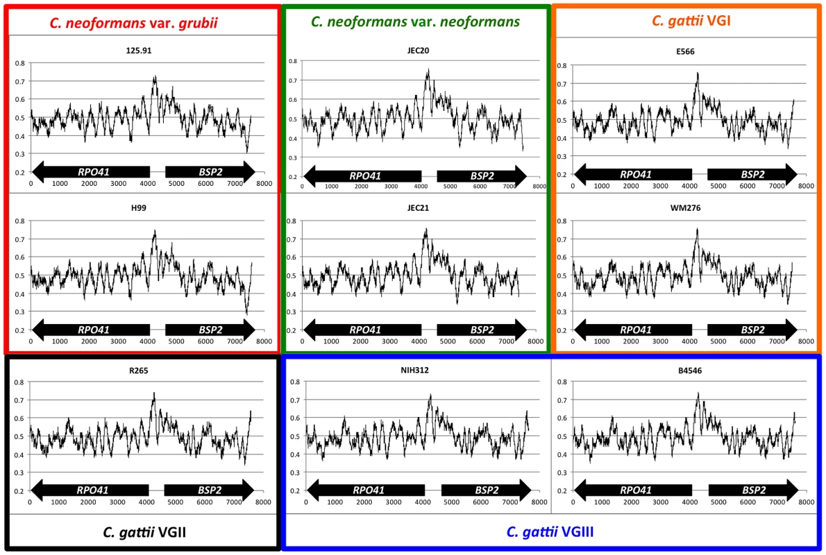
For each figure, the Y axis is the GC%, while the X axis indicates the relative distance (bp) from the 3′ end of the RPO41 gene. Sequences from the MATa and MATα reference strains of the same variety (as in C. neoformans) or the same molecular group (as in C. gattii) are grouped together by rectangles of different colors. The size of the sliding window was 200 bp. Population genetic analyses revealed homogenization between MATa and MATα alleles around the GC-rich intergenic region in natural C. neoformans isolates
We sequenced a region that encompasses the 5′ ends of the RPO41 and BSP2 genes, and the GC rich intergenic region between the RPO41 and BSP2 genes from a group of natural C. neoformans strains, including both var. grubii and var. neoformans (Table 1 and Figure 2). We also PCR amplified and sequenced serotype A and serotype D specific alleles from a group of AD hybrid isolates (Table 1 and Table S1).
Fig. 2. Distribution of polymorphic sites between the two parental strains and the genotypes identified among the meiotic progeny. 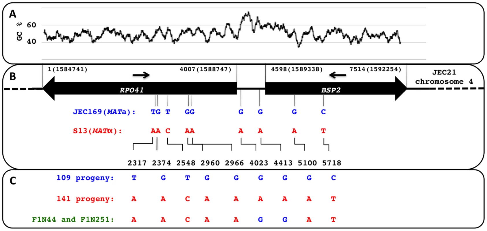
(A) Illustration of the distribution of GC content along the region encompassing the RPO41 and BSP2 genes, as well as the intergenic region between the two genes. (B) Positions of the nine polymorphic sites present within the sequenced region between the two strains, JEC169 (MATa) and S13 (MATα), which were used in the laboratory cross to generate meiotic progeny. The two arrows indicate the approximate locations of the forward and reverse primers used to amplify this region from both natural isolates and progeny generated by laboratory cross. (C) Summary of the genotypes identified among 255 meiotic progeny based on the above mentioned nine polymorphic sites. Tab. 1. List of strains used in this study. 
: Determined in previous studies [51], [52], [53]. Overall, this region showed a serotype specific phylogeny when all of the alleles are compared together, as all of the serotype A alleles (from both haploid var. grubii isolates and serotype AD hybrids) grouped within a well supported cluster that showed considerable divergence from the cluster that included all of the serotype D alleles (Figure 3). This pattern still held when the regions belonging to the three sections (the two genes, RPO41 and BSP2, and the intergenic regions) were analyzed separately (Figure 3), consistent with the view that serotypes A and D are well separated lineages that diverged from each other long ago. Among the three sections, the intergenic region showed the highest level of divergence between the two clusters of serotype A and serotype D alleles (Figure 3). Within each cluster, the level of polymorphism within the intergenic region is comparable to the other two gene coding regions among serotype D alleles, and is higher than the other two sections among serotype A alleles (Table 2). No signal of positive selection (dN/dS>1) was detected in either RPO41 or BSP2 gene coding region for either serotype A or serotype D alleles.
Fig. 3. Phylogenetic trees of alleles existing in natural C. neoformans isolates based on the sequenced RPO41-intergenic-BSP2 region. 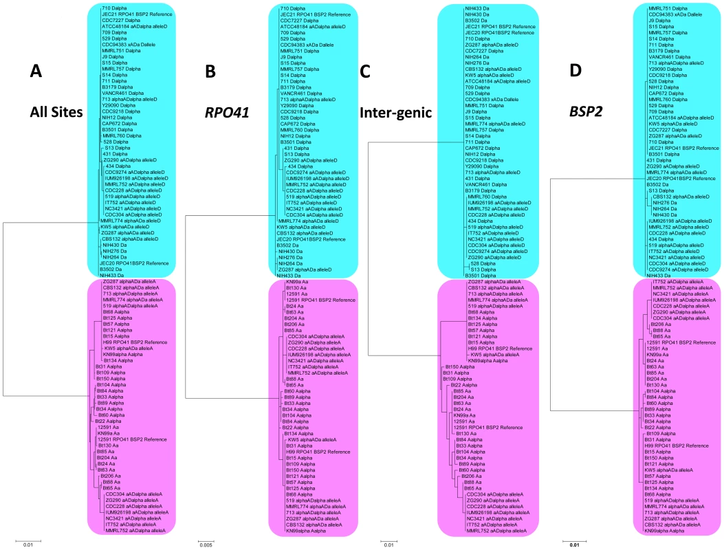
Phylogenetic trees were constructed using maximum likelihood methods with 1000 bootstraps. Alleles shaded with pink color were all serotype A (from both haploid and hybrid isolates), while alleles shaded with blue color were all serotype D (from both haploid and hybrid isolates). A) Sequences from the entire RPO41-intergenic-BSP2 region were analyzed. B) Only sequences within the gene RPO41 were used. C) Only sequences within the intergenic region were studied. D) Only sequences within the gene BSP2 were subjected to analysis. Tab. 2. Genetic distances of different genes/regions within the MAT locus.a 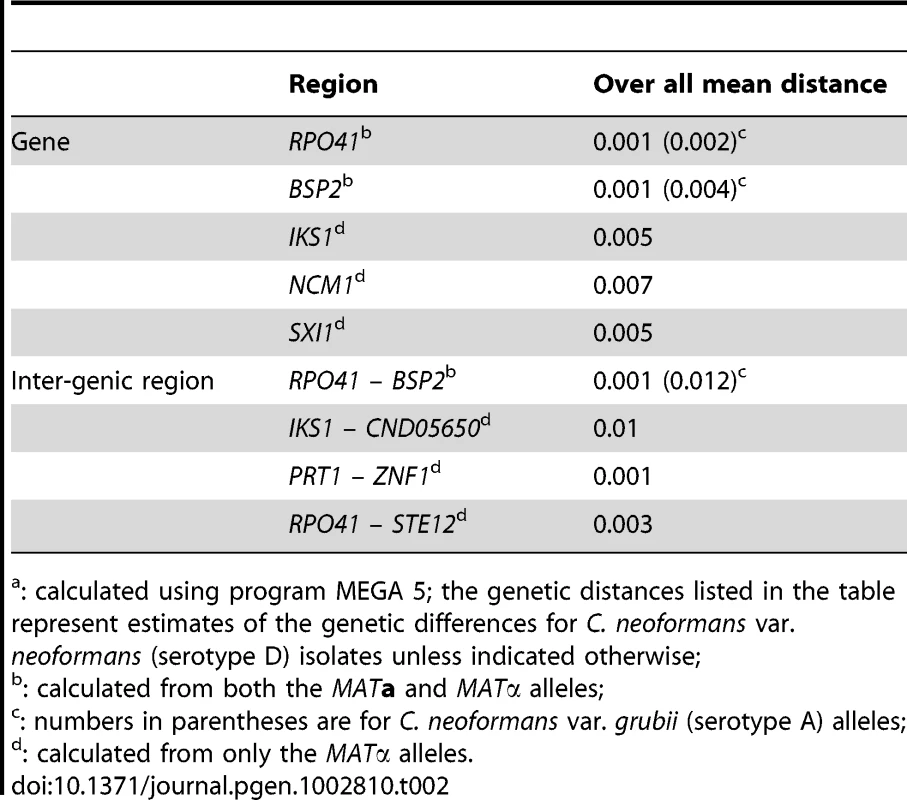
: calculated using program MEGA 5; the genetic distances listed in the table represent estimates of the genetic differences for C. neoformans var. neoformans (serotype D) isolates unless indicated otherwise; When only serotype A alleles were considered, well supported clusters containing all and only the MATa specific alleles were observed when all of the sites were included in the analyses (Figure 4A), as well as when the regions corresponding to the two genic regions were analyzed (Figure 4B and 4D). However, when only the intergenic region was analyzed, this mating type specific topology no longer held. Instead, MATα alleles from six haploid strains were placed within a well supported cluster that otherwise contained only MATa specific alleles (Figure 4C). The phylogeny of the intergenic region was shown to be statistically different (p<0.01) from those of the two flanking genes by the Shimodaira-Hasegawa test [34].
Fig. 4. Phylogenetic trees of serotype A alleles existing in natural C. neoformans isolates based on the sequenced RPO41-intergenic-BSP2 region. 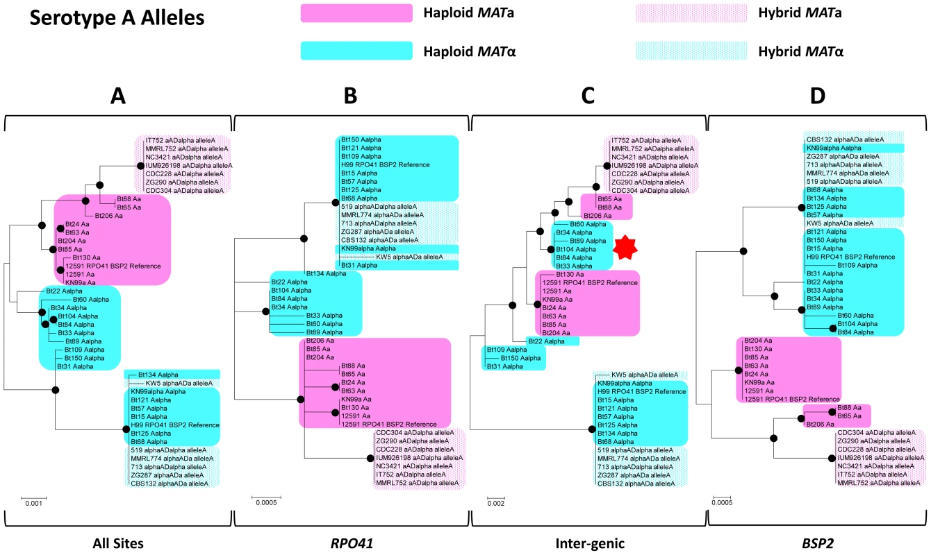
Phylogenetic trees based on whole sequences (A), sequences corresponding to the RPO41 gene (B), the intergenic regions (C), and the BSP2 gene (D) were constructed using the maximum likelihood method with 1000 bootstrap replications. Black dots indicate the nodes supported by bootstrap values of 75 or higher. Solid and dotted pink shades highlight MATa alleles from haploid and hybrid isolates, respectively. Solid and dotted blue shades highlight MATα alleles from haploid and hybrid isolates, respectively. The red star in section (C) highlights the group of haploid MATα alleles that clustered together with MATa alleles. When only serotype D alleles were considered, again a well supported cluster containing all and only the MATa specific alleles was observed when all of the sites were analyzed (Figure 5A), as well as when the region corresponding to the gene RPO41 was analyzed separately (Figure 5B). When the regions corresponding to the intergenic region and the BSP2 gene were analyzed separately, this mating type specific phylogeny was no longer well supported. Specifically, when only the region within the BSP2 gene was analyzed, four MATa specific alleles formed a well supported cluster, while the other five MATa specific alleles were grouped together with most of the MATα specific alleles within a well supported cluster, reflecting a sharing of polymorphisms between alleles from the two mating types in this region. For the intergenic region, other than the two well supported clusters, one containing two MATα haploid alleles and the other containing two haploid MATα alleles and nine MATα alleles from hybrid AD isolates, all of the other MATa and MATα alleles grouped together, indicating a lack of divergence between MATa and MATα alleles at the GC rich intergenic region (Figure 6). Similar to serotype A alleles, the topology of the intergenic region was shown to be statistically different (p<0.01) from those of the RPO41 and BSP2 genes by the Shimodaira-Hasegawa test [34].
Fig. 5. Phylogenetic trees of serotype D alleles existing in natural C. neoformans isolates based on the sequenced RPO41-intergenic-BSP2 region. 
Phylogenetic trees based on whole sequences (A), sequences corresponding to the RPO41 gene (B), the intergenic regions (C), and the BSP2 gene (D) were constructed using the maximum likelihood method with 1000 bootstrap replications. Black dots indicate the nodes supported by bootstrap values of 75 or higher. As in Figure 4, solid and dotted pink shades highlight MATa alleles from haploid and hybrid isolates, respectively, while solid and dotted blue shades highlight MATα alleles from haploid and hybrid isolates, respectively. Fig. 6. Illustration of polymorphic sites among serotype A and D haploid strains. 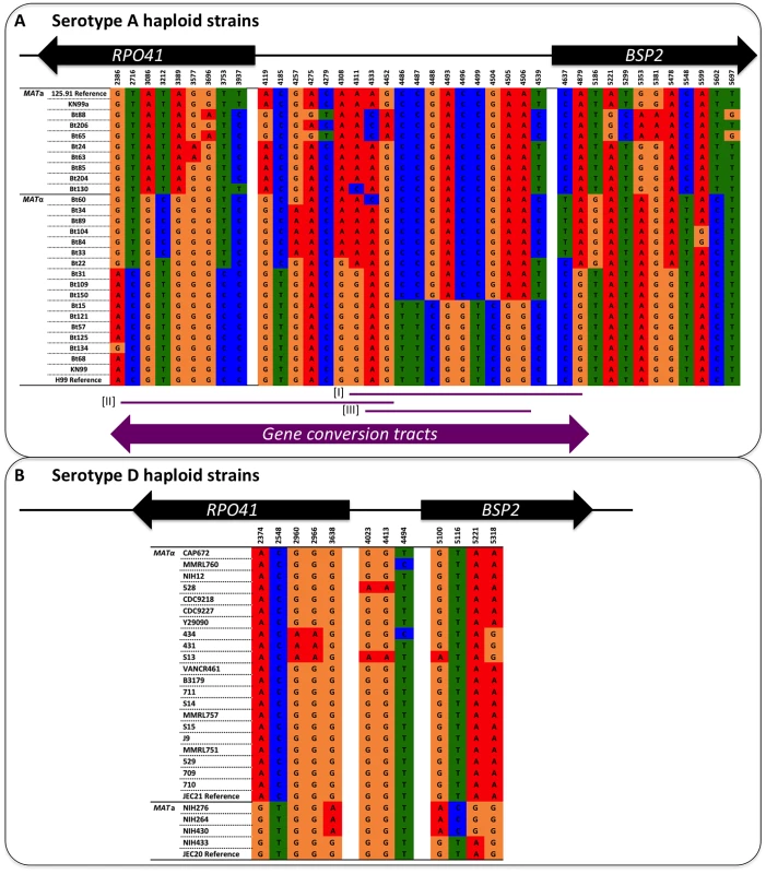
The positions of the polymorphic sites are shown at the top, and they correspond to the distances of the sites from the 3′ end of the RPO41 gene (see also Figure 2). For simplicity, only parsimony informative sites are shown here. (A) Serotype A strains. Sites from 2386 to 3937 are located within the RPO41 gene, sites from 4119 to 4539 are located within the intergenic region, while sites from 4637 to 5697 are located within the BSP2 gene. The purple lines and arrow at the bottom indicate locations and lengths of gene conversion tracts detected by GENECONV software. Examples of strain pairs from which the three gene conversion tracts were detected are: Bt22-Bt150 for tract [I], Bt109-Bt125 for tract [II], and Bt60-Bt31 for tract [III]. (B) Serotype D strains. Sites from 2374 to 3638 are located within the RPO41 gene, sites from 4023 to 4494 are located within the intergenic region, while sites from 5100 to 5318 are located within the BSP2 gene. We further looked for possible gene conversion tracts in this region in the natural population of C. neoformans. For serotype D alleles, we did not detect any statistically significant gene conversion tracts, possibly due to the low level of polymorphism present around the GC rich intergenic region. However, when serotype A alleles were analyzed, we indeed found several statistically significant gene conversion tracts encompassing the GC rich intergenic region (Figure 6; p<0.05 based on 10000 permutations). The lengths of the gene conversion tracts ranged from 560 to 2100 bp, consistent with previous studies in yeast showing most gene conversion tracts were between 1800 to 2000 bp [35]. These gene conversion tracts collectively spanned a 2.5 kb region encompassing the GC rich intergenic region (Figure 6).
Taken together, our population genetic analyses revealed that: 1) compared to the two genic regions, there was higher divergence between serotypes A and D alleles in the intergenic regions; 2) within serotype D alleles, the intergenic region showed a low level of polymorphisms compared to the flanking genic regions, as well as other intergenic regions within the MAT locus, even though no evidence of purifying selection was detected in the flanking genic regions of the RPO41 and BSP2 genes; 3) within serotype A and serotype D alleles, respectively, a mating type specific topology was observed for regions corresponding to the RPO41 and BSP2 genes, albeit the pattern was less well resolved for the serotype D BSP2 region; 4) there was no mating type specific topology at the intergenic regions for both serotypes A and D alleles, and the phylogeny of this GC rich intergenic region is statistically different from those of the flanking genic regions of RPO41 and BSP2, suggesting sequence exchange and homogenization of this GC rich intergenic region in both lineages; and 5) statistically significant gene conversion tracts were detected in serotype A alleles across the GC rich intergenic region. These observed patterns could be explained by a process of ongoing gene conversion at the GC rich intergenic region within each lineage to prevent divergence between the two mating types, while allowing polymorphisms to accumulate independently between the two serotypes.
Meiotic gene conversion occurs around the GC peak within the MAT locus
We hypothesized that if gene conversion at the GC rich intergenic region occurs at a frequency high enough to produce the population structure observed among the natural isolates, we should be able to detect it in the meiotic progeny generated from laboratory crosses. To test this hypothesis, we crossed two fertile serotype D strains of opposite mating type, JEC169 (MATa) and S13 (MATα) and collected 260 recombinant progeny (i.e. the progeny whose phenotype with respect to auxotrophic mutations differed with both parental strains) by screening progeny for auxotrophic mutations present in the two parental strains (see Materials and Methods). We then PCR amplified and sequenced from each of these meiotic progeny the same region analyzed for the natural isolates and looked for gene conversion events that might have occurred around the intergenic region during meiosis.
Among these 260 recombinant progeny recovered, 259 of them had the recombinant genotype ADE2 ura5, while the other one was ade2 URA5. Additionally five of the recombinant progeny were filamentous when grown on YPD solid medium. Further analysis by FACS showed that these five progeny were diploid (data not shown). Thus, these five progeny were likely diploid fusion products of the two parental strains that underwent loss of heterozygosity at the auxotrophic markers (rather than haploid meiotic progeny), and consequently they were excluded from the following analyses.
The remaining 255 recombinant progeny grew as yeast (i.e. no filamentation) on YPD solid medium and our analyses indicated that they were haploid. For chromosome 4, where MAT is located, only the a or α allele was present, but not both. Mating assays by backcrossing each of these 255 progeny to the two parental strains confirmed that the majority are fertile. Specifically, 92 (36.1%) progeny typed as MATa (i.e. successful mating with S13 but not JEC169); 139 (54.5%) progeny typed as MATα (i.e. successful mating with JEC169 but not S13); and 24 (9.4%) progeny were sterile (i.e. no mating was observed with either parental strain).
The RPO41-BSP2 intergenic region of these 255 F1 progeny was then PCR amplified and sequenced. Based on the polymorphic sites within this region between the two parental strains (Figure 2), 109 progeny inherited the alleles from the MATa parent (strain JEC169), and 141 progeny inherited the alleles from the MATα parent (strain S13). These genotypes are in accord with the mating phenotypes of the progeny determined by mating assays (see above). However, two progeny, F1N44 and F1N251, showed evidence of gene conversion at the RPO41-BSP2 intergenic region (Figure 2). Specifically, these two isolates inherited alleles from JEC169 (MATa) at the two markers located within the intergenic region (sites 4023 and 4413, Figure 2), and both inherited alleles from S13 (MATα) at the loci located within the RPO41 and BSP2 genes. The most parsimonious explanation is that gene conversion events occurred across the GC rich intergenic region (Figure 2). In both cases, the two polymorphic sites within the intergenic regions were converted from A to G, consistent with the biased gene conversion in favor of G/C that has been reported previously [27]. Our analyses using PCR-RFLP markers located on other regions of chromosome 4 suggests these two isolates arose from independent gene conversion events, as they inherited different alleles at other markers (Table 3 and Table S2; also see below). Additionally, these two progeny still mate as MATα, suggesting the conversion of the intergenic region from MATα to MATa does not have direct effects on the mating phenotype.
Tab. 3. Genotypes of the two parental strains and representative recombinant progeny at the 18 markers screened in this study. 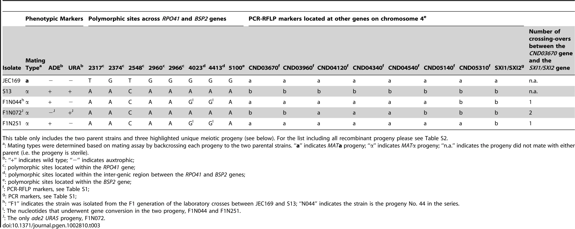
This table only includes the two parent strains and three highlighted unique meiotic progeny (see below). For the list including all recombinant progeny please see Table S2. Assuming DSBs occur only within the intergenic region, the maximum frequency of this gene conversion around the GC rich intergenic region could be estimated to be:In which 0.614 kb is the size of the intergenic region. Alternatively, using the distance between the two polymorphic sites flanking the intergenic region, the minimum gene conversion frequency could be estimated as:in which 2.134 kb is the distance between sites 2966 and 5100 in Figure 2.
The frequency of gene conversion at the GC-rich intergenic region was at least comparable to the meiotic recombination frequencies elsewhere on the same chromosome
To put the observed frequency of gene conversion around the GC rich intergenic region into perspective, we calculated meiotic recombination frequencies at other chromosomal regions by constructing a genetic linkage map using the same meiotic progeny population and markers located on the same chromosome, chromosome 4, as the MAT locus. We then compared these meiotic recombination frequencies directly with the gene conversion frequency observed involving the GC rich intergenic region.
Specifically, we identified eight PCR/PCR-RFLP markers between the two parental strains (Table 3 and Table S2) that were located between ∼100 kb and ∼540 kb away from the SXI1α gene located at one end of the MAT locus. We then used these markers to screen all of the recombinant progeny. Five of the eight markers were co-dominant PCR-RFLP markers (Figure 7A), and we did not observe heterozygosity in any of the 255 recombinant progeny at any of these five markers, further corroborating that these isolates are haploid for chromosome 4.
Fig. 7. Meiotic recombination frequencies in chromosomal regions proximal to the MAT locus. 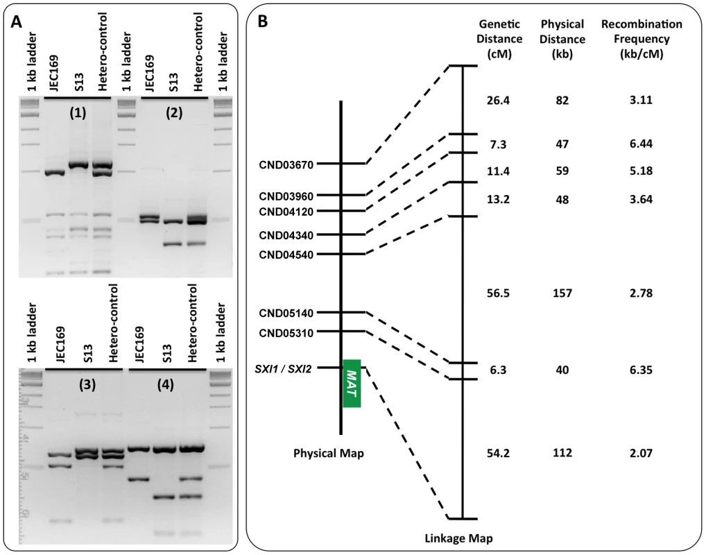
(A) Examples of the co-dominant PCR-RFLP markers used to construct the genetic linkage map of the meiotic progeny shown in Figure 7B. Shown here are the gel separations of the restriction enzyme digestion products of markers CND05310 (A1), CND04540 (A2), CND03960 (A3), and CND04340 (A4). For each marker, from left to right, are the digestions of PCR products using genomic DNA of JEC169, S13, and a mixture of JEC169 and S13 (heterozygous control) as templates, respectively. (B) On the left is the physical map of the eight genetic markers on strain JEC20/JEC21 chromosome 4. The green block indicates the position of the MAT locus. In the middle is the genetic linkage map constructed using the PCR-RFLP data listed in Table 3 and Table S2. The three columns on the right list the genetic distances between adjacent markers, the physical distances between adjacent markers, and the corresponding recombination frequencies (high kb/cM value indicates low recombination frequency). The majority of the F1 progeny (>90%) had 0 to 2 crossing-overs within this region (Table 3, Table 4, and Table S2), and there is extensive genetic diversity among the F1 progeny (Figure 8). The order of the markers in the linkage map is consistent with that in the physical map (Figure 7B). Our results showed that the recombination frequencies (calculated as “kb/(recombination event/100 progeny)”) between adjacent marker pairs ranged between 2.07 and 6.44, and the average recombination frequency within the chromosomal region covered by these markers (calculated as [Total Physical Distance]/[Total Genetic Distance]) was 3.11. Of the seven chromosomal regions, six of them had a recombination frequency that was considerably lower than the frequency of the gene conversion at the GC peak region (estimated to be between 0.78 and 2.72) (Figure 7). Not surprisingly, the only region that showed possibly higher recombination frequency than the gene conversion frequency was between the CND05310 and SXI1/SXI2 genes that encompass the region flanking the MAT locus, which was previously shown to exhibit an elevated recombination frequency during meiosis [13]. Although it is not possible to perform statistical analyses given the small number of gene conversion events observed, these results provide evidence that the frequency of the gene conversion at the GC peak region was at least comparable to, and likely greater than the average recombination frequency in other regions on the same chromosome during meiosis.
Fig. 8. Genetic diversity among meiotic progeny from the laboratory cross between strains JEC169 and S13. 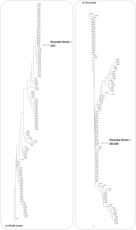
The tree was constructed using the Neighbor-Joining method based on the PCR-RFLP data listed in Table 3 and Table S2. The two parental strains are highlighted by the arrows. Tab. 4. Number of cross-overs among F1 progeny on chromosome 4.a 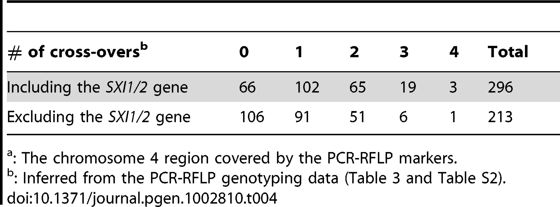
: The chromosome 4 region covered by the PCR-RFLP markers. Discussion
Meiotic recombination is thought to be repressed over the majority of the heterogametic sex chromosomes (e.g. mammalian X and Y chromosomes), as well as the MAT loci in some fungi (e.g. the MAT locus in C. neoformans). Although this ensures the integrity of these highly rearranged chromosomal regions and maintains the divergence between alleles from opposite sexes (mating types), the lack of meiotic recombination also means these regions are effectively asexual, and thus under the threat of gradual deterioration due to 1) the accumulation of deleterious mutations that can no longer be eliminated efficiently through recombination, and 2) the irreversible manner in which the deleterious mutations accumulate within these regions (i.e. Muller's ratchet effect) [36], [37].
In the current study, we showed that within the MAT locus there is a GC rich intergenic region between the RPO41 and BSP2 genes that exists in all of the lineages of C. neoformans and C. gattii (Figure 1). The nucleotide composition of this GC rich region is diverged among different lineages while relatively conserved within each lineage. These observed population genetics patterns reflect the descent from a common ancestor of this GC rich intergenic region in all of the C. neoformans and C. gattii lineages, and the observed divergence among these lineages is the result of independent accumulation of mutations within this intergenic region in different lineages after their split from a common ancestor.
Recombination is thought to be repressed within the MAT locus of C. neoformans, due to sequence divergence and chromosomal rearrangements existing within the MAT locus between the two opposite mating types. If this is the case, the phylogenies of the three sections (the two flanking genes and the intergenic region) should have consistent topologies. In addition, the alleles from opposite mating types should exhibit independent accumulation of mutations, and thus their phylogeny should have a mating type specific topology; that is, alleles cluster together based on the mating types of the strains in which they reside. Furthermore, because no signal of positive selection was detected in the two flanking genic regions, the intergenic region should have accumulated more polymorphisms than regions corresponding to the two flanking genes, in which possible mechanisms such as purifying selection could slow down the accumulation of mutations and maintain sequence identity. However, this is in contrast to the results from our analyses. Specifically, the topologies of the two flanking genes were statistically different from those of the intergenic region when serotype A or serotype D alleles were analyzed. Additionally, among the serotype D alleles, the intergenic GC rich region showed a comparable level of polymorphism when compared to the two flanking genic regions (RPO41 and BSP2), and a lower level of polymorphism when compared to other genes and intergenic regions that are also located within the MAT locus (Table 2). Furthermore, we found evidence of allele sharing between strains of opposite mating types in both the serotype A and D populations (Figure 4, Figure 5), as well as gene conversion tracts around the GC rich intergenic region among serotype A strains (Figure 6). Thus, our results instead support a model in which among serotype A and D C. neoformans strains, respectively, there is still ongoing gene flow between the two mating types at the GC rich intergenic region within the MAT locus.
This hypothesis is supported by observations from the laboratory sexual cross. By collecting and analyzing a large number of meiotic progeny from a laboratory cross, we found that recombination, in the form of gene conversion, occurred around the GC rich intergenic region during sexual reproduction in serotype D C. neoformans. Interestingly, in both of the gene conversion events identified among meiotic progeny, the direction of the gene conversion was A→G, consistent with previous studies indicating that gene conversion at recombination hot spots shows a bias favoring G/C over A/T [25], [27], [38]. However, it could also be that this uni-directional gene conversion is actually favoring MATa alleles over MATα alleles. This could be investigated by analyzing meiotic progeny of a laboratory cross, in which the MATa parent has A or T, while the MATα parent has G or C alleles at the polymorphic sites within the intergenic GC rich region.
The frequency of this gene conversion event was estimated to be between 0.78 and 2.72 kb/(events/100 progeny). This is at least comparable to the meiotic recombination frequencies observed in other regions of chromosome 4 (Figure 7). The genome wide meiotic recombination of serotype D C. neoformans has been previously estimated to be ∼13.2 kb/cM [39], which is considerably lower than the meiotic recombination frequency that we observed at the other regions of chromosome 4 (ranged between 2.07 and 6.44 kb/cM). However, in the study by Marra et al. [39], several of the chromosomes were each composed of multiple linkage groups, suggesting an underestimation of the genetic distances across the genome (those recombination events between the linkage groups), and thus an overestimation of the genomic average recombination frequency. Unfortunately, chromosome 4 was one of the chromosomes that were composed of two separate linkage groups, preventing us from comparing directly the results from the two studies. Another possibility is that chromosome 4 could experience a higher than average recombination frequency during meiosis. It has been shown that in C. neoformans during serotype A and D hybridization, although most of the chromosomes experience significantly reduced recombination frequency, the regions on chromosome 4 still showed levels of recombination that are comparable to those observed in intra-variety mating [40]. It is possible that there is an intrinsic mechanism that promotes recombination along chromosome 4 during sexual reproduction, which could result from the MAT locus residing on chromosome 4. This could be investigated by genome wide analysis of the meiotic recombination frequencies at markers from different chromosomes and with different GC content profiles. Taken together, our analyses support the conclusion that gene conversion is occurring around the GC rich intergenic region at a frequency that is at least comparable to, and likely higher than the typical meiotic recombination frequencies in other genomic regions during sexual reproduction of C. neoformans.
It is not yet clear how gene conversion occurs at this intergenic GC rich region. The two flanking genes, RPO41 and BSP2, are among a group of genes that constitutes a syntenic cluster with the same orientation between MATa and MATα alleles in serotype A, whereas this gene pair is oppositely oriented between MATa and MATα alleles in serotype D (Figure 9). A previous study has hypothesized that this gene cluster represents a strata that was recruited into the MAT locus most recently during evolution [41]. Thus, it could be that the repression of recombination is less severe, and recombination can still be initiated within this region. It has been shown that recombination hot spots flank the MAT locus of C. neoformans, and these hot spots are associated with chromosomal regions with a high GC content [13]. Thus, it is possible that the factors responsible for initiating a high frequency recombination at the flanking regions of MAT locus could also recognize the GC rich intergenic region between the RPO41 and BSP2 genes within the MAT locus, and induce lesions such as double-strand breaks (DSBs) that promote recombination within this region. Additionally, DSBs could also be induced in this region because of the high susceptibility of this region to mutagenic factors due to their high GC content [28]. The DSB could then be repaired through either crossing-over or gene conversion. However, due to the rearranged chromosomal locations within the MAT loci, as well as the opposite orientations (in serotype D) of the MATa and MATα alleles, typical crossing-over within the GC rich intergenic region would result in abnormal chromosomes, such as dicentric or acentric chromosomes, and/or chromosomes with duplications and deletions of a variety of essential genes or genes involved in mating and meiosis (Figure 9). As a consequence, the progeny inheriting these abnormal chromosomes would likely be inviable. On the other hand, DSBs repaired through gene conversion do not produce abnormal chromosomes, thus resulting in the bias that only gene conversion events can be recovered from the meiotic recombination events that had actually occurred at this GC rich intergenic region during sexual reproduction. If this is the case, our estimation of the recombination frequency at this GC rich intergenic region would be an underestimation of the actual recombination frequency (including both nonviable cross-overs and viable gene conversions) within this region.
Fig. 9. Diagrams of the MAT loci of C. neoformans var. grubii and var. neoformans and illustration of meiotic recombination within inverted chromosomal region. 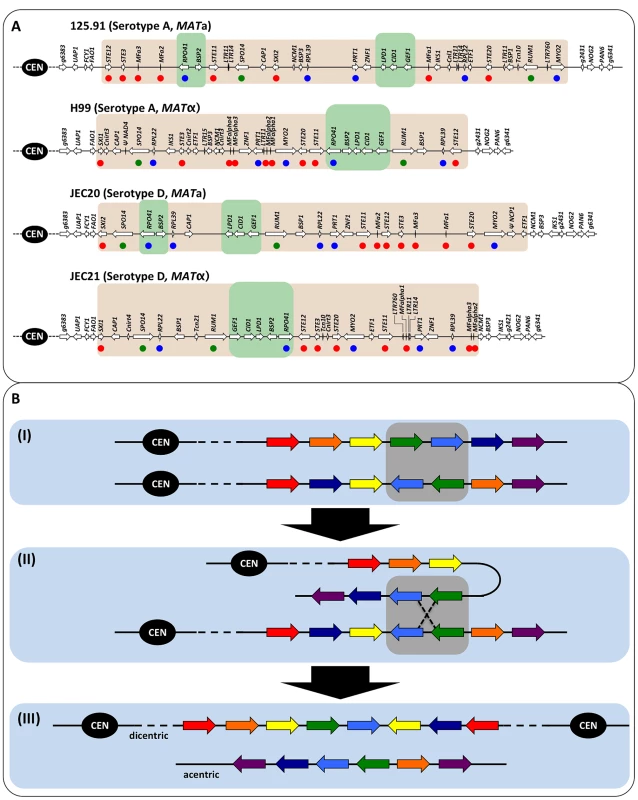
(A) Regions shaded in gray indicate the MAT loci. Green shading indicates the gene clusters, within which the RPO41 and BSP2 genes are located, and which have been hypothesized to be the most recent strata that was recruited into the MAT locus. Red dots denote the genes involved in mating and/or meiosis, green dots indicate hypothetical meiotic genes, and blue dots highlight known essential genes. (B) The black circle labeled “CEN” is the centromere. The seven arrows with different colors indicate seven different genes. The balanced composition of each chromosome at these seven genes is indicated by the presence of one gene of each of the seven colors (e.g., the two chromosomes in (I)). Gray shading highlights the two genes, green and blue, that are inverted. (I) The two chromatids involved in meiotic recombination are shown. (II) Inverted chromosomal regions align during meiosis, and meiotic recombination occurs within the inverted region. (III) Meiotic recombination within the inverted chromosomal region results in two chromosomes that are imbalanced in their gene compositions, and are acentric and dicentric, respectively. It is also possible that recombination, in the form of gene conversion, could actually be initiated and carried out actively during meiosis within the GC rich intergenic region, and maybe even in other regions of the MAT locus of C. neoformans. One of the best studied examples of active initiation of gene conversion during mitosis is mating type switching in the yeast S. cerevisiae through gene conversion initiated by the HO endonuclease, which enables haploid Saccharomyces cells to have the potential to change mating type as often as every mitotic generation [42], [43]. Active initiation of gene conversion within the C. neoformans MAT locus could have two consequences. First, it might help align the opposite mating type alleles during meiosis. The MAT locus of C. neoformans is unusually large (>100 kb), and is highly rearranged between alleles of opposite mating type. Although it has been widely accepted that meiosis in almost all species requires at least one crossover event per chromosome, the highly diverged and rearranged nature between alleles of opposite mating types could still pose a problem for the proper alignment between opposite alleles during meiosis. Thus, a recombination event within the MAT locus could help in generating the tension required between homologous chromosomes for their proper segregation during meiosis I. Second, full repression of recombination within the MAT locus of C. neoformans would render this region asexual, and thus subject the MAT locus to gradual deterioration due to the irreversible accumulation of mutations within the MAT (i.e. Muller's ratchet [36]). It has been shown that five of the 20 genes within the MAT locus are essential (including the RPO41 gene) [41], and although mechanisms such as purifying selection could act to prevent mutations from accumulating within these genes, the maintenance of sequence identity of these essential genes could nonetheless be facilitated by gene conversion events occurring in these regions. This is similar to situations where gene conversion that is biased against new mutations has been proposed to slow the observed mutation rate in plants and bacteria [44], [45]. This is also consistent with recent simulation-based studies that suggest that even low levels of gene conversion are sufficient to maintain the sequence integrity of the genes located on the human Y chromosome [46], [47]. Alternatively, this GC rich intergenic region may represent an ancient recombination hot spot that was adjacent to the essential gene, RPO41, before it was recruited into the MAT locus, as demonstrated by a study showing that purging of deleterious mutations in essential genes may be an important factor driving meiotic crossover [48].
Recombination, including gene conversion, has been reported to occur at chromosomal regions within which recombination has been thought to be repressed, such as centromeric regions [15]. Recent studies provide further empirical evidence that recombination is more universal, and is occurring at considerably higher frequencies within these “non-recombining” regions. It has been shown that in mouse crossing-over within the PAR region located on the otherwise non-recombining sex chromosomes is critical for male meiosis [29], [30]. Additionally, gene conversion has been discovered to be widespread within the centromeric regions of maize [14]. Furthermore, occasional X-Y recombination, as well as gene conversion, has been found to be the most plausible explanation for the observed sex-chromosome homomorphy in European tree frogs [49]. These recombination events play important roles in shaping the evolutionary trajectories of the chromosomal regions that are involved. A recent study by Sloan et al proposed that changes in recombination frequency (including gene conversion) are a central force driving the evolution of mitochondrial genome structure [50]. It is possible that many recombination events occurring in these supposed “non-recombining” regions might have gone unnoticed, partly due to the fact that in many cases, recombination might have taken the form of gene conversion, which can be detected only when sequences involved are non-identical, and thus is less likely to be recorded than crossing-over. Our study provides further evidence that, indeed, some presumed recombination “cold spots” are actually not that cold, after all.
Materials and Methods
Strains and media
The strains used in this study are listed in Table 1, and their VN types have been determined in previous studies [51], [52], [53] Strain JEC169 is an ade2 ura5 auxotrophic isolate derived from JEC20 [54]. The ADE2 and URA5 genes are located on chromosomes 5 and 7, respectively. All of the other strains are clinical or environmental isolates. All of the strains were grown and maintained on YPD agar plates unless mentioned otherwise.
Laboratory cross and screening of recombinant progeny
A cross between strains JEC169 (MATa ade2 ura5) and S13 (MATα ADE2 URA5) was conducted on V8 agar medium plates as previously described [55]. After 2 weeks of incubation at room temperature in the dark, the plate was inspected by light microscopy. The area outside the edge of the mating mixture containing abundant basidiospores was excised, and suspended in water. The suspension was then diluted and spread onto YPD agar plates. After two days of incubation at 30°C, the colonies that appeared on the YPD plates were transferred onto testing plates, SD-ade and SD-ura [56], to confirm their genotypes at the auxotrophic markers. The colonies that only grew on one of the two testing plates were considered to be recombinant, and were then stored in 35% glycerol at −80°C for the following studies.
Mating assays
The mating type of each recombinant progeny was determined by backcrossing the progeny with the two parental strains. The mating assay was performed in the same way as the laboratory cross described above. Mating was determined to be successful if hyphae, basidia, and basidiospore chains were observed after 2 weeks of incubation.
DNA isolation, PCR amplification, enzyme digestion, and sequencing reactions
DNA isolation and PCR amplification were carried out as previously described [57]. Primers used in this study are listed in Table S1. For serotype AD strains, different primer pairs were used to amplify the serotype A and serotype D specific alleles of the RPO41-BSP2 region, respectively (Table S1). All of the PCR reactions were carried out using an annealing temperature of 60°C. Restriction enzyme digestions were performed according to the manufacturer's instructions (New England Laboratory Casework Co., Inc.). Sequencing reactions were conducted at the Genome Sequencing & Analysis Core Facility at the Duke Institute for Genome Sciences & Policy.
Population genetic analyses
Sequence alignments were carried out using ClustalX 2.1 [58]. The aligned sequences were then curated manually using the software MacClade 4.06 [59]. MEGA 5 [60] was used for the phylogeny constructions and population genetics analyses. Bootstrap support for the phylogenetic trees was determined by performing 1000 bootstraps. Kishino-Hasegawa test of topologies was carried using the SHTest implemented in the software PAUP 4.0 [61]. Gene conversion events were detected using program GENECONV v.1.81 following the software's instruction [62]. Significant gene conversion tracts were identified if the Global-P value was smaller than 0.05 based on permutation.
Linkage map construction
A linkage map was constructed using the software program MapMaker [63]. The chromosomal location of each marker was estimated as the position of the midpoint of the PCR product of that marker in the sequence of strain JEC21 chromosome 4 (GenBank: AE017344).
Supporting Information
Zdroje
1. MalkovaASwansonJGermanMMcCuskerJHHousworthEA 2004 Gene conversion and crossing over along the 405-kb left arm of Saccharomyces cerevisiae chromosome VII. Genetics 168 49 63
2. CopenhaverGPHousworthEAStahlFW 2002 Crossover interference in Arabidopsis. Genetics 160 1631 1639
3. HauboldBKroymannJRatzkaAMitchell-OldsTWieheT 2002 Recombination and gene conversion in a 170-kb genomic region of Arabidopsis thaliana. Genetics 161 1269 1278
4. HillikerAJClarkSHChovnickA 1991 The effect of DNA sequence polymorphisms on intragenic recombination in the rosy locus of Drosophila melanogaster. Genetics 129 779 781
5. FrisseLHudsonRRBartoszewiczAWallJDDonfackJ 2001 Gene conversion and different population histories may explain the contrast between polymorphism and linkage disequilibrium levels. The American Journal of Human Genetics 69 831 843
6. HaberJEIraGMalkovaASugawaraN 2004 Repairing a double-strand chromosome break by homologous recombination: revisiting Robin Holliday's model. Philosophical Transactions of the Royal Society of London Series B: Biological Sciences 359 79 86
7. ChenJ-MCooperDNChuzhanovaNFerecCPatrinosGP 2007 Gene conversion: mechanisms, evolution and human disease. Nat Rev Genet 8 762 775
8. ColeFKauppiLLangeJRoigIWangR 2012 Homeostatic control of recombination is implemented progressively in mouse meiosis. Nat Cell Biol 14 424 430
9. NishantKTChenCShinoharaMShinoharaAAlaniE 2010 Genetic analysis of baker's yeast Msh4-Msh5 reveals a threshold crossover level for meiotic viability. PLoS Genet 6 e1001083 doi:10.1371/journal.pgen.1001083
10. PetesTD 2001 Meiotic recombination hot spots and cold spots. Nat Rev Genet 2 360 369
11. JeffreysAJKauppiLNeumannR 2001 Intensely punctate meiotic recombination in the class II region of the major histocompatibility complex. Nat Genet 29 217 222
12. MyersSBottoloLFreemanCMcVeanGDonnellyP 2005 A fine-scale map of recombination rates and hotspots across the human genome. Science 310 321 324
13. HsuehYPIdnurmAHeitmanJ 2006 Recombination hotspots flank the Cryptococcus mating-type locus: implications for the evolution of a fungal sex chromosome. PLoS Genet 2 e184 doi:10.1371/journal.pgen.0020184
14. ShiJWolfSEBurkeJMPrestingGGRoss-IbarraJ 2010 Widespread gene conversion in centromere cores. PLoS Biol 8 e1000327 doi:10.1371/journal.pbio.1000327
15. SymingtonLSPetesTD 1988 Meiotic recombination within the centromere of a yeast chromosome. Cell 52 237 240
16. RozenSSkaletskyHMarszalekJDMinxPJCordumHS 2003 Abundant gene conversion between arms of palindromes in human and ape Y chromosomes. Nature 423 873 876
17. SkaletskyHKuroda-KawaguchiTMinxPJCordumHSHillierL 2003 The male-specific region of the human Y chromosome is a mosaic of discrete sequence classes. Nature 423 825 837
18. Jill PeconSSanner-WachterLO'BrienSJ 2000 Novel gene conversion between X-Y homologues located in the nonrecombining region of the Y chromosome in Felidae (Mammalia). PNAS 97 5307 5312
19. FullertonSMBernardo CarvalhoAClarkAG 2001 Local rates of recombination are positively correlated with GC content in the human genome. Molecular Biology and Evolution 18 1139 1142
20. Eyre-WalkerAHurstLD 2001 The evolution of isochores. Nat Rev Genet 2 549 555
21. Marsolier-KergoatM-CYeramianE 2009 GC content and recombination: Reassessing the causal effects for the Saccharomyces cerevisiae genome. Genetics 183 31 38
22. MeunierJDuretL 2004 Recombination drives the evolution of GC-content in the human genome. Molecular Biology and Evolution 21 984 990
23. PerryJAshworthA 1999 Evolutionary rate of a gene affected by chromosomal position. Current Biology 9 987 S983
24. MaraisG 2003 Biased gene conversion: implications for genome and sex evolution. Trends in Genetics 19 330 338
25. GaltierNPiganeauGMouchiroudDDuretL 2001 GC-content evolution in mammalian genomes: the biased gene conversion hypothesis. Genetics 159 907 911
26. WebsterMTAxelssonEEllegrenH 2006 Strong regional biases in nucleotide substitution in the chicken genome. Molecular Biology and Evolution 23 1203 1216
27. BirdsellJA 2002 Integrating genomics, bioinformatics, and classical genetics to study the effects of recombination on genome evolution. Molecular Biology and Evolution 19 1181 1197
28. Matallana-SurgetSMeadorJAJouxFDoukiT 2008 Effect of the GC content of DNA on the distribution of UVB-induced bipyrimidine photoproducts. Photochemical & Photobiological Sciences 7 794 801
29. KauppiLBarchiMBaudatFRomanienkoPJKeeneyS 2011 Distinct properties of the XY pseudoautosomal region crucial for male meiosis. Science 331 916 920
30. OttoSPPannellJRPeichelCLAshmanT-LCharlesworthD 2011 About PAR: The distinct evolutionary dynamics of the pseudoautosomal region. Trends in Genetics 27 358 367
31. AlexanderI 2011 Sex and speciation: The paradox that non-recombining DNA promotes recombination. Fungal Biology Reviews 25 121 127
32. D'SouzaCAKronstadJWTaylorGWarrenRYuenM 2011 Genome variation in Cryptococcus gattii, an emerging pathogen of immunocompetent hosts. mBio 2 e00342 00310
33. LoftusBJFungERoncagliaPRowleyDAmedeoP 2005 The genome of the basidiomycetous yeast and human pathogen Cryptococcus neoformans. Science 307 1321 1324
34. ShimodairaHHasegawaM 1999 Multiple comparisons of log-likelihoods with applications to phylogenetic inference. Molecular Biology and Evolution 16 1114
35. ManceraEBourgonRBrozziAHuberWSteinmetzLM 2008 High-resolution mapping of meiotic crossovers and non-crossovers in yeast. Nature 454 479 485
36. MullerHJ 1964 The relation of recombination to mutational advance. Mutation Research/Fundamental and Molecular Mechanisms of Mutagenesis 1 2 9
37. FelsensteinJ 1974 The evolutionary advantage of recombination. Genetics 78 737 756
38. GaltierN 2004 Recombination, GC-content and the human pseudoautosomal boundary paradox. Trends in Genetics 20 347 349
39. MarraREHuangJCFungENielsenKHeitmanJ 2004 A genetic linkage map of Cryptococcus neoformans variety neoformans serotype D (Filobasidiella neoformans). Genetics 167 619 631
40. SunSXuJ 2007 Genetic analyses of a hybrid cross between serotypes A and D strains of the human pathogenic fungus Cryptococcus neoformans. Genetics 177 1475 1486
41. FraserJADiezmannSSubaranRLAllenALengelerKB 2004 Convergent evolution of chromosomal sex-determining regions in the animal and fungal kingdoms. PLoS Biol 2 e384 doi:10.1371/journal.pbio.0020384
42. HaberJE 1998 Mating-type gene switching in Saccharomyces cerevisiae. Annual Review of Genetics 32 561 599
43. StrathernJNKlarAJSHicksJBAbrahamJAIvyJM 1982 Homothallic switching of yeast mating type cassettes is initiated by a double-stranded cut in the MAT locus. Cell 31 183 192
44. Birky-JrCWWalshJB 1992 Biased gene conversion, copy number, and apparent mutation rate differences within chloroplast and bacterial genomes. Genetics 130 677 683
45. KhakhlovaOBockR 2006 Elimination of deleterious mutations in plastid genomes by gene conversion. The Plant Journal 46 85 94
46. ConnallonTClarkAG 2010 Gene duplication, gene conversion and the evolution of the Y chromosome. Genetics 186 277 286
47. MaraisGABCamposPRAGordoI 2010 Can intra-Y gene conversion oppose the degeneration of the human Y chromosome? A simulation study. Genome Biology and Evolution 2 347 357
48. KellerPJKnopM 2009 Evolution of mutational robustness in the yeast genome: a link to essential genes and meiotic recombination hotspots. PLoS Genet 5 e1000533 doi:10.1371/journal.pgen.1000533
49. StöckMHornAGrossenCLindtkeDSermierR 2011 Ever-young sex chromosomes in European tree frogs. PLoS Biol 9 e1001062 doi:10.1371/journal.pbio.1001062
50. SloanDBAlversonAJChuckalovcakJPWuMMcCauleyDE 2012 Rapid evolution of enormous, multichromosomal genomes in flowering plant mitochondria with exceptionally high mutation rates. PLoS Biol 10 e1001241 doi:10.1371/journal.pbio.1001241
51. LinXLitvintsevaAPNielsenKPatelSFloydA 2007 αADα hybrids of Cryptococcus neoformans: evidence of same-sex mating in nature and hybrid fitness. PLoS Genet 3 e186 doi:10.1371/journal.pgen.0030186
52. LitvintsevaAPLinXTempletonIHeitmanJMitchellT 2007 Many globally isolated AD hybrid strains of Cryptococcus neoformans originated in Africa. PLoS Pathog 3 e114 doi:10.1371/journal.ppat.0030114
53. LitvintsevaAPMarraRENielsenKHeitmanJVilgalysR 2003 Evidence of sexual recombination among Cryptococcus neoformans serotype A isolates in sub-Saharan Africa. Eukaryot Cell 2 1162 1168
54. MooreTDEEdmanJC 1993 The α-mating type locus of Cryptococcus neoformans contains a peptide pheromone gene. Mol Cell Biol 13 1962 1970
55. YanZHullCMHeitmanJSunSXuJ 2004 SXI1α controls uniparental mitochondrial inheritance in Cryptococcus neoformans. Current Biology 14 R743 744
56. YanZHullCMSunSHeitmanJXuJP 2007 The mating-type specific homeodomain genes SXI1α and SXI2a coordinately control uniparental mitochondrial inheritance in Cryptococcus neoformans. Current Genetics 51 187 195
57. SunSXuJ 2009 Chromosomal rearrangements between serotype A and D strains in Cryptococcus neoformans. PLoS ONE 4 e5524 doi:10.1371/journal.pone.0005524
58. LarkinMABlackshieldsGBrownNPChennaRMcGettiganPA 2007 Clustal W and Clustal X version 2.0. Bioinformatics 23 2947 2948
59. MaddisonDMaddisonW 1989 Interactive analysis of phylogeny and character evolution using the computer program MacClade. Folia Primatol (Basel) 53 190 202
60. TamuraKPetersonDPetersonNStecherGNeiM 2011 MEGA5: Molecular evolutionary genetics analysis using maximum likelihood, evolutionary distance, and maximum parsimony methods. Molecular Biology and Evolution 28 2731 2739
61. SwoffordD 2003 PAUP*. Phylogenetic analysis using parsimony (*and other methods). Version 4. Sinauer Associates, Sunderland, Massachusetts
62. SawyerS 2007 GENECONV [version 1.81a]: A computer package for the statistical detection of gene conversion. Distributed by the author, Department of Mathematics, Washington University in St Louis, available at http://wwwmathwustledu/~sawyer/geneconv/
63. LanderESGreenPAbrahamsonJBarlowADalyMJ 1987 MAPMAKER: An interactive computer package for constructing primary genetic linkage maps of experimental and natural populations. Genomics 1 174 181
Štítky
Genetika Reprodukční medicína
Článek Allelic Heterogeneity and Trade-Off Shape Natural Variation for Response to Soil MicronutrientČlánek The Chicken Frizzle Feather Is Due to an α-Keratin () Mutation That Causes a Defective RachisČlánek A Trans-Species Missense SNP in Is Associated with Sex Determination in the Tiger Pufferfish, (Fugu)
Článek vyšel v časopisePLOS Genetics
Nejčtenější tento týden
2012 Číslo 7- Akutní intermitentní porfyrie
- Růst a vývoj dětí narozených pomocí IVF
- Vliv melatoninu a cirkadiálního rytmu na ženskou reprodukci
- Délka menstruačního cyklu jako marker ženské plodnosti
- Intrauterinní inseminace a její úspěšnost
-
Všechny články tohoto čísla
- Functional Evolution of Mammalian Odorant Receptors
- Oocyte Family Trees: Old Branches or New Stems?
- Allelic Heterogeneity and Trade-Off Shape Natural Variation for Response to Soil Micronutrient
- Guidelines for Genome-Wide Association Studies
- GWAS Identifies Novel Susceptibility Loci on 6p21.32 and 21q21.3 for Hepatocellular Carcinoma in Chronic Hepatitis B Virus Carriers
- DNA Methyltransferases Are Required to Induce Heterochromatic Re-Replication in Arabidopsis
- Genomic Data Reveal a Complex Making of Humans
- Let-7b/c Enhance the Stability of a Tissue-Specific mRNA during Mammalian Organogenesis as Part of a Feedback Loop Involving KSRP
- The Secreted Immunoglobulin Domain Proteins ZIG-5 and ZIG-8 Cooperate with L1CAM/SAX-7 to Maintain Nervous System Integrity
- RsfA (YbeB) Proteins Are Conserved Ribosomal Silencing Factors
- Gene Conversion Occurs within the Mating-Type Locus of during Sexual Reproduction
- The Chicken Frizzle Feather Is Due to an α-Keratin () Mutation That Causes a Defective Rachis
- Meta-Analysis of Genome-Wide Scans for Total Body BMD in Children and Adults Reveals Allelic Heterogeneity and Age-Specific Effects at the Locus
- Balancing Selection at the Tomato Guardee Gene Family Maintains Variation in Strength of Pathogen Defense
- Large-Scale Introgression Shapes the Evolution of the Mating-Type Chromosomes of the Filamentous Ascomycete
- OSD1 Promotes Meiotic Progression via APC/C Inhibition and Forms a Regulatory Network with TDM and CYCA1;2/TAM
- Intact p53-Dependent Responses in miR-34–Deficient Mice
- FANCJ/BACH1 Acetylation at Lysine 1249 Regulates the DNA Damage Response
- CED-10/Rac1 Regulates Endocytic Recycling through the RAB-5 GAP TBC-2
- Histone H2A Mono-Ubiquitination Is a Crucial Step to Mediate PRC1-Dependent Repression of Developmental Genes to Maintain ES Cell Identity
- F-Box Protein Specificity for G1 Cyclins Is Dictated by Subcellular Localization
- The Gene Encodes a Nuclear Protein That Affects Alternative Splicing
- A Key Role for Chd1 in Histone H3 Dynamics at the 3′ Ends of Long Genes in Yeast
- Genome-Wide Association Analysis in Asthma Subjects Identifies as a Novel Bronchodilator Response Gene
- GRHL3/GET1 and Trithorax Group Members Collaborate to Activate the Epidermal Progenitor Differentiation Program
- Brain-Specific Rescue of Reveals System-Driven Transcriptional Rhythms in Peripheral Tissue
- Recent Loss of Self-Incompatibility by Degradation of the Male Component in Allotetraploid
- Pregnancy-Induced Noncoding RNA () Associates with Polycomb Repressive Complex 2 and Regulates Mammary Epithelial Differentiation
- The HEI10 Is a New ZMM Protein Related to Zip3
- The SCF Ubiquitin E3 Ligase Ubiquitylates Sir4 and Functions in Transcriptional Silencing
- Induction of Cytoprotective Pathways Is Central to the Extension of Lifespan Conferred by Multiple Longevity Pathways
- Role of Architecture in the Function and Specificity of Two Notch-Regulated Transcriptional Enhancer Modules
- Loss of ATRX, Genome Instability, and an Altered DNA Damage Response Are Hallmarks of the Alternative Lengthening of Telomeres Pathway
- A Regulatory Loop Involving PAX6, MITF, and WNT Signaling Controls Retinal Pigment Epithelium Development
- The Three Faces of Riboviral Spontaneous Mutation: Spectrum, Mode of Genome Replication, and Mutation Rate
- Unmet Expectations: miR-34 Plays No Role in p53-Mediated Tumor Suppression In Vivo
- A Genome-Wide Association Meta-Analysis of Circulating Sex Hormone–Binding Globulin Reveals Multiple Loci Implicated in Sex Steroid Hormone Regulation
- The Role of Rice HEI10 in the Formation of Meiotic Crossovers
- A Trans-Species Missense SNP in Is Associated with Sex Determination in the Tiger Pufferfish, (Fugu)
- Influences Bone Mineral Density, Cortical Bone Thickness, Bone Strength, and Osteoporotic Fracture Risk
- Evidence of Inbreeding Depression on Human Height
- Comparative Genomics of Plant-Associated spp.: Insights into Diversity and Inheritance of Traits Involved in Multitrophic Interactions
- Detecting Individual Sites Subject to Episodic Diversifying Selection
- Regulates Rhodopsin-1 Metabolism and Is Required for Photoreceptor Neuron Survival
- Identification of Chromatin-Associated Regulators of MSL Complex Targeting in Dosage Compensation
- Three Dopamine Pathways Induce Aversive Odor Memories with Different Stability
- TDP-1/TDP-43 Regulates Stress Signaling and Age-Dependent Proteotoxicity in
- Rapid Turnover of Long Noncoding RNAs and the Evolution of Gene Expression
- The Yeast Rab GTPase Ypt1 Modulates Unfolded Protein Response Dynamics by Regulating the Stability of RNA
- Histone H2B Monoubiquitination Facilitates the Rapid Modulation of Gene Expression during Arabidopsis Photomorphogenesis
- Cellular Variability of RpoS Expression Underlies Subpopulation Activation of an Integrative and Conjugative Element
- Genetic Variants in , , and Influence Male Recombination in Cattle
- Differential Impact of the HEN1 Homolog HENN-1 on 21U and 26G RNAs in the Germline of
- PLOS Genetics
- Archiv čísel
- Aktuální číslo
- Informace o časopisu
Nejčtenější v tomto čísle- Guidelines for Genome-Wide Association Studies
- The Role of Rice HEI10 in the Formation of Meiotic Crossovers
- Identification of Chromatin-Associated Regulators of MSL Complex Targeting in Dosage Compensation
- GWAS Identifies Novel Susceptibility Loci on 6p21.32 and 21q21.3 for Hepatocellular Carcinoma in Chronic Hepatitis B Virus Carriers
Kurzy
Zvyšte si kvalifikaci online z pohodlí domova
Autoři: prof. MUDr. Vladimír Palička, CSc., Dr.h.c., doc. MUDr. Václav Vyskočil, Ph.D., MUDr. Petr Kasalický, CSc., MUDr. Jan Rosa, Ing. Pavel Havlík, Ing. Jan Adam, Hana Hejnová, DiS., Jana Křenková
Autoři: MUDr. Irena Krčmová, CSc.
Autoři: MDDr. Eleonóra Ivančová, PhD., MHA
Autoři: prof. MUDr. Eva Kubala Havrdová, DrSc.
Všechny kurzyPřihlášení#ADS_BOTTOM_SCRIPTS#Zapomenuté hesloZadejte e-mailovou adresu, se kterou jste vytvářel(a) účet, budou Vám na ni zaslány informace k nastavení nového hesla.
- Vzdělávání


