-
Články
- Vzdělávání
- Časopisy
Top články
Nové číslo
- Témata
- Kongresy
- Videa
- Podcasty
Nové podcasty
Reklama- Kariéra
Doporučené pozice
Reklama- Praxe
as an Animal Model for the Study of Biofilm Infections
Pseudomonas aeruginosa is an opportunistic pathogen capable of causing both acute and chronic infections in susceptible hosts. Chronic P. aeruginosa infections are thought to be caused by bacterial biofilms. Biofilms are highly structured, multicellular, microbial communities encased in an extracellular matrix that enable long-term survival in the host. The aim of this research was to develop an animal model that would allow an in vivo study of P. aeruginosa biofilm infections in a Drosophila melanogaster host. At 24 h post oral infection of Drosophila, P. aeruginosa biofilms localized to and were visualized in dissected Drosophila crops. These biofilms had a characteristic aggregate structure and an extracellular matrix composed of DNA and exopolysaccharide. P. aeruginosa cells recovered from in vivo grown biofilms had increased antibiotic resistance relative to planktonically grown cells. In vivo, biofilm formation was dependent on expression of the pel exopolysaccharide genes, as a pelB::lux mutant failed to form biofilms. The pelB::lux mutant was significantly more virulent than PAO1, while a hyperbiofilm strain (PAZHI3) demonstrated significantly less virulence than PAO1, as indicated by survival of infected flies at day 14 postinfection. Biofilm formation, by strains PAO1 and PAZHI3, in the crop was associated with induction of diptericin, cecropin A1 and drosomycin antimicrobial peptide gene expression 24 h postinfection. In contrast, infection with the non-biofilm forming strain pelB::lux resulted in decreased AMP gene expression in the fly. In summary, these results provide novel insights into host-pathogen interactions during P. aeruginosa oral infection of Drosophila and highlight the use of Drosophila as an infection model that permits the study of P. aeruginosa biofilms in vivo.
Published in the journal: . PLoS Pathog 7(10): e32767. doi:10.1371/journal.ppat.1002299
Category: Research Article
doi: https://doi.org/10.1371/journal.ppat.1002299Summary
Pseudomonas aeruginosa is an opportunistic pathogen capable of causing both acute and chronic infections in susceptible hosts. Chronic P. aeruginosa infections are thought to be caused by bacterial biofilms. Biofilms are highly structured, multicellular, microbial communities encased in an extracellular matrix that enable long-term survival in the host. The aim of this research was to develop an animal model that would allow an in vivo study of P. aeruginosa biofilm infections in a Drosophila melanogaster host. At 24 h post oral infection of Drosophila, P. aeruginosa biofilms localized to and were visualized in dissected Drosophila crops. These biofilms had a characteristic aggregate structure and an extracellular matrix composed of DNA and exopolysaccharide. P. aeruginosa cells recovered from in vivo grown biofilms had increased antibiotic resistance relative to planktonically grown cells. In vivo, biofilm formation was dependent on expression of the pel exopolysaccharide genes, as a pelB::lux mutant failed to form biofilms. The pelB::lux mutant was significantly more virulent than PAO1, while a hyperbiofilm strain (PAZHI3) demonstrated significantly less virulence than PAO1, as indicated by survival of infected flies at day 14 postinfection. Biofilm formation, by strains PAO1 and PAZHI3, in the crop was associated with induction of diptericin, cecropin A1 and drosomycin antimicrobial peptide gene expression 24 h postinfection. In contrast, infection with the non-biofilm forming strain pelB::lux resulted in decreased AMP gene expression in the fly. In summary, these results provide novel insights into host-pathogen interactions during P. aeruginosa oral infection of Drosophila and highlight the use of Drosophila as an infection model that permits the study of P. aeruginosa biofilms in vivo.
Introduction
Pseudomonas aeruginosa is an opportunistic pathogen capable of causing both acute and chronic infections in multiple hosts. The characteristics of acute and chronic infections caused by P. aeruginosa are quite distinct and are thought to be associated with co-ordinated expression of a select subset of virulence factors [1]. P. aeruginosa employs a number of strategies that promote chronic infection, including the ability to form microbial communities called biofilms [2]–[4]. Although biofilms have been extensively studied in vitro, the role of P. aeruginosa biofilms in vivo and how the host responds to biofilm infections, has been hindered by the lack of an appropriate model system. We sought to develop a simple biofilm model of infection that would allow the investigation of both the bacterial and host response during biofilm infections.
Biofilms are multicellular microbial communities encased in an extracellular matrix composed of extracellular DNA, multiple exopolysaccharides (EPS), proteins and lipids [5]–[7]. Typically, they display a complex three-dimensional structure and demonstrate increased resistance to antimicrobial compounds, environmental stresses and the host immune response [8], [9]. Exopolysaccharides have been shown to play a structural role in the biofilm as well as being involved in limiting antibiotic diffusion and protecting cells from antibody-mediated killing and phagocytosis by the host immune system [8], [10].
P. aeruginosa infects a wide variety of plants, insects and animals and there are a number of both plant and animal models used to examine bacterial pathogenesis [11]–[19]. Drosophila melanogaster (fruit fly) has gained popularity as a model organism for studying P. aeruginosa infections [20]–[27]. The reasons for this are as follows: (i) D. melanogaster displays evolutionary conservation of innate immune responses and NF-κB signaling cascades [28]; (ii) multiple genetic and molecular tools are available; (iii) the immune response can be measured in multiple ways e.g. clotting, phagocytosis, melanization and antimicrobial peptide (AMP) gene expression [28] and (iv) amenability to high throughput screening, relatively low cost and to date, no requirements for ethical approval.
One of the appealing features of Drosophila immunity is that many of its innate immune defenses display significant functional similarities with the vertebrate immune system (for review see [28]). These immune responses include the use of physical barriers, together with local and systemic responses. The fruit fly epithelia provide the first physical barrier to infection as well as a localized defense by production of AMP and reactive oxygen species (ROS.) Specialized hemocytes acts as phagocytes to engulf invading bacteria. Systemic production of AMPs occurs in the fat body, an organ metabolically similar to the human liver [28].
The Drosophila melanogaster genome encodes seven distinct classes of AMPs: the cecropins [29], diptericin [30], drosocin [31], defensin [32], drosomycin [33], metchnikowin [34] and attacin [35]. The fruit fly can discriminate between various classes of microorganisms [36], resulting in transcriptional activation of AMP genes. Depending on the pathogen associated molecular pattern (PAMP) of the infecting organism, distinct AMPs are induced upon infection by either the Toll or Imd pathway [28]. Although the induction of AMPs was initially thought to be specific to either the Toll or Imd pathway, there is ample evidence that the two pathways overlap [37]–[39].
Two Drosophila infection models have been described; the fly nicking or pricking [20] and fly feeding models [21], which are considered to resemble an acute or chronic infection, respectively. The nicking model consists of pricking flies with a needle dipped into bacterial culture and monitoring rapid fly killing within 1–3 days. In contrast, the fly feeding model results in a longer infection process with survival monitored up to 2 weeks postinfection. In this oral infection model, the main site of P. aeruginosa accumulation at 24h is the food storage organ, known as the crop [25]. In this study we hypothesized that the localized infection and slower killing kinetics observed following oral infection of Drosophila by P. aeruginosa, relative to the kinetics of nicking infection, was a result of the ability of P. aeruginosa to form microcolonies or biofilms in the Drosophila crop.
The aim of this study was to establish a relevant model for the study of P. aeruginosa biofilm infections in vivo. Additionally, we sought to investigate what bacterial genes were important in allowing P. aeruginosa to develop biofilm infections in this model and to identify the host AMP response to both biofilm and non-biofilm infections. The development of a biofilm infection model for studying both the bacterial and host response during infection has the potential to significantly increase our understanding of the relationship between biofilms and the host during infection.
Results/Discussion
P. aeruginosa forms biofilm in the Drosophila crop
Using the fly oral model of infection, 1–3 day old male flies were infected with P. aeruginosa. As we have been previously published, the predominant site of P. aeruginosa accumulation at 24 h was the food storage organ known as the crop [25] with bacteria moving from the crop into other areas of the gut over time (Figure S1). The presence of bacterial cells in areas of the gut outside of the crop is consistent with what has been shown by other groups [40], [41].
We used P. aeruginosa PAO1pCHAP6656 to visualize P. aeruginosa colonization in vivo as these cells produced mCherry as an outer membrane-anchored lipoprotein. PAO1pCHAP6656 was used as it has an easily identifiable membrane-staining pattern [4], [42] and can be used to differentiate true bacterial cells from red autofluorescence in the tissue of the Drosophila crop [25]. The pCHAP6656 plasmid [42] encodes for gentamicin resistance.
To ensure that this was a suitable system for use in vivo, in the absence of antibiotic selection, we monitored plasmid maintenance up to 48 h postinfection. Plating of bacteria recovered from infected flies indicated that pCHAP6656 was maintained up to 48 h postinfection (Figure 1A). At 24 and 48 hours postinfection, flies were sacrificed and crops were surgically removed for microscopic analysis. Imaging of dissected crops indicated that bacteria localized to the periphery of the crop (Figure 1B) and that large aggregates (50–250 µM) or microcolonies were visible at 24 h (Figure 1B–D). These microscopic observations showed P. aeruginosa was present in the Drosophila crop, at high cell densities (∼3×10⁷ CFU/crop, Figure 1A) and organized in microcolonies, indicating the presence of biofilms as early as 24 h postinfection of Drosophila.
Fig. 1. PAO1 (pCHAP6656) infection of the Drosophila crop. 
(A) Plating of bacteria recovered from infected flies on Pseudomonas isolation agar (PIA) +/− Gm 30 (µg/ml) indicated that pCHAP6656 was not lost up to 48 h postinfection. (B) Merged image of brightfield and red fluorescence images from PAO1-infected crops (40x). Red fluorescence images of infected crops at (C) 63x and (D) 100x objectives. White arrows indicate the presence of large bacterial aggregates. Scale bars indicate 200 µM. At least three infected crops were examined from three separate infections and representative images are shown. In vivo biofilms are composed of P. aeruginosa microcolonies, EPS and DNA, that form a characteristic honeycomb-like shape
In vitro grown biofilms are characterized by an extracellular biofilm matrix composed of DNA and EPS [5]–[7]. To determine if the P. aeruginosa microcolonies observed in Drosophila displayed similar characteristics to those of in vitro biofilms, crops were stained for EPS and DNA. We exploited the red and green autofluorescence of the crop (Figure 2A–B), while simultaneously visualizing red fluorescent PAO1pCHAP6656 (Figure 2E), green fluorescent EPS (Figure 2F) and blue fluorescent DNA (Figure 2G). The gross morphology of uninfected crops was compared to PAO1pCHAP6656-infected crops. Uninfected crops had clearly defined muscular fibers and cellular structures (Figure 2A–D). PAO1pCHAP6656-infected crops (Figure 2E–H) demonstrated a loss in the musculature, a blurring of the fibers and an overall lack of the organized structure visible in uninfected crops (Figure 2A–D). FITC conjugated Hippeastrum hybrid Lectin (HHA) [43] (green fluorescence) was used to label exopolysaccharide present in the microcolonies (Figure 2F). DAPI (blue fluorescence) was used to visualize DNA, which is present in the nuclei of Drosophila epithelial cells lining the crop (Figure 2C), in bacterial cells (Figure 2G) and as extracellular DNA surrounding bacterial cells and in the biofilm matrix (Figure 2G). PAO1pCHAP6656 (red) was visible as aggregates in the crop (Figure 2E). An overlap between bacteria (red), EPS (green) and DAPI (blue) was visible (Figure 2H) suggesting that bacterial aggregates stain positively for EPS and DNA. Red and green autofluorescence of the crop itself was also observable (Figure 2A–D). DAPI was used as the stain of choice for DNA. Use of other green or red DNA stains such as TOTO-1 or Sytox red, resulted in very high background due to autofluorescence in the crop. These data indicated that at 24 h postinfection, P. aeruginosa biofilms can be visualized in dissected crops of infected flies and these biofilms stain positively for DNA and EPS, two major characteristics of an in vitro biofilm.
Fig. 2. In vivo P. aeruginosa biofilms stain positively for EPS and DNA. 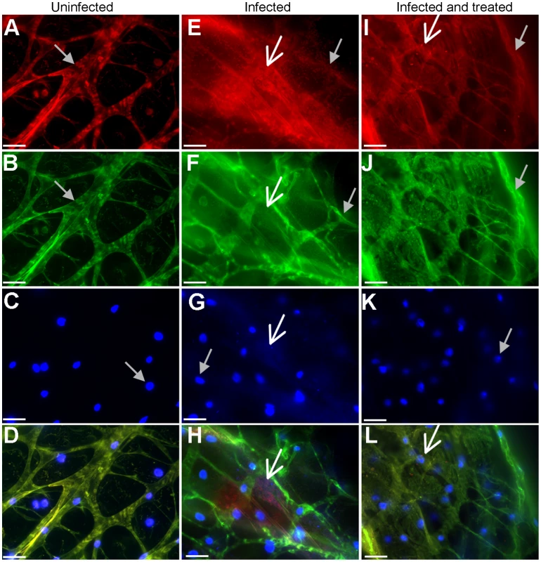
(A) Red autofluorescence, (B) green autofluorescence, (C) DAPI-stained nuclei (all indicated by grey arrow) and (D) merge image of uninfected Drosophila crop. (E) Aggregative red fluorescent PAO1pCHAP6656 (white arrow) along with red autofluorescence (grey arrow), (F) green fluorescent EPS (white arrow) staining and autofluorescence (grey arrow), (G) DAPI staining of bacteria (white arrow) and Drosophila nuclei (grey arrow) and (H) merge image of PAO1-infected crops. DNAse and cellulase treatment of P. aeruginosa-infected crops. (I) Non-aggregative red fluorescent PAO1pCHAP6656 (white arrow) along with red autofluorescence (grey arrow), (J) autofluorescence (grey arrow) and lack of EPS staining with FITC-labeled HHA lectin. (K) DAPI staining of Drosophila nuclei (grey arrow) and absence of bacterial DNA staining and (L) merge image of DNAse and cellulase treated PAO1 in infected crops (white arrow). Scale bars in indicate 100 µM. At least three infected crops were examined from two separate infections and representative images are shown. To show that DNA and EPS were important biofilm components in vivo, infected crops were DNAse - and cellulase-treated prior to EPS and DNA staining (Figure 2I–L) and compared to uninfected (Figure 2A–D) and PAO1pCHAP6656-infected crops without DNAse and cellulase treatment (Figure 2E–H). Aggregates of bacteria, which stained positively for DNA and EPS were visible in infected crops (Figure 2E–H). No aggregates were detected in PAO1pCHAP6656-infected crops treated with DNAse and cellulase, indicating that bacterial biofilms were dissolved following enzymatic treatment (Figure 2I–L). DNAse and cellulase treatment of uninfected crops had no effect on crop structure (data not shown).
On closer inspection of DAPI-stained bacteria in the crop (digital zoom of 4.4X), we observed that the bacteria in the microcolonies were organized into characteristic patterns or clusters (Figure 3). Cells that we previously observed to be positive for DNA and EPS staining (Figure 2A–D) were organized into a cluster of hexagonal bacterial colonies. These structures were found in two orientations, with bacteria lined up side-to-side and stacked one on top of the other or with the pole to pole length of the bacterial cells visible, and each cell attached side-to-side (Figure 3). Because of the size of each of the hexagons, approximately 6–8 µM, we predict that each hexagon is made up of approximately 7 to 15 bacterial cells. This is consistent with what has been observed for mature honeycomb structures produced by S. epidermidis [44]. These honeycomb-like shaped microcolonies were not firmly attached to the epithelial cell surface but were floating inside the enclosed fly crop (Video S1).
Fig. 3. Visualization and staining of in vivo microcolonies in the Drosophila crop. 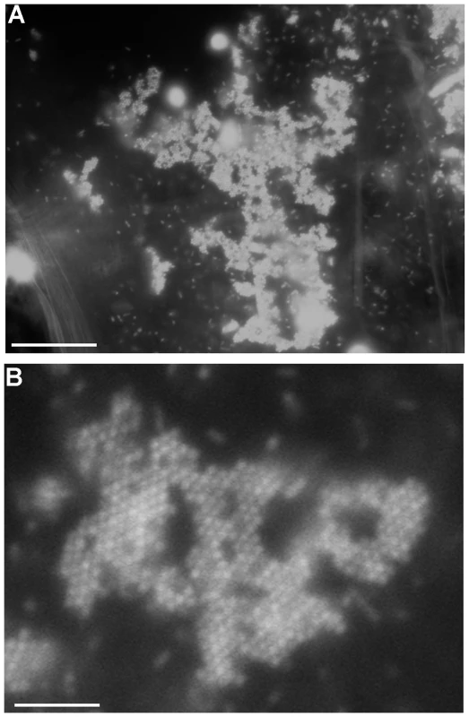
(A) DAPI-stained bacterial cells (100X) and (B) digitally zoomed images (4.4X) of DAPI stained microcolonies in the crop, demonstrating a honeycomb-like structure (white arrow). Scale bars in A indicate 200 µM. Scale bars in B indicate 45.4 µM. At least three infected crops were examined from three separate infections and representative images are shown. Honeycomb-like structures were visualized in 2 out of every three PAO1-infected crops examined. Microbial species, including Sinorhizobium meliloti, Rhizobium leguminosarum [45], [46], Staphlococcus epidermidis and P. aeruginosa [44] were previously shown to form complex biofilm structures, organised in honeycomb - and veil-like patterns. Honeycomb structures are one of the most densely packed structures found in nature and similar to what is observed with bee honeycombs [47], it is predicted that these structures enable close packing together of cells with the least amount of matrix components, including energy-expensive EPS. To our knowledge, bacterial microcolonies that resemble honeycomb structures have not previously been visualized in vivo in an animal model and highlight the use of Drosophila as an infection model amenable to microscopic analysis of infected tissues.
P. aeruginosa biofilm infection results in loss of integrity of the fly crop structure
To investigate the potential for detrimental consequences of P. aeruginosa biofilm infections on host tissue we examined the architecture of infected fly crops during a biofilm infection. Excised crops were visualized macroscopically for gross changes in shape and size at 10X magnification (Figure 4A and B). Brightfield imaging of uninfected (Figure 4A) and PAO1-infected (Figure 4B) crops indicated significant changes in the size and gross morphology of the crop in response to infection. Infected crops were smaller in size, softer in texture and were more sensitive to breaking apart upon handling (data not shown). This observation is consistent with tissue damage however it is also possible that a smaller softer crop may be as a result of an empty crop as P. aeruginosa infection may interfere with the ability of Drosophila to feed and drink.
Fig. 4. Crop integrity in response to P. aeruginosa infection. 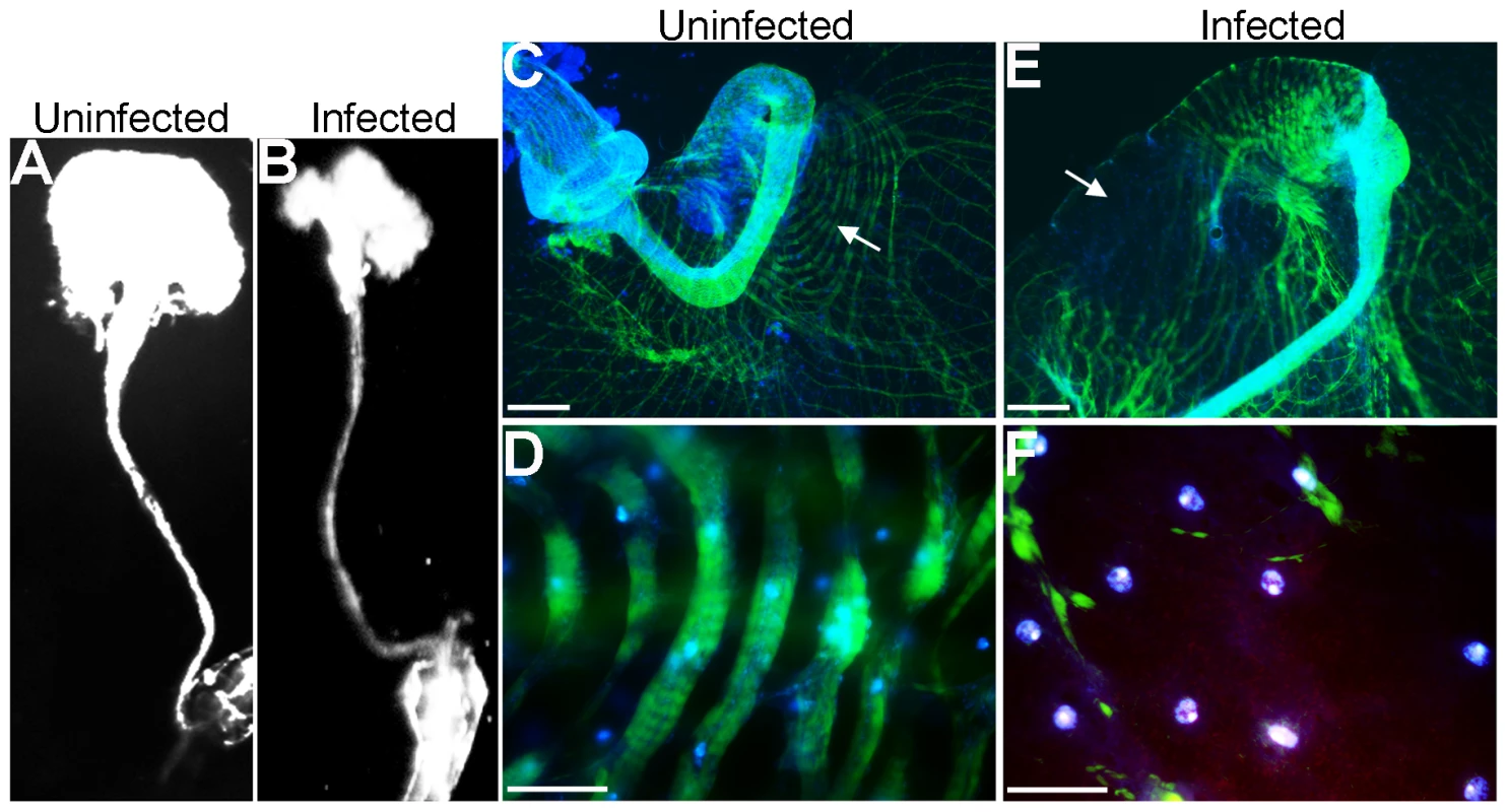
(A) The macroscopic structure of (A), uninfected and (B), PAO1pCHAP6656-infected Drosophila crops (Olympus OV100 intravital observation system). Merged fluorescent image of phallodin 488-stained actin (green) and DAPI-stained nuclei (blue) in uninfected crops using (C) 10x and (D) 63x objectives. PAO1pCHAP6656-infected crops (red) at (E) low and high (F) magnification. Scale bars in C and E indicate 400 µM; scale bars in D and E indicate 100 µM. White arrows in C and E indicate the area of the crop where higher magnification images were taken. At least five infected crops were examined from two separate infections and representative images are shown. To investigate the morphological structure of the musculature of the crop in greater detail in the presence and absence of biofilm infection, F-actin staining using Phallodin 488 was performed. Drosophila nuclei were counterstained with DAPI and crops examined by fluorescence microscopy. The musculature of uninfected crops consisted of wide ribbons of circular muscles covering the crop wall with a intricate network of branched and interconnecting fibers (Figure 4CD) This architecture was severely compromised or absent in infected crops (Figure 4EF). P. aeruginosa (red) localized predominantly to the crop edge, which was where the most disorganised actin staining was detected, including depolymerisation and degradation of actin filaments (Figure 4F).
Expression of EPS is essential for in vivo biofilm formation
We examined in vivo biofilm phenotypes of P. aeruginosa mutants known to exhibit altered EPS production and biofilm formation phenotypes in vitro. The pelB::lux mutant [48] is defective for biofilm formation in vitro, as mutants in the pel operon are known to have decreased EPS production and biofilm formation [49], while strain PAZHI3 (a mutant in the posttranscriptional regulatory protein RsmA) displayed increased production of both pel and psl EPS (Figure S2) and is a hyperbiofilm former [50]. Flies were infected with PAO1, pelB::lux or PAZHI3, all carrying pCHAP6656, and 24 h postinfection crops were excised and examined for the presence of microcolonies. Microscopy was performed on PAO1pCHAP6656, pelB::luxpCHAP6656, and PAZH13pCHAP6656-infected crops. Twelve fields of view were captured, from a minimum of 3 crops infected with each strain (Figure 5A–C), for quantification of microcolony formation (Figure 5D). Image analysis (ImageJ) was performed to differentiate and count the frequency of individual cells, as well as small and large microcolonies (Figure 5A) (See materials and methods for additional information). Large microcolonies (>20 cells, Figure 5A,C) were present only in PAO1 - or PAZHI3-infected crops and were absent from pelB::lux-infected crops (Figure 5B). PAZHI3-infected crops had more microcolonies (n = 15) present than those seen in PAO1-infected crops (n = 9) (Figure 4D). Furthermore, the large microcolonies (categorized as those microcolonies consisting of >20 cells) observed in PAZH13-infected crops were significantly larger (p<0.001) in size (approx 17-fold) that those microcolonies observed in PAO1-infected crops indicating the hyper-biofilm features of PAZHI3 detectable in vitro were also observed in vivo. No large microcolonies were detected in pelB::lux-infected flies (Figure 4B).
Fig. 5. The role of Pel EPS during in vivo biofilm formation in Drosophila. 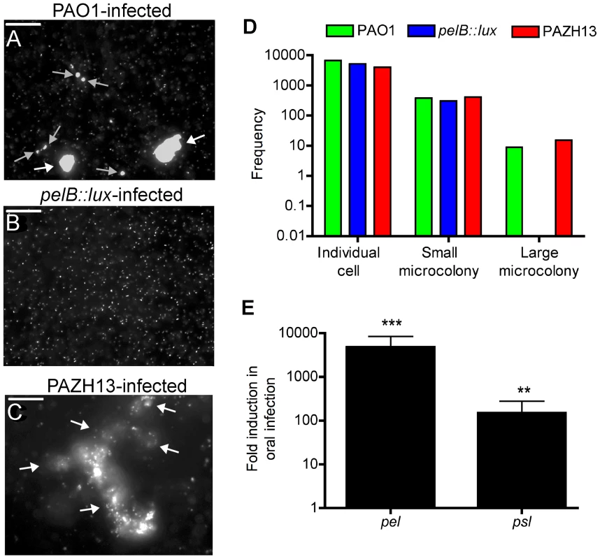
Representative images of P. aeruginosa pCHAP6656-infected crops. (A) PAO1pCHAP6656-infected crops contain individual bacterial cells, a number of small microcolonies (grey arrows) and two large microcolonies (white arrows). (B) PelB::luxpCHAP6656-infected crops contain individual bacterial cells and no small or large microcolonies. (C) PAZH13pCHAP6656-infected crops contain some individual bacterial cells, and five large microcolonies (white arrows). At least 3 infected crops were examined for each strain. Scale bar equals 100 µM. (D) Quantitative analysis of microcolony formation in response to infection with PAO1 and relevant mutant strains. At least 3 infected crops were examined for each strain. Data presented is the frequency of individual bacterial cells, small or large bacterial microcolonies in a total of 12 fields of view. (E) Expression of pel and psl EPS genes during oral infection relative to acute infection. Values are mean +/− SEM from triplicate qRT-PCR experiments on RNA isolated from two independent Drosophila infections. To further confirm the importance of EPS during oral Drosophila infection, qRT-PCR was used to measure expression of pel and psl during infection. Psl expression was significantly induced, approximately 150 fold (p<0.01). Pel expression was highest during oral infection of flies, induced approximately 2200 fold (p<0.001), relative to acute infection of flies (Figure 5E). These data indicate that Pel may play a more important role during oral infection of Drosophila, since it is more highly expressed. These data highlight the importance of Pel EPS as a biofilm matrix component for the establishment and/or maintenance of biofilms in vivo, in addition to its well-characterized importance for attachment and maturation, during the early and later stages of biofilm formation in vitro [51]–[53].
Non-biofilm forming strains disseminate at a faster rate than biofilm forming strains following oral infection
We hypothesized that biofilm forming and non-biofilm forming strains would differ in their ability and/or timing to disseminate and that ultimately the kinetics of bacterial dissemination may play a role in fly survival. Initial experiments were performed to compare in vivo localization of PAO1, pelB::lux and PAZHI3 strains in infected flies. Results of viable plate counts indicated a slightly lower bacterial load was recovered from the GI tract of pelB::lux-infected flies (3.8×10⁵±1.82×10⁴ CFU/fly; mean ± SEM), compared to that of PAO1-infected flies (4.8×10⁵±2.95×10⁴ CFU/fly). There was a corresponding increase in the number of viable pelB::lux bacteria (2.8×10⁴±1.83×10³ CFU/fly), recovered from the fly body, excluding the GI system, compared to that of PAO1-infected flies (1.03×10³±1.38×10² CFU/fly) 5 days postinfection (Figure 6A). Similar numbers of bacteria were isolated from the GI system or fly body of PAZH13-infected flies compared to PAO1-infected flies. To provide evidence of altered dissemination between biofilm and non-biofilm forming strains, hemolymph was recovered from infected flies at day 2 and day 5 postinfection. The pelB::lux mutant was present in the hemolymph at significantly higher numbers than PAO1 or PAZH13 at two days postinfection, while no significant difference in dissemination was observed five days postinfection (Figure 6B). Previous studies have shown that pelA mutants demonstrated increased rates of swarming motility [49], which in combination with reduced biofilm formation and may contribute to the increased rate of dissemination observed during infection of Drosophila 2 days postinfection. The fact that significantly increased numbers of pelB::lux are observed in the hemolymph 2 days postinfection relative to PAO1, while no significant difference is observed 5 days postinfection, suggests that upon detection by the host immune system in the hemolymph, pelB::lux bacteria are unable to persist or are cleared by the immune system.
Fig. 6. In vivo localization and antibiotic resistance profiling of biofilm and non-biofilm infections. 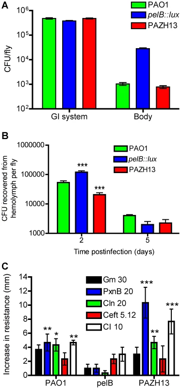
(A) Localization of bacteria in the fly 5 days postinfection. The GI tract including the crop, was dissected out, crushed and plated on PIA agar to determine CFU per GI tract/fly. The remainder of the fly body, including the head, was crushed separately and plated on PIA to determine CFU/rest of body per fly. (B) The number of CFU recovered from Drosophila hemolymph 2- and 5-days postinfection with PAO1, PAZH13 or pelB::lux. Two biological replicate experiments were performed, each containing 20 Drosophila, and values represented are mean +/−SEM. (C) Antibiotic resistance profiling of biofilm and non-biofilm infections. Increase in antibiotic resistance, as measured by zone of inhibition in disk diffusion assay, in P. aeruginosa strains recovered for Drosophila after oral infection relative to planktonic cultures. Antibiotic concentration indicated in µg/ml. Two biological replicate experiments were performed in triplicate and mean +/− SEM is shown. * p<0.05, ** p<0.01, ***p<0.001. P. aeruginosa recovered from biofilm infections in vivo have increased resistance to antimicrobials
Resistance to antimicrobials is a general feature of all biofilms. We hypothesized that PAO1 recovered from a biofilm infection of Drosophila would display increased resistance to antimicrobials. Antimicrobial sensitivities were compared in PAO1 directly recovered from flies relative to PAO1 planktonic cultures or PAO1 planktonic cultures exposed to pulverized fly tissues, termed “mock-infected” PAO1. Identical bacterial inocula from these three conditions were swabbed onto Pseudomonas Isolation Agar (PIA) and antimicrobial sensitivity was measured by disk diffusion. Antimicrobial resistance of PAO1 recovered from mock-infected cultures was not significantly different relative to the resistance profiles of PAO1 recovered from planktonic cultures (data not shown). PAO1 directly recovered from infected flies had significantly increased resistance to polymyxin B, colistin, and ciprofloxacin, but not to gentamicin or ceftazidime relative to planktonic PAO1 cultures (Figure 6C). PAZHI3 recovered from infected flies was also significantly more resistant to polymyxin B, colistin, and ciprofloxacin than planktonic PAZHI3 (Figure 6C). In contrast, antimicrobial resistance profiles of the pelB::lux mutant (which failed to form biofilms in vivo) were not significantly different in cells directly recovered from infected flies relative to planktonic or mock-infected cells (Figure 6C).
Polymyxin B and colistin are cationic AMPs: short, amphipathic peptides that bind to and disrupt both the outer and cytoplasmic membranes resulting in bacterial cell death [54]. Ciprofloxacin is a member of the fluoroquinolone drug class which inhibits DNA gyrase and hence DNA replication. The increased antibiotic resistance phenotype of P. aeruginosa recovered from the flies compared to planktonic cultures, is analogous to the increased resistance observed in in vitro biofilm populations compared to planktonic cultures [4], [55].
Drosophila infected with biofilm and non-biofilm forming P. aeruginosa have altered survival kinetics
To assess the comparative abilities of biofilm and non-biofilm forming P. aeruginosa strains for their ability to cause disease in Drosophila, we monitored fly survival over 14 days in response to oral infection. The non-biofilm forming pelB::lux mutant was significantly more virulent compared to PAO1, having a significantly increased rate of Drosophila killing (Figure 7A). In contrast, hyperbiofilm-forming PAZHI3 demonstrated significantly reduced virulence compared to PAO1, as indicated by a greater survival of infected flies up to 14 days postinfection (Figure 7A). There was no difference in the bacterial load (CFU) in biofilm and non-biofilm infected flies (data not shown). PAO1 mutants in psl showed similar killing kinetics to PAO1-infected flies (Figure S3) indicating that Pel EPS contributes to pathogenesis during infection of Drosophila while Psl EPS does not. Previous in vitro studies have indicated that both Pel and Psl are important in P. aeruginosa biofilm formation [6], [52] and that Pel EPS also contributes to antibiotic resistance [56]. Our data highlight a unique role for Pel EPS in P. aeruginosa biofilm formation in vivo, as well as a role in dissemination and virulence.
Fig. 7. Kaplan-Meier survival curves post P. aeruginosa infection. 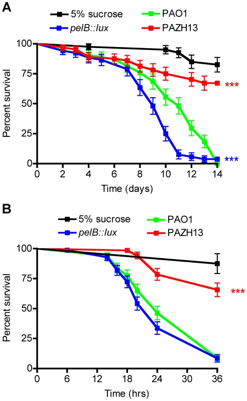
Survival curves of (A) oral and (B) acute infection with PAO1, pelB::lux, PAZH13 or 5% sucrose control. Experiments were performed at least 3 times each with a minimum of 80 flies and representative curves (mean +/− standard deviation) are shown. *** p<0.001. The production of Pel EPS and biofilm formation inversely correlated with virulence and the ability of P. aeruginosa to cause death in Drosophila after oral infection (Figure 7A). To determine if EPS production also affected the outcome of acute P. aeruginosa infections, Drosophila killing kinetics were compared in male flies nicked in the thoracic region, with the relevant P. aeruginosa strains, up to 36 h. Pel production was not found to be important factor during acute infection as PAO1 and pelB::lux infections resulted in similar killing kinetics in acutely-infected Drosophila up to 36 h postinfection (Figure 7B). PAZHI3 was attenuated for virulence during acute infection, similar to what was seen for oral infection (Figure 7). Reduced killing of Drosophila by PAZHI3 is similar to reduced virulence previously observed for PAZHI3 in a mouse model of acute infection [50].
Biofilm infections induce AMP gene expression
To monitor the AMP response to biofilm and non-biofilm infections in Drosophila, we assessed the expression of the AMP genes cecropin A1, diptericin and drosomycin using qRT-PCR during oral infection with PAO1, pelB::lux and PAZHI3 (Figure 8A–C). As no difference in killing kinetics were observed between flies acutely infected with biofilm forming PAO1 and non-biofilm forming pelB::lux, AMP gene expression was not monitored following acute infection.
Fig. 8. Biofilm infections induce antimicrobial peptide gene expression in Drosophila. 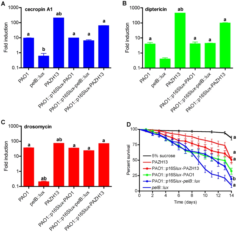
Real time RT-PCR analysis of (A) cecropin A1 (B) diptericin and (C) drosomycin following oral infection with PAO1, pelB::lux, PAZH13 or following oral co-infection with a 1∶1 ratio of PAO1-PAO1p16Slux, PAO1p16Slux-pelB::lux or PAO1p16Slux-PAZH13. For co-infection experiments (last 3 bars) the strains used for each infection are listed, separated by a hyphen. The levels of AMP gene expression was represented as fold change relative to uninfected flies. Values are mean +/− SEM from triplicate qPCR experiments on RNA isolated from two independent Drosophila infections. a, significant fold change (p<0.05, ANOVA) relative to uninfected flies; b, significant fold change (p<0.05, ANOVA) relative to PAO1-infected flies. (D) Kaplan-Meier survival curves of Drosophila following oral co-infection with a 1∶1 ratio of PAO1-PAO1p16Slux, PAO1p16Slux-pelB::lux, PAO1p16Slux-PAZH13 and relevant controls. Experiments were performed at least 3 times each with a minimum of 80 flies and representative curves (mean +/− standard deviated) are shown. a, significant difference (p<0.05, ANOVA) relative to PAO1-PAO1p16Slux-infected flies (green); b, significant difference (p<0.05, ANOVA) relative to pelB::lux-infected flies. PAO1 oral infection induced the expression of cecropin A1, diptericin and drosomycin between 4 - and 36-fold relative to uninfected flies (Figure 8A–C). Increased gene expression was also detected in PAZHI3-infected flies at levels between 72 - and 446-fold. While PAZH13 is hyperbiofilm former in vitro [50] and in vivo (Figure 5C), it is also a pleiotrophic mutant [57]–[61]. Thus, while there is a correlation between biofilm formation and increased AMP expression, we cannot rule out the possibility that the higher levels of AMP induction seen in response to PAZH13 infection may not be solely attributable to increased biofilm formation. In response to pelB::lux infection, we observed lower expression of all three AMP genes, between 1.6 - and 5-fold, compared to uninfected flies. Suppression of AMP gene expression is thought to be one of the main mechanisms whereby commensal bacteria fail to elicit an immune response in the host [62]. However, virulent strains of P. aeruginosa have also been documented to suppress AMP gene expression and the Drosophila immune response during an acute infection [24]. In this study, decreased expression of AMP gene expression by the pelB::lux mutant (Figure 8) appears to be associated with increased fly death following oral infection as flies die at a significantly faster rate compared to those infected with the biofilm forming Pel positive strains PAO1 or PAZH13 (Figure 7A). While this study does not demonstrate active suppression, it is possible that increased fly mortality post oral infection resulted from decreased expression of AMP gene expression in the fly and/or a more rapid (within 2 days) dissemination of pelB::lux to the hemolymph, resulting in systemic infection and fly death. However in addition to difference in localization of pelB::lux (Figure 6A), it may also be that pelB::lux is more toxic, eliciting pathological changes in Drosophila resulting in more rapid death.
As EPS can be a cell-surface or secreted product, we hypothesized that co-infection of Drosophila with a 1-1 mixture of P. aeruginosa wildtype and pelB::lux would restore AMP gene expression and killing, similar to levels observed in orally PAO1-infected flies. In these experiments, PAO1::p16Slux [63] was used instead of PAO1 as the wildtype strain as bacterial load and AMP gene expression did not differ significantly in PAO1::p16Slux-infected flies compared to PAO1-infected flies (data not shown). Use of PAO1::p16Slux allowed us to differentiate between wildtype and mutant strains for quantitative bacteriology using erythromycin resistance in PAO1::p16Slux as the differentiating marker. Relative to uninfected flies, AMP gene expression was measured in flies co-infected with PAO1::p16Slux and PAO1, pelB::lux or PAZHI3. There was no significant difference in AMP gene expression following co-infection with PAO1::p16Slux and PAO1 (Figure 8A–C). Co-infection with PAO1::p16Slux and pelB::lux resulted in induction of 6.3-, 4.3-, and 23-fold for cecropin A1, diptericin and drosomycin, respectively (Figure 8A–C), induction levels similar to those observed for wildtype infections. For PAO1::p16Slux and PAZHI3 co-infected flies, AMP genes were induced at levels between 62 to 97 fold (Figure 8A–C). These data indicated that co-infection of pelB::lux and PAO1 restored AMP gene expression to levels similar to those observed in PAO1-infected flies. In all co-infection experiments, quantitative bacteriology was performed at T0, T24 and T120 (hours) to ensure that the bacterial load was at a ratio of approximately 1-1 at the initial stage of infection (T0), at the time of RNA extraction (T24), and at later time points during infection (T120) (Figure S4). No significant differences were observed in the growth of different bacterial strains in Drosophila following co-infection at any of the time points investigated, indicating that pelB::lux or PAZH13 mutants were not altered in their ability to compete with PAO1 for colonization during infection of Drosophila.
Drosophila survival was also monitored following co-infection experiments. Co-infection of flies with pelB::lux and PAO1::p16Slux resulted in significantly increased fly survival relative to pelB::lux-infected flies, increasing fly survival to levels similar to those seen during wildtype infection (PAO1 and PAO1::p16Slux) (Figure 8D). Co-infection of flies with PAZH13 and PAO1::p16Slux had no significant effect on fly survival with flies dying at similar rates regardless of whether they were infected with PAZH13 alone or co-infected with PAZH13 and PAO1::p16Slux. Single infection with PAO1::p16Slux or PAO1, or co-infection with PAO1 and PAO1::p16Slux, had similar killing kinetics (data not shown).
Prior to this study, it was not known if the fly immune system responded differently to biofilm and non-biofilm forming bacteria. Drosomycin expression is regulated through the Toll pathway [64]; Diptericin is regulated via the Imd pathway [65] and both pathways overlap to regulate cecropin A1 expression [66]. Our data indicates that both of the central immune pathways in Drosophila are activated in response to biofilms. In addition this data indicates that it is Pel positive biofilms, and possibly Pel EPS itself, that may act as a specific host immune signal inducing AMP gene expression in Drosophila as a psl mutant has no effect on Drosophila killing (Figure S3). Future work will focus on identifying the specific bacterial components involved in AMP gene expression and other host signalling pathways in response to Pel and Psl positive biofilms and non-biofilm P. aeruginosa infections.
In the Drosophila oral infection model, our data suggests that Pel positive biofilms induced AMP gene expression in the fly. Although biofilm infections induce AMP gene expression (Figure 8A–C), biofilm-forming bacteria isolated from fly crops postinfection are more resistant to the AMPs polymyxin B and colistin than those recovered from planktonic cultures (Figure 6). Bacterial Pel EPS may be a cue to the host to increase AMP gene expression thus serving to slow dissemination of the bacteria, and in this way slow systemic infection which would rapidly kill the host. On the other hand, EPS may also induce inflammation in the crop/GI system resulting in a localized damage to the host. Strains incapable of forming Pel positive biofilms in vivo resulted in a decreased AMP response but disseminated earlier, resulting in a systemic infection associated with faster host killing. These interpretations are supported by the Drosophila survival data obtained from co-infection experiments, where co-infection of flies with pelB::lux and PAO1 significantly increases Drosophila survival compared to infection with pelB::lux alone.
Biofilm infections do not alter kinetics of subsequent acute infection but modify fly survival in response to subsequent oral challenge
It has previously been shown that P. aeruginosa eludes host defenses by suppressing AMP gene expression in a Drosophila model of acute infection [24]. This study also demonstrated that infection with a less virulent P. aeruginosa strain resulted in immune potentiation and protected flies from subsequent acute infection with a more virulent P. aeruginosa strain [24]. To determine if oral infection, biofilm formation and induction of AMPs in Drosophila could alter the kinetics of fly survival following subsequent acute infection, we performed the following experiment. Male flies were orally infected with PAO1 (biofilm, AMP induction), pelB::lux (non-biofilm, AMP repression) or PAZHI3 (hyperbiofilm, AMP induction) for 24 h. After 24 h, orally infected flies from each of the three groups above and uninfected flies were nicked with PAO1 (acute infection), LB (sterile nicking) or not treated. Oral infection with PAO1, pelB::lux or PAZHI3 had no significant effect on the rate of fly survival during subsequent acute infection (nicking) with PAO1 (Figure 9).
Fig. 9. Kaplan-Meier survival curves of Drosophila orally infected (feeding) for 24h followed by subsequent acute (nicking) or secondary oral infection. 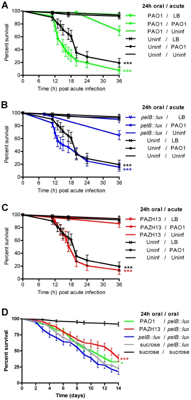
Survival following oral infection with (A) PAO1, (B) pelB::lux or (C) PAZH13 and relevant controls followed by acute infection with PAO1 or relevant controls. (D) Drosophila survival following oral infection with PAO1, pelB::lux, PAZH13 or uninfected (sucrose control) followed by oral infection with pelB::lux or uninfected (sucrose control). Strain name preceding the forward slash "/" indicates the strain or uninfected sucrose control used for oral infection of Drosophila; the strain name following the forward slash "/" indicates the strain or nicked (LB) or uninfected sucrose controls used for subsequent acute or secondary oral infection of Drosophila. Experiments were performed at least twice, each with a minimum of 100 flies and representative curves (mean +/- standard deviated) are shown. * p<0.05, ***p<0.001. To determine if oral PAO1 or PAZH13 biofilm infections altered Drosophila survival following subsequent oral infection with pelB::lux, the following experiment was performed. Drosophila were allowed to feed on PAO1, PAZH13, pelB::lux or a sucrose control for 24 h (primary infection), which is sufficient for biofilm formation to occur in the crop (Figure 1). After 24 h, all flies were transferred to new vials containing pelB::lux as the food source (secondary infection). Survival was monitored up to 14 days after the primary infection. Primary infection with PAO1 or PAZH13, followed by secondary infection with pelB::lux significantly increased fly survival compared to flies who were infected with pelB::lux for both the primary and secondary infection. Increased Drosophila survival following primary infection with PAO1 or PAZH13 was not due to failure of the secondary infecting pelB::lux strain to infect Drosophila, as pelB::lux tetracycline resistant colonies (the antibiotic marker of the lux transposon) were recovered (at ≥3.8×10⁶ CFU/fly or 76–99% of total bacterial load) from all secondary pelB::lux infected flies 5 days postinfection.
Primary oral infection with a biofilm-forming strain protected Drosophila from secondary oral infection with pelB::lux. Oral infection with a biofilm forming strain induced AMP gene expression, which may explain why increased fly survival was observed against secondary oral infection with pelB::lux. However the AMPs induced following oral infection may not be sufficient to alter Drosophila survival against subsequent acute infection. A possible reason for this is that AMP induction following biofilm infection is localized to the gut and does not protect Drosophila from death as a result of pricking and acute systemic infection. It is also possible that the pathology resulting from tissue damage following oral infection (Figure 4) may prevent Drosophila from responding to and coping with subsequent acute infection.
Conclusions
P. aeruginosa infections are associated with the highest case fatality rate of all Gram-negative infections [67]. This is partly due to the ability of P. aeruginosa to resist antimicrobial therapy. One of the main evasion strategies used by P. aeruginosa, and other microbes, is the formation of multicellular, dense aggregates called biofilms. We have shown that specific antibiotic resistance mechanisms are induced in P. aeruginosa biofilms [4]. Biofilm infections are estimated to account for 65% of all bacterial infections [68]. While some studies have investigated the host response to P. aeruginosa infection [20], [69], [70], little is known regarding the bacterial and/or host factors involved in the pathogenesis of biofilm infections. The aim of this research was to develop a Drosophila infection model that enables biofilms to be intricately studied in vivo.
In this work we present evidence that oral infection of Drosophila by P. aeruginosa PAO1 resulted in biofilm formation in the Drosophila crop (Figure 1). We demonstrated that biofilms formed in vivo retain the typical characteristics of in vitro grown biofilms, including DNA and EPS staining (Figure 2) and increased resistance to antibiotics (Figure 6C). We also showed that biofilm infections resulted in significantly decreased numbers of bacteria disseminating to the hemolymph 2 days postinfection, and contributed to increased AMP gene expression in the fly (Figures 6, 8). Non-biofilm forming pelB::lux infections, on the other hand, resulted in decreased AMP gene expression in the fly, significantly increased numbers of bacteria disseminating to the hemolymph 2 days postinfection, as well as early and increased fly mortality (Figures 6–8). The increased virulence of the pelB::lux mutant was attenuated by co-infection of Drosophila with biofilm-forming and AMP-inducing strains PAO1 or PAZH13 (Figure 8D). Furthermore, primary infection with either of these AMP-inducing strains altered the survival kinetics of Drosophila from secondary oral infection with the more virulent pelB::lux but not from subsequent acute infection (Figure 9). In summary, we have developed a novel P. aeruginosa biofilm model of infection that can be used for studying both the bacterial and host response during infection. This model has the potential to significantly increase our understanding of the relationship between biofilms and the host during infection and also to tease out fundamental differences between the host response to biofilm and non-biofilm P. aeruginosa infections.
Materials and Methods
Bacterial strains and plasmids
Pseudomonas aeruginosa PAO1 and PAO1::p16Slux [63] were used as wildtype P. aeruginosa strains. The pelB::lux mutant is from a mini-Tn5-lux transposon mutant library that was previously constructed and mapped [48]. PAZHI3 is an rsmA mutant in the PAO1 background [61]. The plasmid pCHAP6656 encodes mCherry fluorescent outer membrane-anchored lipoproteins [42].
P. aeruginosa oral infection of Drosophila
Drosophila were maintained routinely on medium containing corn meal, agar, sucrose, glucose, brewers' yeast, propionic acid, and phosphoric acid [71]. Infections were performed as previously described [25]. Mid-log phase LB cultures of P. aeruginosa were spun down and resuspended in 5% sucrose. Cultures were adjusted to an OD600 = 25 (2.5×10¹⁰ CFU per ml) in sucrose. The resuspended cells (0.12 mls) were spotted onto a sterile filter (Whatman) that was placed on the surface of 5 ml of solidified 5% sucrose agar in a plastic vial (VWR). The vials were allowed to dry at room temperature for approximately 30 minutes prior to addition of Drosophila. Because of the high concentration of bacteria on the feeding discs and the possibility of bacteria forming aggregates on the feeding discs over time, male Canton S flies (1–3 days old) were starved for 3 hours prior to being added to vials (10–14 flies per vial). This ensured that Drosophila fed heavily on P. aeruginosa within the first couple of hours. It is therefore unlikely that the P. aeruginosa strains on the filters had sufficient time to form biofilms prior to being eaten by Drosophila and causing an infection. Male flies were used as the infection lasts up to 14 days. During this time period females would have laid eggs, which if hatched, would interfere with the experimental results. Flies were anaesthetized by placing them on an ice-cold tile throughout the sorting and transferring process. Infection vials were stored at 26°C in a humidity controlled environment. The number of live flies to start the experiment was documented and live flies were counted at 24 hour intervals.
Acute P. aeruginosa infection of Drosophila
Healthy 3 day-old male flies were used in the fly nicking assays according to a modified method of [20]. Flies were sorted following anesthesis on a cold tile. The male flies were nicked in the dorsal thorax with a 27.5-gauge needle (BD Biosciences), which was dipped in bacterial culture normalized to an optical density at 600 nm of 1.0 in LB broth. After nicking, 10–14 flies were placed into a vial of 5% sucrose agar and maintained at room temperature. Fly survival was monitored and recorded from 12 to 36 h postinoculation.
Excision of gastrointestinal tract, crop and live cell imaging
Flies were sacrificed after which the inferior region of the abdomen was dissected under a dissecting microscope and the entire gastrointestinal (GI) system gently pulled through the resulting opening. The crop was separated from the rest of the GI system. For visualization of whole crop morphology, an Olympus OV100 intravital observation system was used and image analysis performed using Adobe Photoshop. For staining of bacteria and matrix components, crops were placed in phosphate buffered saline (PBS) and permeabilized with 0.1% Triton X100 for 15 mins. Following a wash step in PBS, crops were stained with fluorescent dyes of interest: 10 µg/ml FITC labelled HAA lectin (EY Laboratories, Inc) for exopolysaccharide, 80 µg/µl DAPI (Sigma) for Drosophila nuclei and DNA in the biofilm, and 1/40 (75 units) phallodin 488 (molecular probes) for F-actin. Crops were placed on a drop of PBS on a microscope slide, sealed with a coverslip and clear nail varnish and allowed to dry prior to viewing on a Leica DMIREB2 inverted, epifluorescence microscope. Crops were visualized using the 10, 40, 63 or 100x objective. Red, green and blue fluorescent images were merged using Adobe Photoshop.
Quantification of biofilm formation in the crop
Image analysis using ImageJ was performed to identify and count the frequency of each individual cell, as well as small and large aggregates or biofilms present in the Drosophila crop. Using the ‘analyse particle’ function, the integrated density (sum of the grey values of the pixels in the object) of each event was measured. Data was organized into bins, depending on the integrated density of the cell/aggregate, counted and the frequency of each bin was calculated from 12 fields of view taken from at least 3 crops infected with either the wild-type PAO1 or the pelB::lux and rsmA mutants (Figure 4D). Bins were separated into three groups, individual cells, small microcolonies or large microcolonies, which had integrated density values >100,000, between 100,000–500,000 and >500,000, respectively. Images representing each of 3 strains are represented in Figure 5.
Quantitative bacteriology from whole flies
Five infected live flies for each infection were crushed using a pellet pestle (Krackeler Scientific Inc.) in 300 µl PBS, serially diluted and plated onto Pseudomonas isolation agar (PIA; Difco) for PAO1 enumeration. To enumerate the CFUs in different regions of the fly, the GI systems of 3 flies was excised as previously described, crushed and plated. Flies with their GI tract removed were also pooled and plated. PIA plates were incubated at 37°C for 24 hours. Colonies were counted following incubation and CFU/fly was calculated.
Hemolymph isolation
Hemolymph was isolated from 20 infected or uninfected flies in triplicate according to the method of Frydman, 2006 [72], yielding approximately 2 µl of hemolymph per replicate experiment. Hemolymph was serially diluted in PBS and plated on PIA agar to determine CFU per fly.
Antimicrobial disc diffusion assay
Drosophila 5 days postinfection with PAO1 were crushed (5 flies per treatment) to give a predicted innoculum of 1×10⁷ CFU/ml. This was later verified by plate counts. Mock-infected flies consisted of planktonically grown PAO1 that was added to crushed uninfected flies prior to inoculation on PIA. This control was included to ensure any Drosophila product present in crushed flies did not alter the antibiotic resistance phenotype of PAO1. Plates were inoculated using a sterile swab. One µl of the following antibiotics were dispensed onto the agar plate: gentamicin (Gm) 30 µg/ml, polymyxin B (PxnB) 20 µg/ml, colistin (Coln) 20 µg/ml, ceftazadine (Ceft) 5.12 µg/ml and ciprofloxacin (CI) 10 µg/ml. Plates were incubated for 24 h at 37°C, after which zone sizes (mm) were determined. Zones of inhibition were measured for overnight cultures of planktonically grown PAO1, Drosophila 5 days postinfection with PAO1 and mock-infected PAO1.
RNA isolation, reverse transcription and qPCR
Total RNA was extracted from five flies from each infection 24 hours postinfection using TRIzol (Invitrogen), as previously described [73] RNA was DNAsed using DNAfree (Ambion) and cDNA synthesized with a High Capacity cDNA synthesis kit (ABI Biosystems). 100 ng of cDNA was used as template in the Real-time PCR reactions. Custom TaqMan probes and for diptericin (Dm01841768_s1), cecropin A1 (Dm02609400_s1) and drosomycin (Dm01822006_s1) and TaqMan Gene Expression Mastermix were used as recommended by the manufacturer (ABI Biosystems). RpL32 (Dm02151827_g1) was used as the constitutive control. Prokaryotic gene expression was measured using the iQ SYBR green supermix (Biorad) and bacterial specific primers to pel, psl and the 16S housekeeping gene (pelrtF 5′atcaagccctatccgttcct 3′, pelrtR 5′ aacggatggctgaaggtatg 3′, pslrtF 5′ agcagcaagctggtgatctt 3′, pslrtR 5′ggttgcgtaccaggtattcg 3′, 16SrtF 5′ gaaatccccgggctcaacctg 3′, 16SrtR 5′ccccacgctttcgcacctca3′). For quantitative RT-PCR (qRT-PCR), quantification and melting curve analyses were performed with an iQ5 (Bio-Rad) according to manufacturer's instructions. Each reaction is done in triplicate and standard deviations used to calculate a range of fold activation using the 2ΔΔCt method [74].
Statistical analysis
Survival curves were plotted and statistical analysis was performed using GraphPad Prism 5 software. 2-way ANOVA was used to calculate significant differences between PAO1 and mutant strains.
Drosophila gene identification
The FlyBase gene identification numbers for Drosophila genes are as follows: Drosomycin FBgn0010381; Diptericin FBgn0034407; Cecropin A1 FBgn0000276.
Supporting Information
Zdroje
1. YahrTLGreenbergEP 2004 The genetic basis for the commitment to chronic versus acute infection in Pseudomonas aeruginosa. Mol Cell 16 497 498
2. ParsekMRSinghPK 2003 Bacterial biofilms: An emerging link to disease pathogenesis. Annu Rev Microbiol 57 677 701
3. Moreau-MarquisSStantonBAO'TooleGA 2008 Pseudomonas aeruginosa biofilm formation in the cystic fibrosis airway. Pulm Pharmacol Ther 21 595 9
4. MulcahyHCharron-MazenodLLewenzaS 2008 Extracellular DNA chelates cations and induces antibiotic resistance in Pseudomonas aeruginosa biofilms. PLoS Pathog 4 e1000213
5. WhitchurchCBTolker-NielsenTRagasPCMattickJS 2002 Extracellular DNA required for bacterial biofilm formation. Science 295 1487
6. RyderCByrdMWozniakDJ 2007 Role of polysaccharides in Pseudomonas aeruginosa biofilm development. Curr Opin Microbiol 10 644 648
7. SutherlandIW 2001 The biofilm matrix–an immobilized but dynamic microbial environment. Trends Microbiol 9 222 227
8. DaviesD 2003 Understanding biofilm resistance to antibacterial agents. Nat Rev Drug Discov 2 114 122
9. CostertonJWStewartPSGreenbergEP 1999 Bacterial biofilms: A common cause of persistent infections. Science 284 1318 1322
10. DaveyMEO'tooleGA 2000 Microbial biofilms: From ecology to molecular genetics. Microbiol Mol Biol Rev 64 847 867
11. RahmeLGStevensEJWolfortSFShaoJTompkinsRG 1995 Common virulence factors for bacterial pathogenicity in plants and animals. Science 268 1899 1902
12. RahmeLGTanMWLeLWongSMTompkinsRG 1997 Use of model plant hosts to identify Pseudomonas aeruginosa virulence factors. Proc Natl Acad Sci U S A 94 13245 13250
13. Mahajan-MiklosSTanMWRahmeLGAusubelFM 1999 Molecular mechanisms of bacterial virulence elucidated using a Pseudomonas aeruginosa -Caenorhabditis elegans pathogenesis model. Cell 96 47 56
14. ComolliJCHauserARWaiteLWhitchurchCBMattickJS 1999 Pseudomonas aeruginosa gene products PilT and PilU are required for cytotoxicity in vitro and virulence in a mouse model of acute pneumonia. Infect Immun 67 3625 3630
15. van HeeckerenAMSchluchterMD 2002 Murine models of chronic Pseudomonas aeruginosa lung infection. Lab Anim 36 291 312
16. KimDHFeinbaumRAlloingGEmersonFEGarsinDA 2002 A conserved p38 MAP kinase pathway in Caenorhabditis elegans innate immunity. Science 297 623 626
17. CossonPZulianelloLJoin-LambertOFaurissonFGebbieL 2002 Pseudomonas aeruginosa virulence analyzed in a Dictyostelium discoideum host system. J Bacteriol 184 3027 3033
18. KurzCLEwbankJJ 2007 Infection in a dish: High-throughput analyses of bacterial pathogenesis. Curr Opin Microbiol 10 10 16
19. Kukavica-IbruljIBragonziAParoniMWinstanleyCSanschagrinF 2008 In vivo growth of Pseudomonas aeruginosa strains PAO1 and PA14 and the hypervirulent strain LESB58 in a rat model of chronic lung infection. J Bacteriol 190 2804 2813
20. D'ArgenioDAGallagherLABergCAManoilC 2001 Drosophila as a model host for Pseudomonas aeruginosa infection. J Bacteriol 183 1466 1471
21. ChuganiSAWhiteleyMLeeKMD'ArgenioDManoilC 2001 QscR, a modulator of quorum-sensing signal synthesis and virulence in Pseudomonas aeruginosa. Proc Natl Acad Sci U S A 98 2752 2757
22. EricksonDLLinesJLPesciECVenturiVStoreyDG 2004 Pseudomonas aeruginosa relA contributes to virulence in Drosophila melanogaster. Infect Immun 72 5638 5645
23. SalunkhePSmartCHMorganJAPanageaSWalshawMJ 2005 A cystic fibrosis epidemic strain of Pseudomonas aeruginosa displays enhanced virulence and antimicrobial resistance. J Bacteriol 187 4908 4920
24. ApidianakisYMindrinosMNXiaoWLauGWBaldiniRL 2005 Profiling early infection responses: Pseudomonas aeruginosa eludes host defenses by suppressing antimicrobial peptide gene expression. Proc Natl Acad Sci U S A 102 2573 2578
25. SibleyCDDuanKFischerCParkinsMDStoreyDG 2008 Discerning the complexity of community interactions using a Drosophila model of polymicrobial infections. PLoS Pathog 4 e1000184
26. LutterEIFariaMMRabinHRStoreyDG 2008 Pseudomonas aeruginosa cystic fibrosis isolates from individual patients demonstrate a range of levels of lethality in two Drosophila melanogaster infection models. Infect Immun 76 1877 1888
27. ApidianakisYRahmeLG 2009 Drosophila melanogaster as a model host for studying Pseudomonas aeruginosa infection. Nat Protoc 4 1285 1294
28. LemaitreBHoffmannJ 2007 The host defense of Drosophila melanogaster. Annu Rev Immunol 25 697 743
29. KylstenPSamakovlisCHultmarkD 1990 The cecropin locus in Drosophila; a compact gene cluster involved in the response to infection. EMBO J 9 217 224
30. WickerCReichhartJMHoffmannDHultmarkDSamakovlisC 1990 Insect immunity. characterization of a Drosophila cDNA encoding a novel member of the diptericin family of immune peptides. J Biol Chem 265 22493 22498
31. BuletPDimarcqJLHetruCLagueuxMCharletM 1993 A novel inducible antibacterial peptide of Drosophila carries an O-glycosylated substitution. J Biol Chem 268 14893 14897
32. DimarcqJLHoffmannDMeisterMBuletPLanotR 1994 Characterization and transcriptional profiles of a Drosophila gene encoding an insect defensin. A study in insect immunity. Eur J Biochem 221 201 209
33. FehlbaumPBuletPMichautLLagueuxMBroekaertWF 1994 Insect immunity. Septic injury of Drosophila induces the synthesis of a potent antifungal peptide with sequence homology to plant antifungal peptides. J Biol Chem 269 33159 33163
34. LevashinaEAOhresserSBuletPReichhartJMHetruC 1995 Metchnikowin, a novel immune-inducible proline-rich peptide from Drosophila with antibacterial and antifungal properties. Eur J Biochem 233 694 700
35. AslingBDushayMSHultmarkD 1995 Identification of early genes in the Drosophila immune response by PCR-based differential display: The attacin A gene and the evolution of attacin-like proteins. Insect Biochem Mol Biol 25 511 518
36. LeulierFParquetCPili-FlourySRyuJHCaroffM 2003 The Drosophila immune system detects bacteria through specific peptidoglycan recognition. Nat Immunol 4 478 484
37. LevashinaEAOhresserSLemaitreBImlerJL 1998 Two distinct pathways can control expression of the gene encoding the Drosophila antimicrobial peptide metchnikowin. J Mol Biol 278 515 527
38. De GregorioESpellmanPTTzouPRubinGMLemaitreB 2002 The toll and imd pathways are the major regulators of the immune response in Drosophila. EMBO J 21 2568 2579
39. TanjiTHuXWeberANIpYT 2007 Toll and IMD pathways synergistically activate an innate immune response in Drosophila melanogaster. Mol Cell Biol 27 4578 4588
40. ChatterjeeMIpYT 2009 Pathogenic stimulation of intestinal stem cell response in Drosophila. J Cell Physiol 220 664 671
41. ApidianakisYPitsouliCPerrimonNRahmeL 2009 Synergy between bacterial infection and genetic predisposition in intestinal dysplasia. Proc Natl Acad Sci U S A 106 20883 20888
42. LewenzaSMhlangaMMPugsleyAP 2008 Novel inner membrane retention signals in Pseudomonas aeruginosa lipoproteins. J Bacteriol 190 6119 6125
43. MaLLuHSprinkleAParsekMRWozniakDJ 2007 Pseudomonas aeruginosa psl is a galactose - and mannose-rich exopolysaccharide. J Bacteriol 189 8353 8356
44. SchaudinnCStoodleyPKainovicAOkeeffeTCostertonB 2007 Bacterial biofilms, other structures seen as mainstream concepts. Microbe 2 231 6
45. RinaudiLVGonzalezJE 2009 The low-molecular-weight fraction of exopolysaccharide II from Sinorhizobium meliloti is a crucial determinant of biofilm formation. J Bacteriol 191 7216 7224
46. RussoDMWilliamsAEdwardsAPosadasDMFinnieC 2006 Proteins exported via the PrsD-PrsE type I secretion system and the acidic exopolysaccharide are involved in biofilm formation by Rhizobium leguminosarum. J Bacteriol 188 4474 4486
47. PearceP 1978 Structure in nature is a strategy for design. Cambridge MIT Press 245
48. GoodmanALMerighiMHyodoMVentreIFillouxA 2009 Direct interaction between sensor kinase proteins mediates acute and chronic disease phenotypes in a bacterial pathogen. Genes Dev 23 249 259
49. CaiazzaNCMerrittJHBrothersKMO'TooleGA 2007 Inverse regulation of biofilm formation and swarming motility by Pseudomonas aeruginosa PA14. J Bacteriol 189 3603 3612
50. MulcahyHO'CallaghanJO'GradyEPMaciaMDBorrellN 2008 Pseudomonas aeruginosa RsmA plays an important role during murine infection by influencing colonization, virulence, persistence, and pulmonary inflammation. Infect Immun 76 632 638
51. VasseurPVallet-GelyISosciaCGeninSFillouxA 2005 The pel genes of the Pseudomonas aeruginosa PAK strain are involved at early and late stages of biofilm formation. Microbiology 151 985 997
52. FriedmanLKolterR 2004 Two genetic loci produce distinct carbohydrate-rich structural components of the Pseudomonas aeruginosa biofilm matrix. J Bacteriol 186 4457 4465
53. MatsukawaMGreenbergEP 2004 Putative exopolysaccharide synthesis genes influence Pseudomonas aeruginosa biofilm development. J Bacteriol 186 4449 4456
54. ZhangLDhillonPYanHFarmerSHancockRE 2000 Interactions of bacterial cationic peptide antibiotics with outer and cytoplasmic membranes of Pseudomonas aeruginosa. Antimicrob Agents Chemother 44 3317 3321
55. CeriHOlsonMEStremickCReadRRMorckD 1999 The calgary biofilm device: New technology for rapid determination of antibiotic susceptibilities of bacterial biofilms. J Clin Microbiol 37 1771 1776
56. ColvinKMGordonVDMurakamiKBorleeBRWozniakDJ 2011 The pel polysaccharide can serve a structural and protective role in the biofilm matrix of Pseudomonas aeruginosa. PLoS Pathog 7 e1001264
57. BrencicALoryS 2009 Determination of the regulon and identification of novel mRNA targets of Pseudomonas aeruginosa RsmA. Mol Microbiol 72 612 632
58. MulcahyHO'CallaghanJO'GradyEPAdamsCO'GaraF 2006 The posttranscriptional regulator RsmA plays a role in the interaction between Pseudomonas aeruginosa and human airway epithelial cells by positively regulating the type III secretion system. Infect Immun 74 3012 3015
59. BurrowesEBaysseCAdamsCO'GaraF 2006 Influence of the regulatory protein RsmA on cellular functions in Pseudomonas aeruginosa PAO1, as revealed by transcriptome analysis. Microbiology 152 405 418
60. HeurlierKWilliamsFHeebSDormondCPessiG 2004 Positive control of swarming, rhamnolipid synthesis, and lipase production by the posttranscriptional RsmA/RsmZ system in Pseudomonas aeruginosa PAO1. J Bacteriol 186 2936 2945
61. PessiGWilliamsFHindleZHeurlierKHoldenMT 2001 The global posttranscriptional regulator RsmA modulates production of virulence determinants and N-acylhomoserine lactones in Pseudomonas aeruginosa. J Bacteriol 183 6676 6683
62. RyuJHKimSHLeeHYBaiJYNamYD 2008 Innate immune homeostasis by the homeobox gene caudal and commensal-gut mutualism in Drosophila. Science 319 777 782
63. RiedelCUCaseyPGMulcahyHO'GaraFGahanCG 2007 Construction of p16Slux, a novel vector for improved bioluminescent labeling of gram-negative bacteria. Appl Environ Microbiol 73 7092 7095
64. LemaitreBNicolasEMichautLReichhartJMHoffmannJA 1996 The dorsoventral regulatory gene cassette spatzle/Toll/cactus controls the potent antifungal response in Drosophila adults. Cell 86 973 983
65. HedengrenMAslingBDushayMSAndoIEkengrenS 1999 Relish, a central factor in the control of humoral but not cellular immunity in Drosophila Mol Cell 4 827 837
66. Hedengren-OlcottMOlcottMCMooneyDTEkengrenSGellerBL 2004 Differential activation of the NF-kappaB-like factors relish and dif in Drosophila melanogaster by fungi and gram-positive bacteria. J Biol Chem 279 21121 21127
67. AliagaLMediavillaJDCoboF 2002 A clinical index predicting mortality with Pseudomonas aeruginosa bacteraemia. J Med Microbiol 51 615 619
68. PoteraC 1999 Forging a link between biofilms and disease. Science 2831837, 1839
69. SadikotRTBlackwellTSChristmanJWPrinceAS 2005 Pathogen-host interactions in Pseudomonas aeruginosa pneumonia. Am J Respir Crit Care Med 171 1209 1223
70. IrazoquiJETroemelERFeinbaumRLLuhachackLGCezairliyanBO 2010 Distinct pathogenesis and host responses during infection of C. elegans by P P. aeruginosa aeruginosa and S. aureus. PLoS Pathog 6 e1000982
71. MeadCG 1964 A deoxyribonucleic acid-associated ribonucleic acid from Drosophila melanogaster. J Biol Chem 239 550 554
72. FrydmanMH 2006 Isolation of live bacteria from adult insects. Protocol Exchange doi:10.1038/nprot.2006.131
73. LiehlPBlightMVodovarNBoccardFLemaitreB 2006 Prevalence of local immune response against oral infection in a Drosophila/Pseudomonas infection model. PLoS Pathog 2 e56
74. LivakKJSchmittgenTD 2001 Analysis of relative gene expression data using real-time quantitative PCR and the 2(-delta delta C(T)) method. Methods 25 402 408
75. MaLJacksonKDLandryRMParsekMRWozniakDJ 2006 Analysis of Pseudomonas aeruginosa conditional psl variants reveals roles for the psl polysaccharide in adhesion and maintaining biofilm structure postattachment. J Bacteriol 188 8213 8221
Štítky
Hygiena a epidemiologie Infekční lékařství Laboratoř
Článek Quorum Sensing in Fungi: Q&AČlánek Blood Feeding and Insulin-like Peptide 3 Stimulate Proliferation of Hemocytes in the MosquitoČlánek The DEAD-box RNA Helicase DDX6 is Required for Efficient Encapsidation of a Retroviral GenomeČlánek A Phenome-Based Functional Analysis of Transcription Factors in the Cereal Head Blight Fungus,Článek A Wide Extent of Inter-Strain Diversity in Virulent and Vaccine Strains of AlphaherpesvirusesČlánek The Anti-Sigma Factor TcdC Modulates Hypervirulence in an Epidemic BI/NAP1/027 Clinical Isolate ofČlánek Critical Roles for LIGHT and Its Receptors in Generating T Cell-Mediated Immunity during InfectionČlánek Frequent and Recent Human Acquisition of Simian Foamy Viruses Through Apes' Bites in Central Africa
Článek vyšel v časopisePLOS Pathogens
Nejčtenější tento týden
2011 Číslo 10- Stillova choroba: vzácné a závažné systémové onemocnění
- Jak souvisí postcovidový syndrom s poškozením mozku?
- Perorální antivirotika jako vysoce efektivní nástroj prevence hospitalizací kvůli COVID-19 − otázky a odpovědi pro praxi
- Diagnostika virových hepatitid v kostce – zorientujte se (nejen) v sérologii
- Infekční komplikace virových respiračních infekcí – sekundární bakteriální a aspergilové pneumonie
-
Všechny články tohoto čísla
- Quorum Sensing in Fungi: Q&A
- Discovery of an Ebolavirus-Like Filovirus in Europe
- Toll-like Receptor 7 Controls the Anti-Retroviral Germinal Center Response
- Tubule-Guided Cell-to-Cell Movement of a Plant Virus Requires Class XI Myosin Motors
- Herpesvirus Telomerase RNA (vTR) with a Mutated Template Sequence Abrogates Herpesvirus-Induced Lymphomagenesis
- Mitochondrial Peroxiredoxin Plays a Crucial Peroxidase-Unrelated Role during Infection: Insight into Its Novel Chaperone Activity
- Sustained CD8+ T Cell Memory Inflation after Infection with a Single-Cycle Cytomegalovirus
- Novel Mouse Xenograft Models Reveal a Critical Role of CD4 T Cells in the Proliferation of EBV-Infected T and NK Cells
- Toll-8/Tollo Negatively Regulates Antimicrobial Response in the Respiratory Epithelium
- Exhausted Cytotoxic Control of Epstein-Barr Virus in Human Lupus
- Structural and Functional Analysis of Laninamivir and its Octanoate Prodrug Reveals Group Specific Mechanisms for Influenza NA Inhibition
- Infection Drives IL-17-Mediated Neutrophilic Allergic Airways Disease
- Blood Feeding and Insulin-like Peptide 3 Stimulate Proliferation of Hemocytes in the Mosquito
- HIV-1 Replication in the Central Nervous System Occurs in Two Distinct Cell Types
- Deep Molecular Characterization of HIV-1 Dynamics under Suppressive HAART
- Fitness Landscape of Antibiotic Tolerance in Biofilms
- The DEAD-box RNA Helicase DDX6 is Required for Efficient Encapsidation of a Retroviral Genome
- Preventing Sepsis through the Inhibition of Its Agglutination in Blood
- A Phenome-Based Functional Analysis of Transcription Factors in the Cereal Head Blight Fungus,
- IFITM3 Inhibits Influenza A Virus Infection by Preventing Cytosolic Entry
- Targeting Cattle-Borne Zoonoses and Cattle Pathogens Using a Novel Trypanosomatid-Based Delivery System
- A Wide Extent of Inter-Strain Diversity in Virulent and Vaccine Strains of Alphaherpesviruses
- Coordinated Destruction of Cellular Messages in Translation Complexes by the Gammaherpesvirus Host Shutoff Factor and the Mammalian Exonuclease Xrn1
- Signal Transduction through CsrRS Confers an Invasive Phenotype in Group A
- Biochemical and Structural Insights into the Mechanisms of SARS Coronavirus RNA Ribose 2′-O-Methylation by nsp16/nsp10 Protein Complex
- Histone Deacetylase 8 Is Required for Centrosome Cohesion and Influenza A Virus Entry
- Severe Acute Respiratory Syndrome Coronavirus Envelope Protein Regulates Cell Stress Response and Apoptosis
- Co-opts the FGF2 Signaling Pathway to Enhance Infection
- IRAK-2 Regulates IL-1-Mediated Pathogenic Th17 Cell Development in Helminthic Infection
- Trafficking of Hepatitis C Virus Core Protein during Virus Particle Assembly
- The Anti-interferon Activity of Conserved Viral dUTPase ORF54 is Essential for an Effective MHV-68 Infection
- A Viral Nuclear Noncoding RNA Binds Re-localized Poly(A) Binding Protein and Is Required for Late KSHV Gene Expression
- Suppression of Methylation-Mediated Transcriptional Gene Silencing by βC1-SAHH Protein Interaction during Geminivirus-Betasatellite Infection
- ISG15 Is Critical in the Control of Chikungunya Virus Infection Independent of UbE1L Mediated Conjugation
- Non-Hematopoietic Cells in Lymph Nodes Drive Memory CD8 T Cell Inflation during Murine Cytomegalovirus Infection
- RNA Polymerase II Stalling Promotes Nucleosome Occlusion and pTEFb Recruitment to Drive Immortalization by Epstein-Barr Virus
- Noninfectious Retrovirus Particles Drive the / Dependent Neutralizing Antibody Response
- Endophytic Life Strategies Decoded by Genome and Transcriptome Analyses of the Mutualistic Root Symbiont
- An Integrated Approach to Elucidate the Intra-Viral and Viral-Cellular Protein Interaction Networks of a Gamma-Herpesvirus
- as an Animal Model for the Study of Biofilm Infections
- Homeostatic Proliferation Fails to Efficiently Reactivate HIV-1 Latently Infected Central Memory CD4+ T Cells
- The Anti-Sigma Factor TcdC Modulates Hypervirulence in an Epidemic BI/NAP1/027 Clinical Isolate of
- Enhances Protective and Detrimental HLA Class I-Mediated Immunity in Chronic Viral Infection
- The Mouse IAPE Endogenous Retrovirus Can Infect Cells through Any of the Five GPI-Anchored EphrinA Proteins
- The Urgent Need for Robust Coral Disease Diagnostics
- HacA-Independent Functions of the ER Stress Sensor IreA Synergize with the Canonical UPR to Influence Virulence Traits in
- A Novel Core Genome-Encoded Superantigen Contributes to Lethality of Community-Associated MRSA Necrotizing Pneumonia
- Critical Roles for LIGHT and Its Receptors in Generating T Cell-Mediated Immunity during Infection
- The SARS-Coronavirus-Host Interactome: Identification of Cyclophilins as Target for Pan-Coronavirus Inhibitors
- Frequent and Recent Human Acquisition of Simian Foamy Viruses Through Apes' Bites in Central Africa
- Mechanisms of Trafficking to the Brain
- Defining Emerging Roles for NF-κB in Antivirus Responses: Revisiting the Enhanceosome Paradigm
- The Role of Sialyl Glycan Recognition in Host Tissue Tropism of the Avian Parasite
- Evolutionarily Divergent, Unstable Filamentous Actin Is Essential for Gliding Motility in Apicomplexan Parasites
- The Herpes Simplex Virus-1 Transactivator Infected Cell Protein-4 Drives VEGF-A Dependent Neovascularization
- Distinct Single Amino Acid Replacements in the Control of Virulence Regulator Protein Differentially Impact Streptococcal Pathogenesis
- Soluble Rhesus Lymphocryptovirus gp350 Protects against Infection and Reduces Viral Loads in Animals that Become Infected with Virus after Challenge
- A Genetic Screen Reveals Arabidopsis Stomatal and/or Apoplastic Defenses against pv. DC3000
- Hepatitis C Virus Reveals a Novel Early Control in Acute Immune Response
- Fumarate Reductase Activity Maintains an Energized Membrane in Anaerobic
- PLOS Pathogens
- Archiv čísel
- Aktuální číslo
- Informace o časopisu
Nejčtenější v tomto čísle- Severe Acute Respiratory Syndrome Coronavirus Envelope Protein Regulates Cell Stress Response and Apoptosis
- The SARS-Coronavirus-Host Interactome: Identification of Cyclophilins as Target for Pan-Coronavirus Inhibitors
- Biochemical and Structural Insights into the Mechanisms of SARS Coronavirus RNA Ribose 2′-O-Methylation by nsp16/nsp10 Protein Complex
- Evolutionarily Divergent, Unstable Filamentous Actin Is Essential for Gliding Motility in Apicomplexan Parasites
Kurzy
Zvyšte si kvalifikaci online z pohodlí domova
Autoři: prof. MUDr. Vladimír Palička, CSc., Dr.h.c., doc. MUDr. Václav Vyskočil, Ph.D., MUDr. Petr Kasalický, CSc., MUDr. Jan Rosa, Ing. Pavel Havlík, Ing. Jan Adam, Hana Hejnová, DiS., Jana Křenková
Autoři: MUDr. Irena Krčmová, CSc.
Autoři: MDDr. Eleonóra Ivančová, PhD., MHA
Autoři: prof. MUDr. Eva Kubala Havrdová, DrSc.
Všechny kurzyPřihlášení#ADS_BOTTOM_SCRIPTS#Zapomenuté hesloZadejte e-mailovou adresu, se kterou jste vytvářel(a) účet, budou Vám na ni zaslány informace k nastavení nového hesla.
- Vzdělávání


