-
Články
- Vzdělávání
- Časopisy
Top články
Nové číslo
- Témata
- Kongresy
- Videa
- Podcasty
Nové podcasty
Reklama- Kariéra
Doporučené pozice
Reklama- Praxe
Tetraspanin Is Required for Generation of Reactive Oxygen Species by the Dual Oxidase System in
Reactive oxygen species (ROS) are toxic but essential molecules responsible for host defense and cellular signaling. Conserved NADPH oxidase (NOX) family enzymes direct the regulated production of ROS. Hydrogen peroxide (H2O2) generated by dual oxidases (DUOXs), a member of the NOX family, is crucial for innate mucosal immunity. In addition, H2O2 is required for cellular signaling mediated by protein modifications, such as the thyroid hormone biosynthetic pathway in mammals. In contrast to other NOX isozymes, the regulatory mechanisms of DUOX activity are less understood. Using Caenorhabditis elegans as a model, we demonstrate that the tetraspanin protein is required for induction of the DUOX signaling pathway in conjunction with the dual oxidase maturation factor (DUOXA). In the current study, we show that genetic mutation of DUOX (bli-3), DUOXA (doxa-1), and peroxidase (mlt-7) in C. elegans causes the same defects as a tetraspanin tsp-15 mutant, represented by exoskeletal deficiencies due to the failure of tyrosine cross-linking of collagen. The deficiency in the tsp-15 mutant was restored by co-expression of bli-3 and doxa-1, indicating the involvement of tsp-15 in the generation of ROS. H2O2 generation by BLI-3 was completely dependent on TSP-15 when reconstituted in mammalian cells. We also demonstrated that TSP-15, BLI-3, and DOXA-1 form complexes in vitro and in vivo. Cell-fusion-based analysis suggested that association with TSP-15 at the cell surface is crucial for BLI-3 activation to release H2O2. This study provides the first evidence for an essential role of tetraspanin in ROS generation.
Published in the journal: . PLoS Genet 8(9): e32767. doi:10.1371/journal.pgen.1002957
Category: Research Article
doi: https://doi.org/10.1371/journal.pgen.1002957Summary
Reactive oxygen species (ROS) are toxic but essential molecules responsible for host defense and cellular signaling. Conserved NADPH oxidase (NOX) family enzymes direct the regulated production of ROS. Hydrogen peroxide (H2O2) generated by dual oxidases (DUOXs), a member of the NOX family, is crucial for innate mucosal immunity. In addition, H2O2 is required for cellular signaling mediated by protein modifications, such as the thyroid hormone biosynthetic pathway in mammals. In contrast to other NOX isozymes, the regulatory mechanisms of DUOX activity are less understood. Using Caenorhabditis elegans as a model, we demonstrate that the tetraspanin protein is required for induction of the DUOX signaling pathway in conjunction with the dual oxidase maturation factor (DUOXA). In the current study, we show that genetic mutation of DUOX (bli-3), DUOXA (doxa-1), and peroxidase (mlt-7) in C. elegans causes the same defects as a tetraspanin tsp-15 mutant, represented by exoskeletal deficiencies due to the failure of tyrosine cross-linking of collagen. The deficiency in the tsp-15 mutant was restored by co-expression of bli-3 and doxa-1, indicating the involvement of tsp-15 in the generation of ROS. H2O2 generation by BLI-3 was completely dependent on TSP-15 when reconstituted in mammalian cells. We also demonstrated that TSP-15, BLI-3, and DOXA-1 form complexes in vitro and in vivo. Cell-fusion-based analysis suggested that association with TSP-15 at the cell surface is crucial for BLI-3 activation to release H2O2. This study provides the first evidence for an essential role of tetraspanin in ROS generation.
Introduction
Reactive oxygen species (ROS) are considered deleterious by-products of aerobic metabolism that inflict oxidative damage in organisms, and have been associated with numerous diseases and aging. ROS are produced in phagocytic and non-phagocytic cells and function to eliminate invading microbes [1], [2]. The physiological generation of ROS is directed by the NADPH oxidase (NOX) family of enzymes, which are highly conserved integral membrane proteins comprising seven members in mammals (NOX1–NOX5, DUOX1, and DUOX2) [3]–[5]. Studies of the NOX family have uncovered multiple biological functions of ROS in developmental processes, apoptosis, protein modification, cellular signaling and are well documented in host defense mechanisms [1], [6]–[8]. Dual oxidases (DUOX) were originally identified as thyroid oxidases, key H2O2 generators for the iodination of tyrosine in thyroid hormone precursors during thyroid hormone biosynthesis [9]–[11]. Whereas most NOX enzymes release superoxide, DUOXs release only H2O2 at the cell surface in physiological conditions, by rapid dismutation of intermediate superoxide [12], [13]. Mutations in the DUOX2 gene are linked to congenital hypothyroidism in humans and mice [14], [15]. DUOX-mediated H2O2 production is also crucial for other biological processes, such as extracellular matrix formation [16]–[18], innate immunity [19]–[22], and wound healing [23], [24]. In C. elegans, BLI-3 encodes a nematode orthologue of DUOXs that is essential for exoskeletal development via tyrosine cross-linking [17], [25]–[27], but which also functions in pathogen-induced ROS production [28]–[30].
The activity of the catalytic core of NOX enzymes is post-translationally controlled by the recruitment of regulatory subunits to the plasma membrane [5], [31], [32]. In contrast to NOX isozymes, the current understanding of the regulation of DUOX proteins is unclear, despite the identification of maturation factors (DUOXA) [33]. Dual oxidase maturation factors (DUOXA1 and DUOXA2) heterodimerize with DUOX and contribute to its intracellular trafficking [34]–[36]. In the absence of DUOXA, DUOX is not recruited to the plasma membrane and is inactive [37], [38]. DUOX1 preferentially dimerizes with DUOXA1, while DUOX2 preferentially forms dimers with DUOXA2 to achieve maximum activity [35], [36]. Similar to DUOX2, missense mutations in the DUOXA2 gene were found in patients with congenital hypothyroidism [39]. In addition to regulation by DUOXA, DUOX contains EF-hand motifs in the cytoplasmic region, and calcium (Ca2+) stimulation is essential for H2O2 production.
Tetraspanins are integral membrane proteins defined by conserved secondary structures including four transmembrane regions, short cytoplasmic tails at the N - and C-termini, and small and large extracellular loops containing conserved cysteine residues [40], [41]. They constitute a widely expressed protein superfamily with 33 members in humans. Tetraspanin acts as a molecular facilitator by association and orchestrates a number of other proteins and tetraspanins in specialized membrane microdomains, termed tetraspanin-enriched microdomains (TEMs). TEMs are a distinct class of membrane microdomains with their own biochemical characteristics. TEMs are reportedly a new type of signaling platform involved in cell-cell communication [42]–[44]. Numerous studies have shown the functional relevance of tetraspanins in cell adhesion, motility, membrane fusion and antigen presentation. Additionally, tetraspanins are implicated in pathological processes such as tumor malignancy and infectious diseases [45], [46]. In addition to modification of integrin-mediated cellular functions [47], tetraspanins are important for the proteolytic regulation of β-amyloid precursor protein (β-APP) and Notch, and the specificity of Norrin/β-catenin signaling by regulating its receptor, Frizzled-4 [48]–[50].
Evidence from model organisms and inherited human diseases has provided insight into tetraspanin functions in vivo. Previously we demonstrated that a reduction in tetraspanin tsp-15 function, led to exoskeletal deficiencies and lesions in the maintenance of barrier function [51]. The exoskeleton (cuticle) of C. elegans is mainly composed of collagen, synthesized and secreted from the apical surface of underlying epidermal cells (hypodermis) [52]. In the current study, we have identified a series of mutations in genes that are components of the nematode DUOX system. Based on our evidence, we propose that tetraspanin is a newly identified regulatory component of the DUOX system for H2O2 production.
Results
Identification of DUOX system mutants resembling the tsp-15 mutant
The splicing error mutation, sv15, within tsp-15 causes a reduction in function of tsp-15 [51]. We characterized other tsp-15 mutants and found that those with deletions within tsp-15 coding regions were lethal to embryos (Table 1, Figure S1, Table S1, Video S1). We screened for novel mutants similar to the tsp-15 hypomorph mutant to obtain clues for the tsp-15 mutant phenotype. We have shown that tsp-15(sv15) mutants have a distinct blister phenotype compared with classical bli mutants, that were classified by dpy-7p::gfp expression (Figure 1B) [51]. Both N2 and OB43 imIs1[dpy-7p::gfp] strains were mutated and screened to exclude typical bli mutants [53]. We isolated thirteen alleles classified in four independent complementation groups (CG1–CG4; Table S2). Through single nucleotide polymorphism (SNP) mapping, DNA sequencing, RNAi, and complementation assays, we identified five mutations (Table S2). All identified responsible genes encoded DUOX or related proteins (Figure 1). The im10 mutation in CG1 is a missense mutation in the F56C11.1 gene encoding bli-3/CeDuox-1, a homologue of mammalian dual oxidases (Figure 1A) [17]. A conserved proline at position 1311 in the NOX domain was changed to leucine (P1311L) in im10 mutants. The gk141 was thought to have a deletion in the bli-3 region, and hT2-balanced heterozygotes produced gk141/hT2 adults, indicating that gk141/gk141 homozygotes were embryonically lethal (Table 1, Figure S2A). The im21 and im32 mutations in CG3 were located in the splicing site of the C06E1.3 gene (Figure 1A), possibly causing premature termination. Amino acid comparisons implied that C06E1.3 is a nematode homologue of DUOXA and essential for maturation and membrane targeting of DUOX (Figure S3) [33]; we named this gene doxa-1. Both im38 and im39 in CG4 were identified as missense mutations in ZK430.8, reported as mlt-7 (Figure 1A, Figure S2C) [25]. Mutations im38 and im39 caused a change in the conserved isoleucine at 343 to serine, and phenylalanine at 375 to serine in the peroxidase domain. MLT-7 is a heme peroxidase and essential for cuticle biogenesis in combination with BLI-3 [25].
Fig. 1. bli-3 and doxa-1 mutants are similar to the tsp-15 mutant. 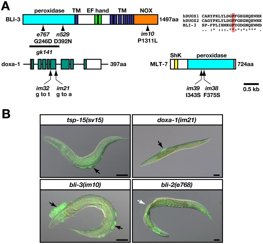
(A) The structure of the gene/proteins related to tsp-15 function. Schematic representation of the BLI-3 and MLT-7 protein with functional domain, and the genomic structure of the doxa-1 gene. The im10, im21, im32, im38 and im39 mutations are indicated. Previously identified missense mutations in the bli-3 gene including e767 (glycine to aspartic acid at 246) and n529 (aspartic acid to asparagine at 392) are shown. The bold line indicates the region of the gk141 deletion allele. The im10 mutation has a leucine instead of a proline at position 1311 within the NOX domain. TM and NOX refer to the transmembrane and NOX domains, respectively. The im21 mutation is characterized by a G to A transition in the splice donor site at the fifth intron. The im32 mutation is a G to T transversion in the splice acceptor site at the fourth intron. The im38 and im39 alleles are indicated in the MLT-7 protein. Both alleles contain the missense mutations in the peroxidase domain. (B) bli-3(im10) and doxa-1(im21), but not bli-2(e768) are similar to tsp-15(sv15). Hypodermal expression of GFP driven by dpy-7p::gfp in the mutants revealed an unusual accumulation of cellular materials in the blisters of bli-3, doxa-1 and tsp-15 mutants (indicated by black arrows), but not in bli-2 mutants (indicated by the white arrow). The scale bars represent 50 µm. Tab. 1. The tsp-15, doxa-1, and bli-3 genes are in the same genetic pathway. 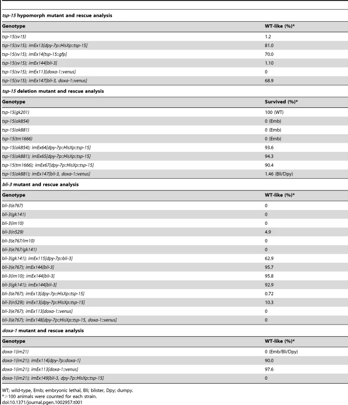
WT; wild-type, Emb; embryonic lethal, Bli; blister, Dpy; dumpy. The bli-3, doxa-1, and mlt-7 mutants were rescued by their own cDNA driven by a hypodermis-specific dpy-7 promoter (Table 1, Figure S2). For im21, Venus-tagged doxa-1 at the C-termini (doxa-1::venus, Figure S4A) driven by the doxa-1 promoter effectively rescued the phenotype (Table 1, Figure S2B, Figure S4B). The doxa-1::venus transgene was expressed in the hypodermis, and other tissues such as the pharynx, uterus, gonad, and vulva (Figure S4B).
Involvement of tsp-15 in bli-3 pathway
To investigate the genetic relationship between tsp-15 and newly isolated mutants, we performed mutant rescue assays (Figure 2, Table 1). Over-expression of tsp-15 did not restore the defects of the bli-3 and doxa-1 mutants. Over-expression of bli-3 as well as doxa-1 alone did not rescue the tsp-15 mutant. Co-expression of bli-3 and doxa-1 in the tsp-15 hypomorph mutant effectively rescued the cuticle deficiency (Figure 2, Table 1). In contrast, bli-3 and doxa-1 co-expression in the tsp-15 null mutant resulted in partial rescue of the lethal phenotype. In approximately 1.5% of transgenic animals, embryonic lethality was recovered and the larvae showed a tsp-15 hypomorph mutant-like morphology (Figure 2), which has not been observed in tsp-15 null mutants previously.
Fig. 2. TSP-15 function is compensated with BLI-3 system. 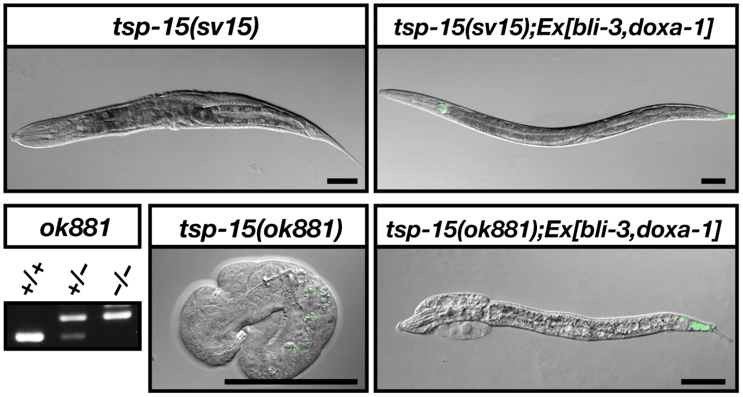
Restoration of the tsp-15 defect by co-expression of bli-3 and doxa-1. Co-expression of bli-3 and doxa-1 in the tsp-15(sv15) hypomorph mutant effectively rescued the phenotype of sv15. The embryonically lethal tsp-15(ok881) null mutant was partially rescued and developed into larvae via bli-3 and doxa-1 co-expression. Green fluorescence at the tail tip represents expression of the lin-44::gfp injection marker. The ok881 deletion homozygosity of surviving transgenic larvae was confirmed by genomic PCR. The scale bars represent 50 µm. Decreased dityrosine levels in tsp-15 mutants
BLI-3 is reported as a key enzyme for the generation of H2O2 for tyrosine cross-linking in the cuticle since the level of di - and tri-tyrosine formation is reduced in the cuticle of bli-3(RNAi) animals [17]. To examine distribution of cross-linked tyrosine in the exoskeleton of tsp-15 mutants, we carried out immunohistochemical analysis using anti-di-tyrosine antibody. We also checked the endogenous distribution of DPY-7 (a nematode collagen) in tsp-15 mutants by immunostaining. In the normal embryo, di-tyrosine was distributed over the entire cuticle representing the body surface structure, whereas di-tyrosine formation was severely reduced in tsp-15 null embryos (Figure 3A). In contrast, the level of collagen found in tsp-15 null embryos, examined via DPY-7, was comparable to tsp-15(+) embryos, although distribution was severely disturbed (Figure 3B). Thus, cuticle collagen is likely synthesized and secreted from the hypodermis correctly, but a failure of cross-linking of secreted collagen results in fragility of the cuticle in tsp-15 mutants.
Fig. 3. Deterioration of dityrosine in the tsp-15 mutant. 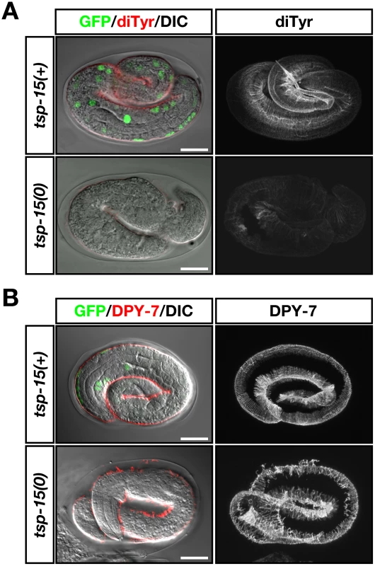
(A) Representative immunofluorescent images showing the distribution of dityrosine in embryos of the tsp-15(ok854) null mutant. Embryos were obtained from the OB129 strain, which was the tsp-15(ok854) mutant rescued by a tsp-15p::(His)6Xp::tsp-15 extrachromosomal array. Nuclear GFP fluorescence by sur-5::gfp defined the rescued (tsp-15(+)) or spontaneously array-lost (null; tsp-15(0)) embryo. Micrographs on the left show merged Nomarski images exhibiting GFP and dityrosine immunolocalization. Right panels show the reconstruction of confocal images for dityrosine distribution in the same embryo that is displayed on the left. In tsp-15(+) normal embryos, dityrosine localization showed a regular pattern representing the cuticle surface structure. Fluorescence intensity was severely deteriorated in tsp-15(0) embryos. Scale bars indicate 10 µm. (B) DPY-7 localization was compared under the same conditions as in (A). In tsp-15(+) embryos, DPY-7 localized as regular bands in the cuticle. In the tsp-15(0) embryo, the expression of DPY-7 was comparable to the normal embryo despite its disorganized pattern. Scale bars indicate 10 µm. BLI-3 activity depends on both TSP-15 and DOXA-1
DUOX was originally identified as a hydrogen peroxide generator for thyroid hormone synthesis [9], [11] and DUOXA is essential for DUOX targeting to the plasma membrane [33]. We produced stable transfectants of TSP-15, BLI-3 and DOXA-1 in human HT1080 cells (Figure 4A) to confirm the roles of TSP-15 and DOXA-1 for BLI-3. Release of H2O2 into the culture medium was measured in the absence of other C. elegans proteins. Extracellular H2O2 from HT1080B and HT1080TB cells was almost equivalent to basal activity (see Materials and Methods for description of stable transfectants). Unlike the mammalian DUOX system, BLI-3 was only slightly activated by co-expression of DOXA-1 in HT1080DB cells. In addition to DOXA-1, concomitant expression of TSP-15 strongly enhanced production of H2O2 in HT1080TDB cells (Figure 4B). The generation of H2O2 was blocked by the flavoprotein inhibitor diphenyleneiodonium (DPI), indicating that DUOX was involved in enhanced H2O2 production in TSP-15-transduced cells. We concluded that BLI-3 requires TSP-15 and DOXA-1 for proper function. BLI-3P1311L and BLI-3G246D identical to the im10 or e767 mutation, respectively, resulted in decreased H2O2 production (Figure 4B). The same results were observed in other independently established stable cell lines, and by transient expression in COS-7 and HeLa cells (data not shown). Regulation of Ca2+ characteristically elicits DUOX activity as a thyroid hormone synthesizer. BLI-3 did not require calcium stimulation to produce H2O2 in HT1080TDB cells, and HT1080DB cells were not activated by calcium stimulation either (Figure 4C). This may be due to the fact that the critical amino acid residues for Ca2+-binding are poorly conserved in the EF-hand motifs of BLI-3 proteins [17]. Furthermore, BLI-3 was not activated by forskolin (fsk) and phorbol 12-myristate 13-acetate (PMA), which have previously been reported to be mammalian DUOX stimulators (Figure 4C) [54].
Fig. 4. Both TSP-15 and DOXA-1 are required for H2O2 production by BLI-3 in mammalian cells. 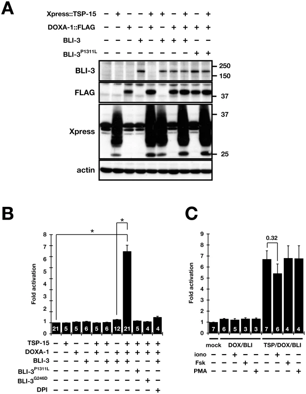
(A) Immunoblot analysis of the expression of Xpress-tagged TSP-15, FLAG-tagged DOXA-1, BLI-3 and BLI-3P1311L in HT1080 stable transfectants. Xpress-tagged TSP-15 (30 kDa) is highly glycosylated (Figure S5). (B) Extracellular H2O2 production from stable transfectants. Fold-activation compared with non-transfected HT1080 cells was determined. Only cells expressing TSP-15, DOXA-1, and BLI-3 (HT1080TDB) significantly generated H2O2. A 10 µM concentration of DPI inhibited H2O2 production in HT1080TDB cells. BLI-3 carrying the G246D or P1311L mutation did not release H2O2. The graph shows the means ± SEM. The numbers in the bars indicate the number of independent experiments (n). *P<10−9. (C) BLI-3 was not activated by mammalian DUOX stimulators. Ionomycin (iono, 1 µM), forskolin (Fsk, 1 µM), or phorbol 12-myristate 13-acetate (PMA, 1 µM) was added to HT1080DB and HT1080TDB cells during culture. The graph shows the means ± SEM. The numbers in the bars indicate the number of independent experiments (n). TSP-15 and DOXA-1 associate with BLI-3
Tetraspanins form protein complexes with a number of other molecules. We performed co-immunoprecipitation assays to determine whether TSP-15 associates with BLI-3 and DOXA-1. BLI-3 was transiently expressed in COS-7 cells where TSP-15 and/or DOXA-1 was stably expressed. As a result, BLI-3 co-immunoprecipitated with DOXA-1 (Figure 5A; lanes 17 and 18) and TSP-15 (Figure 5A, lanes 12 and 14). Co-immunoprecipitation of BLI-3 and DOXA-1 was independent of TSP-15 expression (Figure 5A; lane 17). TSP-15 co-immunoprecipitated with BLI-3 in the absence of DOXA-1 (Figure 5A; lane 12). In addition, TSP-15 and DOXA-1 association was also observed (Figure 5A; lane 14 and 18, Figure S6), indicating that TSP-15, DOXA-1, and BLI-3 form protein complexes. TSP-15 was not co-immunoprecipitated with over-expressed EGF receptor under the same conditions (data not shown). We also verified the same molecular interaction in transgenic animals. In doxa-1::venus transgenic worms (Figure S4B), BLI-3 co-immunoprecipitated with the DOXA-1::Venus fusion protein (Figure 5B; lane 6). Endogenous and tagged TSP-15 associated with BLI-3 (Figure 5B; lanes 2 and 4) and with DOXA-1::Venus (Figure 5B; lane 8). We concluded that TSP-15, DOXA-1 and BLI-3 form a complex.
Fig. 5. Direct association of BLI-3 with TSP-15 and DOXA-1. 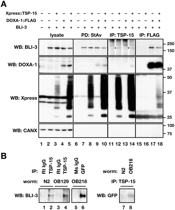
(A) Direct association of BLI-3 with TSP-15, and BLI-3 with DOXA-1. BLI-3 was transiently expressed in COS-7 stable transfectants expressing Xpress::TSP-15, DOXA-1::FLAG, or both. Cell surface proteins were labeled with biotin. A 1% CHAPS cell lysate was used for immunoprecipitation or pull-down assay with anti-TSP-15 antibody, anti-FLAG antibody or streptavidin. BLI-3 was co-immunoprecipitated with both TSP-15 (indicated by asterisks) and DOXA-1, and TSP-15 and DOXA-1 were co-immunoprecipitated. Bands above asterisks are non-specific. Cell surface localization of BLI-3 was independent of TSP-15 and DOXA-1. ER-resident calnexin (CANX) was not detected on the cell surface. BLI-3 expression was not affected by TSP-15 and DOXA-1. (B) Direct association of BLI-3 with TSP-15, BLI-3 with DOXA-1, and TSP-15 with DOXA-1 was confirmed in C. elegans. Xpress::tsp-15 was expressed in OB129, and doxa-1::venus is expressed in the OB218 transgenic strain. Venus-tagged DOXA-1 was analyzed with anti-GFP antibody. BLI-3 was co-immunoprecipitated with endogenous and Xpress-tagged TSP-15 and Venus-tagged DOXA-1 in 1% Triton-X100 cell lysates. Endogenous TSP-15 also associated with DOXA-1::Venus. Normal rat and mouse IgG was used as a negative control. BLI-3 expression levels in total cell lysates were unaffected by DOXA-1 and TSP-15 expression (Figure 5A; lanes 2–5). We examined surface expression of BLI-3 using biotin labeling. The amount of BLI-3 protein at the cell surface was not augmented by TSP-15 (Figure 5A; lanes 6–10). BLI-3 was also recruited to the plasma membrane without TSP-15. Therefore, TSP-15 is dispensable for BLI-3 targeting. It is reported that membrane targeting of mammalian DUOX depends on DUOXA in a system using non-thyroidal cells [33]–[36]; however, BLI-3 was recruited to the plasma membrane without DOXA-1 in our system.
BLI-3 activation by TSP-15 at the cell surface
The molecular requirement of TSP-15 in the BLI-3 system is possibly due to formation of a complex at the plasma membrane, therefore BLI-3 could be activated by TSP-15 localized at the cell surface. We assessed this hypothesis through cell fusion-based analysis (Figure 6A). TSP-15-expressing cells (HT1080T) and DOXA-1/BLI-3-expressing cells (HT1080DB), both of which did not produce H2O2 (Figure 4B), were fused utilizing Sendai virus (HVJ). After the cell fusion reaction, extracellular H2O2 production from fused cells was measured. The fused cells (T::DB) produced H2O2, and the production was inhibited by DPI treatment (Figure 6B). In contrast, BLI-3 carrying the P1311L mutation did not result in H2O2 production. Inhibition of de novo protein synthesis by cycloheximide (CHX) resulted in a slight decrease in H2O2 producing activity in T::DB fusion cells (Figure 6B). These results indicate that TSP-15 did not promote BLI-3 protein expression, but existing TSP-15 at the cell surface was sufficient to activate BLI-3 for H2O2 production. The capability of H2O2 production was rapidly acquired after cell fusion. H2O2 was immediately detectable in T::DB fusion cells 15 min after the fusion reaction (Figure 6C), indicating that alternatively derived TSP-15, BLI-3 and DOXA-1 rapidly formed functional units at the cell surface.
Fig. 6. Requirement of TSP-15 for reconstitution of BLI-3 function at cell surface. 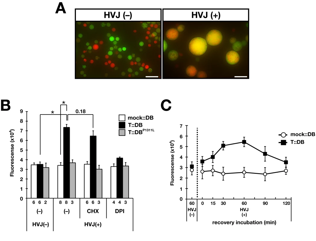
(A) HVJ-mediated cell fusion. GFP-expressing HT1080 cells and HT1080DB cells labeled with Cell Tracker Orange were fused with HVJ. Under HVJ(+) conditions, fused cells were large compared with HVJ(−) cells and exhibited yellow/orange fluorescence. Scale bar indicates 50 µm. (B) HT1080DB cells fused with HT1080T cells (T::DB) produced H2O2, which was inhibited by DPI. Mock::DB and T::DBP1311L fusion cells did not produce H2O2. Treatment of T::DB fusion cells with 10 µg/ml cycloheximide (CHX) did not inhibit H2O2 production. The graph shows the means ± SEM. The number of independent experiments is indicated. *P<10−6. (C) Rapid H2O2 production from T::DB fusion cells. The recovery time after the fusion event was examined to determine when fusion cells acquired the ability to produce H2O2. Maximum H2O2 production was observed at 30–60 min, although production was observed at 15 min post-fusion. The graph shows the means ± SEM (n = 3). Discussion
Organisms have developed regulatory systems to control ROS generation in host defense and cellular signaling. For mammalian DUOX proteins, association with a maturation factor (DUOXA) for targeting to the plasma membrane, Ca2+ regulation via EF-hand motifs, and PKA - or PKC-mediated phosphorylation were identified as regulatory systems. We propose that the tetraspanin protein is a novel component of the DUOX system for ROS generation (Figure 7). Using C. elegans as a model organism, we identified a series of genes for ROS generation in which mutants exhibited a phenotype resembling the tetraspanin tsp-15 mutant. The genes bli-3, doxa-1, and mlt-7 were, respectively, the homologues of mammalian DUOX, DUOXA and peroxidase, and mutants of these displayed the same cuticle deficiency (Figure 1, Figure 2, Figure S1, Figure S2). The reason for this cuticle disorganization is deterioration of tyrosine cross-linking in cuticle development as shown in bli-3 knockdown-animals (Figure 3) [17]. We showed that the tsp-15 mutant was rescued by simultaneous over-expression of bli-3 and doxa-1, implying that these three genes are part of the same genetic pathway (Figure 1, Figure 2). Reconstitution of BLI-3, TSP-15 and DOXA-1 in mammalian cells demonstrated that H2O2 generation by BLI-3 was dependent on TSP-15 as well as DOXA-1 (Figure 4).
Fig. 7. Molecular regulation of BLI-3 by TSP-15 and DOXA-1. 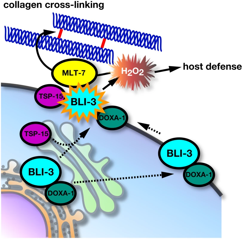
TSP-15 and DOXA-1 are essential for H2O2 production by BLI-3. TSP-15 associates with BLI-3 at the cell surface or during trafficking. The role of DOXA-1 in BLI-3 targeting to the plasma membrane remains elusive. BLI-3/DOXA-1 complexes at the cell surface are inactive, but recruiting to the tetraspanin-microdomain facilitates the formation of a functional unit for generation of H2O2 that is utilized by innate host immunity, and cross-linking of extracellular matrix with peroxidase (MLT-7). It was hypothesized that TSP-15 might enhance BLI-3 protein levels by elevating protein expression or promoting targeting to the cell surface; however, BLI-3 and DOXA-1 protein expression levels were comparable, with or without TSP-15 expression. TSP-15 expression did not affect BLI-3 expression at the cell surface, and did not enhance the association of BLI-3 and DOXA-1. This implies that the role of TSP-15 in the BLI-3 system is not just augmentation of its expression. Our observations support this notion, since over-expression of bli-3 and doxa-1 resulted in incomplete rescue of tsp-15 null mutants. In the sv15 hypomorph mutant, tsp-15 expression was reduced, and expressed at 10% of wild-type levels (data not shown), therefore the BLI-3 system recovered to produce adequate H2O2 by concomitant over-expression of bli-3 and doxa-1. If the up-regulation of BLI-3 activity by TSP-15 is quantitative, tsp-15 null mutants should be completely rescued by BLI-3/DOXA-1, however this was not the case. The molecular role of tetraspanin in the DUOX system is likely qualitative. We believe that TSP-15 up-regulates the activity of BLI-3 at the plasma membrane. Cell fusion-based analysis strongly supports this idea, since cells that acquired TSP-15 from other cells rapidly produced H2O2 even when protein synthesis was inhibited. During the HVJ-mediated fusion process, intracellular organelles were morphologically altered and repaired within 30 min [55]. We observed that H2O2 generation was initiated 15 min after recovery, suggesting that individually derived BLI-3/DOXA-1 and TSP-15 rapidly assembled at the cell surface, forming functional complexes. The lipid raft marker protein, flotillin, was rapidly assembled during cell fusion [56]. Inhibition of de novo protein synthesis did not affect H2O2 production in fusion cells, indicating that the existing TSP-15 at the cell surface is sufficient for facilitation of BLI-3 activity.
The molecular mechanisms of up-regulation are still unclear, but we showed that TSP-15, BLI-3 and DOXA-1 form complexes in vitro and in vivo (Figure 5). BLI-3 directly associates with DOXA-1, as demonstrated in mammalian DUOX and DUOXA. We also demonstrated the association between BLI-3 and TSP-15, and that this was independent of DOXA-1 expression. It is known that tetraspanin associates with a number of membrane proteins and forms large protein complexes at certain membrane microdomains. We speculate that TSP-15 may establish or maintain a specialized membrane microdomain that facilitates generation of H2O2 in conjunction with BLI-3. As reported for other NOX isozymes and their subunits, association with TSP-15 might induce a conformational change in BLI-3 to function properly. Alternatively, TSP-15 may support the recruitment of unknown factors at the membrane microdomain that are essential for BLI-3 activity. Although DOXA-1 is essential for H2O2 production by BLI-3, the role of DOXA-1 in the BLI-3 system is unclear. Unlike mammalian DUOX, BLI-3 was unexpectedly recruited to the plasma membrane in the absence of DOXA-1. DOXA-1 might not regulate BLI-3 trafficking in the DUOX system in C. elegans, but we cannot exclude the possibility that expression of C. elegans proteins in mammalian cells may cause dysregulation in BLI-3 trafficking. Further investigation is needed to clarify the molecular functions of DOXA-1 in the BLI-3 system. In addition, unlike the mammalian DUOX system, BLI-3 did not respond to various stimuli when reconstituted in mammalian cells (Figure 4C). Absence of negative regulatory factors may explain the constitutive active state of BLI-3 in the heterologous system. For instance, NOXA1 has an inhibitory role in stabilizing the inactive state of mammalian DUOX1 [57]. No NOXA1-like sequence has been found in C. elegans, but further investigation would clarify this hypothesis.
Reciprocal homology searches suggested that several human tetraspanins are related to TSP-15 with CD151 (TSPAN24) and TSPAN11 being the most closely related. However, we have not identified any mammalian tetraspanins that could be functionally substituted for TSP-15 in the tsp-15 mutant [51], or for H2O2 production in the BLI-3/DOXA-1 reconstitution system (data not shown). It is also uncertain whether mammalian tetraspanins have a pivotal role in mammalian DUOX system. Mutations in tetraspanin genes have not been identified in patients suffering from congenital hypothyroidism. In contrast to other NOX isozymes, understanding the regulation of DUOX proteins is emerging. Our data clearly shows that tetraspanin is a new component for directing DUOX activity, contributing to greater understanding of the molecular mechanisms of ROS generation and disorders caused by impairment of ROS generation systems [58], [59].
Materials and Methods
Worm strains and culture
C. elegans was grown at 20°C on NGM plates as described previously [60]. The Bristol N2 strain was used as the wild type. Strains and their genotype used in this study are listed in Table S1.
Mutant screening and identification
N2 or OB43 were mutated with 50 mM ethyl methanesulfonate or 5 mM ethyl nitrosourea for 4 h. F2 recessive mutants showing a sv15 mutant-like phenotype were screened. SNPs between N2 and Hawaiian CB4856 strains were used for physical mapping of the alleles [61]. Mutations were determined by further DNA sequencing and confirmed by complementation tests, rescue assays by DNA transformation, and RNAi analyses (see Text S1). Mutants were outcrossed at least five times with N2.
cDNA cloning and construction of vectors
Total RNA was isolated from mixed stages of N2 or mutant worms using TRIzol reagent (Invitrogen, Carlsbad, CA, USA). First strand cDNA was synthesized by ReverTra Ace (Toyobo, Japan). A 4.6 kb fragment of bli-3, 1.2 kb of doxa-1, and 2.2 kb of mlt-7 full length cDNA was prepared by RT-PCR. The bli-3P1311L and bli-3G246D vectors were constructed by PCR-based site-directed mutagenesis. doxa-1::venus translational fusion construct (Figure S4A) contains a 3.1 kb genomic PCR fragment of the doxa-1 5′ flanking region and 2.2 kb doxa-1 genomic coding region without a termination codon, which was cloned into the Venus translational fusion vector (a gift from Takeshi Ishihara, Kyusyu University). The (His)6Xpress-tagged tsp-15 (HisXp::tsp-15), bli-3, FLAG-tagged doxa-1 (doxa-1::FLAG) and mlt-7 were sub-cloned under the control of the dpy-7 promoter for hypodermis-specific expression in the mutant rescue assay [51] or into the pCx4 retroviral vector for transfection into mammalian cells [62]. A 34.2 kb fosmid clone, WRM065cD06, was purchased from Geneservice (Cambridge, UK). A 14.9 kb HaeII restriction fragment (nt 16368–31233) which contained the full bli-3 coding region and also the 5′ flanking region was used for the rescue assay.
Immunohistochemistry and microscopic imaging of embryos
Embryos were collected from gravid hermaphrodites and were immunostained as previously described [63]. Mouse anti-dityrosine (1C3; Nikken Seil Co., Ltd. Shizuoka, Japan), mouse anti-DPY-7 (a gift from Iain L. Johnstone, University of Glasgow) [64] were used at 1∶200 and 1∶500 dilutions, respectively. Alexa Fluor 488 or Alexa Fluor 546 (Invitrogen) conjugated anti-mouse IgG antibodies were used as secondary antibodies at 1∶500 dilutions. Confocal images were acquired with a LSM5 Pascal microscope (Zeiss, Germany). The three-dimensional projections were reconstructed using images of serial Z-section (1–1.5 µm). Micrographs of fluorescence microscopy were captured using a BX50 microscope (Olympus, Tokyo, Japan) equipped with a VB7010 digital CCD camera (Keyence, Osaka, Japan). Image processing and movie construction was performed with Adobe Photoshop CS4 and Image J 1.34, respectively.
Cell culture and transfection
HT1080, HeLa, and COS-7 cells were cultured in Dulbecco's modified Eagle's medium (DMEM) supplemented with 10% fetal calf serum. Transfection for transient expression was performed using FuGENE HD reagent (Roche, Germany) according to the manufacturer's protocol. Stable transfectants were obtained by retrovirus transfection [62]. Plasmids were transfected into Plat-E packaging cells with FuGENE 6 reagent (Roche). Culture supernatant was added to HT1080 ecoR, HeLa ecoR and COS-7 ecoR cells which are stable transfectants of the ecotropic retrovirus receptor, mCAT-1 (gifts from Hiroto Mizushima, Osaka University). Transfected cells were selected by incubation with a combination of 1 µg/ml puromycin, 10 µg/ml blastcidine S, and/or 300 µg/ml zeocin for at least two weeks. Stable transfectants of C. elegans genes were named after the transgene that was transduced. T, D, and B refer to tsp-15, doxa-1, and bli-3, respectively (e. g. HT1080TDB cells express tsp-15, doxa-1, and bli-3).
Antibodies
Both BLI-3 and DOXA-1 rabbit antiserum was prepared by SCRUM Inc. (Tokyo, Japan). BLI-3 rabbit antiserum was raised against keyhole limpet hemocyanin (KLH)-coupled BLI-3 peptides corresponding to residues 254–269 (N1) and a mixture of residues 1232–1245 and 1478–1490 (Cmix). DOXA-1 rabbit antiserum was raised against a mixture of KLH-coupled DOXA-1 peptides corresponding to residues 170–184 and 326–340. Rabbit serum was purified using a peptide-conjugated sepharose column. Anti TSP-15 monoclonal antibody (2C2) was obtained by immunizing rats with HA-tagged TSP-15 protein into footpads. Hybridoma supernatant was purified by ion-exchange chromatography followed by anti-rat IgG-conjugated Sepharose column chromatography. The 2C2 monoclonal antibody is available for immunoprecipitation.
Western blot, co-immunoprecipitation, and cell surface biotinylation
Cells were harvested and suspended in lysis buffer (20 mM Tris-HCl, 150 mM NaCl, 2 mM EDTA, pH 7.5) with 1% CHAPS. For surface labeling, cells were incubated with 0.2 mg/ml sulfo-NHS-LC-biotin (Thermo Fisher Scientific, Rockford, IL, USA) at 4°C for 30 min and then lysed. For immunoprecipitation and pull-down assays, cleared cell lysates were incubated with anti-FLAG M2 beads (Sigma-Aldrich, St Louis, MO, USA), rat anti-TSP-15 antibody (2C2)-conjugated agarose, or streptavidin agarose beads (Thermo Fisher Scientific). Cleared cell lysate and immunoprecipitates were blotted with mouse anti-Omni (Xpress) (D-8; Santa Cruz Biotechnology, Santa Cruz, CA, USA), mouse anti-FLAG (M2; Sigma-Aldrich), rabbit anti-BLI-3 (N1), rabbit anti-DOXA-1, mouse anti-actin (MAB1501; Millipore, Bedford, MA, USA), or mouse anti-calnexin (AF18; Abcam, Cambridge, UK) antibodies. HRP-conjugated goat anti-rabbit IgG (Zymed, San Francisco, CA, USA), and donkey anti-mouse IgG (Millipore) were used as secondary antibodies. For the immunoblotting of worms, mixed stages of N2, OB129 or OB218 strains were cultured on 150 mm dishes and harvested. Worms were homogenized by sonication and lysed in 1% Triton X-100. Rat anti-TSP-15 or mouse anti-GFP antibody (3E6, Wako chemicals, Osaka, Japan), and anti-rat IgG - or anti-mouse IgG-conjugated Sepharose was added to the cleared worm lysate. Normal rat IgG or mouse IgG was used as a negative control for specific antibodies. Immunoprecipitates were blotted with anti BLI-3 (Cmix) antibody.
Monitoring H2O2 production in mammalian cells
The H2O2 release into culture supernatants was measured using Amplex Red reagent (10-acetyl-3,7-dihydroxyphenoxazine; Invitrogen), which reacts with H2O2 and is transformed into fluorescent resorufin in the presence of peroxidase [65]. Stable or transient transfectants were plated on 96-well plates at 1–5×10⁴ cells/well, and cultured for 24–48 h. Cells were incubated in 100 µl Hanks' balanced salt solution containing 50 µM Amplex Red, and 0.1 U/ml HRP (Nacalai Tesque, Kyoto, Japan), with or without 10 µM diphenyleneiodonium (DPI), 1 µM ionomycin, 1 µM forskolin (Fsk), or 1 µM phorbol 12-myristate 13-acetate (PMA) at 37°C for 1 h. The fluorescence (530 nm excitation, 590 nm emission) was measured by a Power Scan HT (DS Pharma Biomedicals, Osaka, Japan). Fold-increase was compared with the basal activity of non-transfected HT1080 cells and was determined from least four independent experiments.
HVJ–mediated cell fusion
HVJ (Sendai virus)-mediated cell fusion was performed using GenomONE-CF (Ishihara Sangyo Co. Ltd., Osaka, Japan). Cells were harvested using trypsin. HT1080DB or HT1080DBP1311L cells (8×10⁵) were mixed with HT1080 ecoR or HT1080T cells (8×10⁵) in a total volume of 200 µl reaction buffer. A 1 µl volume of inactivated HVJ was then added to the cell mixture. After incubation for 15 min at 37°C, 1 ml of DMEM was added and further incubated for 1 h at 37°C for recovery. Cells were treated with 10 µg/ml cycloheximide (Sigma-Aldrich) during incubation for inhibition of protein synthesis. Cells were washed, and H2O2 production was measured as described above. For the time-course experiment, recovery incubation was examined from 0–120 min, and H2O2 production was measured after a 30 min incubation. For visualization of the fusion event, HT1080DB cells were pre-stained with 10 µM Cell Tracker Orange (Invitrogen) for 20 min at 37°C, and then fused with GFP-expressing HT1080 cells. Each experiment was tested in duplicate and performed at least three times.
Statistical analysis
Data are presented as mean value and error bars indicate the standard error of the mean (SEM) from multiple independent assays. Significance was determined using a two-tailed Student's t-test.
Supporting Information
Zdroje
1. BedardK, KrauseK-H (2007) The NOX family of ROS-generating NADPH oxidases: physiology and pathophysiology. Physiol Rev 87 : 245–313.
2. LetoTL, GeisztM (2006) Role of Nox family NADPH oxidases in host defense. Antioxid Redox Signal 8 : 1549–1561.
3. LambethJD (2004) NOX enzymes and the biology of reactive oxygen. Nat Rev Immunol 4 : 181–189.
4. LetoTL, MorandS, HurtD, UeyamaT (2009) Targeting and regulation of reactive oxygen species generation by Nox family NADPH oxidases. Antioxid Redox Signal 11 : 2607–2619.
5. SumimotoH (2008) Structure, regulation and evolution of Nox-family NADPH oxidases that produce reactive oxygen species. FEBS J 275 : 3249–3277.
6. AguirreJ, LambethJD (2010) Nox enzymes from fungus to fly to fish and what they tell us about Nox function in mammals. Free Radic Biol Med 49 : 1342–1353.
7. BedardK, LardyB, KrauseK-H (2007) NOX family NADPH oxidases: not just in mammals. Biochimie 89 : 1107–1112.
8. FinkelT (2011) Signal transduction by reactive oxygen species. J Cell Biol 194 : 7–15.
9. De DekenX, WangD, ManyMC, CostagliolaS, LibertF, et al. (2000) Cloning of two human thyroid cDNAs encoding new members of the NADPH oxidase family. J Biol Chem 275 : 23227–23233.
10. DonkóA, PéterfiZ, SumA, LetoT, GeisztM (2005) Dual oxidases. Philos Trans R Soc Lond, B, Biol Sci 360 : 2301–2308.
11. DupuyC, OhayonR, ValentA, Noël-HudsonMS, DèmeD, et al. (1999) Purification of a novel flavoprotein involved in the thyroid NADPH oxidase. Cloning of the porcine and human cDNAs. J Biol Chem 274 : 37265–37269.
12. DupuyC, KaniewskiJ, DèmeD, PommierJ, VirionA (1989) NADPH-dependent H2O2 generation catalyzed by thyroid plasma membranes. Studies with electron scavengers. Eur J Biochem 185 : 597–603.
13. Ameziane-El-HassaniR, MorandS, BoucherJ-L, FrapartY-M, ApostolouD, et al. (2005) Dual oxidase-2 has an intrinsic Ca2+-dependent H2O2-generating activity. J Biol Chem 280 : 30046–30054.
14. JohnsonKR, MardenCC, Ward-BaileyP, GagnonLH, BronsonRT, et al. (2007) Congenital hypothyroidism, dwarfism, and hearing impairment caused by a missense mutation in the mouse dual oxidase 2 gene, Duox2. Mol Endocrinol 21 : 1593–1602.
15. MorenoJC, BikkerH, KempersMJE, van TrotsenburgASP, BaasF, et al. (2002) Inactivating mutations in the gene for thyroid oxidase 2 (THOX2) and congenital hypothyroidism. N Engl J Med 347 : 95–102.
16. AnhNTT, NishitaniM, HaradaS, YamaguchiM, KameiK (2011) Essential role of Duox in stabilization of Drosophila wing. J Biol Chem 286 : 33244–33251.
17. EdensWA, SharlingL, ChengG, ShapiraR, KinkadeJM, et al. (2001) Tyrosine cross-linking of extracellular matrix is catalyzed by Duox, a multidomain oxidase/peroxidase with homology to the phagocyte oxidase subunit gp91phox. J Cell Biol 154 : 879–891.
18. WongJL, CrétonR, WesselGM (2004) The oxidative burst at fertilization is dependent upon activation of the dual oxidase Udx1. Dev Cell 7 : 801–814.
19. AllaouiA, BotteauxA, DumontJE, HosteC, De DekenX (2009) Dual oxidases and hydrogen peroxide in a complex dialogue between host mucosae and bacteria. Trends in Molecular Medicine 15 : 571–579.
20. BaeYS, ChoiMK, LeeW-J (2010) Dual oxidase in mucosal immunity and host-microbe homeostasis. Trends Immunol 31 : 278–287.
21. GeisztM, WittaJ, BaffiJ, LekstromK, LetoTL (2003) Dual oxidases represent novel hydrogen peroxide sources supporting mucosal surface host defense. FASEB J 17 : 1502–1504.
22. HaE-M, OhC-T, BaeYS, LeeW-J (2005) A direct role for dual oxidase in Drosophila gut immunity. Science 310 : 847–850.
23. JuarezMT, PattersonRA, Sandoval-GuillenE, McGinnisW (2011) Duox, Flotillin-2, and Src42A are required to activate or delimit the spread of the transcriptional response to epidermal wounds in Drosophila. PLoS Genet 7: e1002424 doi:10.1371/journal.pgen.1002424.
24. NiethammerP, GrabherC, LookAT, MitchisonTJ (2009) A tissue-scale gradient of hydrogen peroxide mediates rapid wound detection in zebrafish. Nature 459 : 996–999.
25. TheinM, WinterA, StepekG, MccormackG, StapletonG, et al. (2009) The combined extracellular matrix cross-linking activity of the peroxidase MLT-7 and the duox BLI-3 are critical for post-embryonic viability in Caenorhabditis elegans. J Biol Chem 284 : 17549–17563.
26. MeitzlerJL, BrandmanR, Ortiz de MontellanoPR (2010) Perturbed heme binding is responsible for the blistering phenotype associated with mutations in the Caenorhabditis elegans dual oxidase 1 (DUOX1) peroxidase domain. J Biol Chem 285 : 40991–41000.
27. BenedettoA, AuC, AvilaDS, MilatovicD, AschnerM (2010) Extracellular dopamine potentiates Mn-induced oxidative stress, lifespan reduction, and dopaminergic neurodegeneration in a BLI-3-dependent manner in Caenorhabditis elegans. PLoS Genet 6: e1001084 doi:10.1371/journal.pgen.1001084.
28. ChávezV, Mohri-ShiomiA, GarsinDA (2009) Ce-Duox1/BLI-3 generates reactive oxygen species as a protective innate immune mechanism in Caenorhabditis elegans. Infect Immun 77 : 4983–4989.
29. van der HoevenR, McCallumKC, CruzMR, GarsinDA (2011) Ce-Duox1/BLI-3 generated reactive oxygen species trigger protective SKN-1 activity via p38 MAPK signaling during infection in C. elegans. PLoS Pathog 7: e1002453 doi:10.1371/journal.ppat.1002453.
30. JainC, YunM, PolitzSM, RaoRP (2009) A pathogenesis assay using Saccharomyces cerevisiae and Caenorhabditis elegans reveals novel roles for yeast AP-1, Yap1, and host dual oxidase BLI-3 in fungal pathogenesis. Eukaryotic Cell 8 : 1218–1227.
31. BrownDI, GriendlingKK (2009) Nox proteins in signal transduction. Free Radic Biol Med 47 : 1239–1253.
32. LambethJD, KawaharaT, DieboldB (2007) Regulation of Nox and Duox enzymatic activity and expression. Free Radic Biol Med 43 : 319–331.
33. GrasbergerH, RefetoffS (2006) Identification of the maturation factor for dual oxidase. Evolution of an eukaryotic operon equivalent. J Biol Chem 281 : 18269–18272.
34. GrasbergerH, De DekenX, MiotF, PohlenzJ, RefetoffS (2007) Missense mutations of dual oxidase 2 (DUOX2) implicated in congenital hypothyroidism have impaired trafficking in cells reconstituted with DUOX2 maturation factor. Mol Endocrinol 21 : 1408–1421.
35. LuxenS, NoackD, FraustoM, DavantureS, TorbettBE, et al. (2009) Heterodimerization controls localization of Duox-DuoxA NADPH oxidases in airway cells. J Cell Sci 122 : 1238–1247.
36. MorandS, UeyamaT, TsujibeS, SaitoN, KorzeniowskaA, et al. (2009) Duox maturation factors form cell surface complexes with Duox affecting the specificity of reactive oxygen species generation. FASEB J 23 : 1205–1218.
37. De DekenX, WangD, DumontJE, MiotF (2002) Characterization of ThOX proteins as components of the thyroid H2O2-generating system. Exp Cell Res 273 : 187–196.
38. MorandS, AgnandjiD, Noel-HudsonM-S, NicolasV, BuissonS, et al. (2004) Targeting of the dual oxidase 2 N-terminal region to the plasma membrane. J Biol Chem 279 : 30244–30251.
39. ZamproniI, GrasbergerH, CortinovisF, VigoneMC, ChiumelloG, et al. (2008) Biallelic inactivation of the dual oxidase maturation factor 2 (DUOXA2) gene as a novel cause of congenital hypothyroidism. J Clin Endocrinol Metab 93 : 605–610.
40. BoucheixC, RubinsteinE (2001) Tetraspanins. Cell Mol Life Sci 58 : 1189–1205.
41. HemlerME (2003) Tetraspanin proteins mediate cellular penetration, invasion, and fusion events and define a novel type of membrane microdomain. Annu Rev Cell Dev Biol 19 : 397–422.
42. CharrinS, Le NaourF, SilvieO, MilhietP-E, BoucheixC, et al. (2009) Lateral organization of membrane proteins: tetraspanins spin their web. Biochem J 420 : 133–154.
43. HemlerME (2005) Tetraspanin functions and associated microdomains. Nat Rev Mol Cell Biol 6 : 801–811.
44. Yáñez-MóM, BarreiroO, Gordon-AlonsoM, Sala-ValdésM, Sánchez-MadridF (2009) Tetraspanin-enriched microdomains: a functional unit in cell plasma membranes. Trends Cell Biol 19 : 434–446.
45. HemlerME (2008) Targeting of tetraspanin proteins — potential benefits and strategies. Nat Rev Drug Discov 7 : 747–758.
46. ZöllerM (2009) Tetraspanins: push and pull in suppressing and promoting metastasis. Nat Rev Cancer 9 : 40–55.
47. BerditchevskiF (2001) Complexes of tetraspanins with integrins: more than meets the eye. J Cell Sci 114 : 4143–4151.
48. DunnCD, SulisML, FerrandoAA, GreenwaldI (2010) A conserved tetraspanin subfamily promotes Notch signaling in Caenorhabditis elegans and in human cells. Proc Natl Acad Sci USA 107 : 5907–5912.
49. JungeHJ, YangS, BurtonJB, PaesK, ShuX, et al. (2009) TSPAN12 regulates retinal vascular development by promoting Norrin - but not Wnt-induced FZD4/β-catenin signaling. Cell 139 : 299–311.
50. WakabayashiT, CraessaertsK, BammensL, BentahirM, BorgionsF, et al. (2009) Analysis of the γ-secretase interactome and validation of its association with tetraspanin-enriched microdomains. Nat Cell Biol 11 : 1340–1346.
51. MoribeH, YochemJ, YamadaH, TabuseY, FujimotoT, et al. (2004) Tetraspanin protein (TSP-15) is required for epidermal integrity in Caenorhabditis elegans. J Cell Sci 117 : 5209–5220.
52. Page AP, Johnstone IL (2007) The cuticle. In: The C. elgans Research Community, editor. WormBook. doi/10.1895/wormbook. 1.138.1, http://www.wormbook.org.
53. JohnstoneIL (2000) Cuticle collagen genes. Expression in Caenorhabditis elegans. Trends Genet 16 : 21–27.
54. RiguttoS, HosteC, GrasbergerH, MilenkovicM, CommuniD, et al. (2009) Activation of dual oxidases Duox1 and Duox2: differential regulation mediated by cAMP-dependent protein kinase and protein kinase C-dependent phosphorylation. J Biol Chem 284 : 6725–6734.
55. KimJ, OkadaY (1980) Morphological changes in Ehrlich ascites tumor cells during the cell fusion reaction with HVJ (Sendai Virus). I. Alterations of cytoplasmic organelles and their reversion. Exp Cell Res 130 : 191–202.
56. RientoK, FrickM, SchaferI, NicholsBJ (2009) Endocytosis of flotillin-1 and flotillin-2 is regulated by Fyn kinase. J Cell Sci 122 : 912–918.
57. PacqueletS, LehmannM, LuxenS, RegazzoniK, FraustoM, et al. (2008) Inhibitory action of NoxA1 on dual oxidase activity in airway cells. J Biol Chem 283 : 24649–24658.
58. GrasbergerH (2010) Defects of thyroidal hydrogen peroxide generation in congenital hypothyroidism. Mol Cell Endocrinol 322 : 99–106.
59. LambethJD, KrauseK-H, ClarkRA (2008) NOX enzymes as novel targets for drug development. Semin Immunopathol 30 : 339–363.
60. BrennerS (1974) The genetics of Caenorhabditis elegans. Genetics 77 : 71–94.
61. WicksSR, YehRT, GishWR, WaterstonRH, PlasterkRH (2001) Rapid gene mapping in Caenorhabditis elegans using a high density polymorphism map. Nat Genet 28 : 160–164.
62. AkagiT, SasaiK, HanafusaH (2003) Refractory nature of normal human diploid fibroblasts with respect to oncogene-mediated transformation. Proceedings of the National Academy of Sciences of the USA 100 : 13567–13572.
63. MillerDM, ShakesDC (1995) Immunofluorescence microscopy. Methods Cell Biol 48 : 365–394.
64. McMahonL, MurielJM, RobertsB, QuinnM, JohnstoneIL (2003) Two sets of interacting collagens form functionally distinct substructures within a Caenorhabditis elegans extracellular matrix. Mol Biol Cell 14 : 1366–1378.
65. ZhouM, DiwuZ, Panchuk-VoloshinaN, HauglandRP (1997) A stable nonfluorescent derivative of resorufin for the fluorometric determination of trace hydrogen peroxide: applications in detecting the activity of phagocyte NADPH oxidase and other oxidases. Anal Biochem 253 : 162–168.
Štítky
Genetika Reprodukční medicína
Článek Genome-Wide Association Study for Serum Complement C3 and C4 Levels in Healthy Chinese SubjectsČlánek Role of Transposon-Derived Small RNAs in the Interplay between Genomes and Parasitic DNA in RiceČlánek An Essential Role of Variant Histone H3.3 for Ectomesenchyme Potential of the Cranial Neural CrestČlánek A Mimicking-of-DNA-Methylation-Patterns Pipeline for Overcoming the Restriction Barrier of BacteriaČlánek A Genome-Wide Association Study Identifies Five Loci Influencing Facial Morphology in Europeans
Článek vyšel v časopisePLOS Genetics
Nejčtenější tento týden
2012 Číslo 9- Akutní intermitentní porfyrie
- Růst a vývoj dětí narozených pomocí IVF
- Vliv melatoninu a cirkadiálního rytmu na ženskou reprodukci
- Intrauterinní inseminace a její úspěšnost
- Délka menstruačního cyklu jako marker ženské plodnosti
-
Všechny články tohoto čísla
- Heterozygous Mutations in DNA Repair Genes and Hereditary Breast Cancer: A Question of Power
- GWAS of Diabetic Nephropathy: Is the GENIE out of the Bottle?
- The Conflict within and the Escalating War between the Sex Chromosomes
- Proteome-Wide Analysis of Disease-Associated SNPs That Show Allele-Specific Transcription Factor Binding
- Exome Sequencing Identifies Rare Deleterious Mutations in DNA Repair Genes and as Potential Breast Cancer Susceptibility Alleles
- A Gene Family Derived from Transposable Elements during Early Angiosperm Evolution Has Reproductive Fitness Benefits in
- Genome-Wide Association Study for Serum Complement C3 and C4 Levels in Healthy Chinese Subjects
- Role of Transposon-Derived Small RNAs in the Interplay between Genomes and Parasitic DNA in Rice
- Co-Evolution of Mitochondrial tRNA Import and Codon Usage Determines Translational Efficiency in the Green Alga
- SIRT6/7 Homolog SIR-2.4 Promotes DAF-16 Relocalization and Function during Stress
- CNV Formation in Mouse Embryonic Stem Cells Occurs in the Absence of Xrcc4-Dependent Nonhomologous End Joining
- Tetraspanin Is Required for Generation of Reactive Oxygen Species by the Dual Oxidase System in
- Citrullination of Histone H3 Interferes with HP1-Mediated Transcriptional Repression
- Variation in Genes Related to Cochlear Biology Is Strongly Associated with Adult-Onset Deafness in Border Collies
- The Long Non-Coding RNA Affects Chromatin Conformation and Expression of , but Does Not Regulate Its Imprinting in the Developing Heart
- Rif2 Promotes a Telomere Fold-Back Structure through Rpd3L Recruitment in Budding Yeast
- Is a Metastasis Susceptibility Gene That Suppresses Metastasis by Modifying Tumor Interaction with the Cell-Mediated Immunity
- The p38/MK2-Driven Exchange between Tristetraprolin and HuR Regulates AU–Rich Element–Dependent Translation
- Rare Copy Number Variants Contribute to Congenital Left-Sided Heart Disease
- A Genetic Basis for a Postmeiotic X Versus Y Chromosome Intragenomic Conflict in the Mouse
- An Essential Role of Variant Histone H3.3 for Ectomesenchyme Potential of the Cranial Neural Crest
- Characterization of Inducible Models of Tay-Sachs and Related Disease
- Hominoid-Specific Protein-Coding Genes Originating from Long Non-Coding RNAs
- Transcriptional Repression of Hox Genes by HP1/HPL and H1/HIS-24
- Integrative Genomic Analysis Identifies Isoleucine and CodY as Regulators of Virulence
- Convergence of the Transcriptional Responses to Heat Shock and Singlet Oxygen Stresses
- Genomics of Adaptation during Experimental Evolution of the Opportunistic Pathogen
- Enrichment of HP1a on Drosophila Chromosome 4 Genes Creates an Alternate Chromatin Structure Critical for Regulation in this Heterochromatic Domain
- Vsx2 Controls Eye Organogenesis and Retinal Progenitor Identity Via Homeodomain and Non-Homeodomain Residues Required for High Affinity DNA Binding
- The Long Path from QTL to Gene
- TCF7L2 Modulates Glucose Homeostasis by Regulating CREB- and FoxO1-Dependent Transcriptional Pathway in the Liver
- The Non-Flagellar Type III Secretion System Evolved from the Bacterial Flagellum and Diversified into Host-Cell Adapted Systems
- Complex Chromosomal Rearrangements Mediated by Break-Induced Replication Involve Structure-Selective Endonucleases
- Factors That Promote H3 Chromatin Integrity during Transcription Prevent Promiscuous Deposition of CENP-A in Fission Yeast
- A Mimicking-of-DNA-Methylation-Patterns Pipeline for Overcoming the Restriction Barrier of Bacteria
- Determinants of Human Adipose Tissue Gene Expression: Impact of Diet, Sex, Metabolic Status, and Genetic Regulation
- Genome-Wide Association Studies Identify Heavy Metal ATPase3 as the Primary Determinant of Natural Variation in Leaf Cadmium in
- Tethering of the Conserved piggyBac Transposase Fusion Protein CSB-PGBD3 to Chromosomal AP-1 Proteins Regulates Expression of Nearby Genes in Humans
- A Genome-Wide Association Study Identifies Five Loci Influencing Facial Morphology in Europeans
- Normal DNA Methylation Dynamics in DICER1-Deficient Mouse Embryonic Stem Cells
- H4K20me1 Contributes to Downregulation of X-Linked Genes for Dosage Compensation
- The NDR Kinase Scaffold HYM1/MO25 Is Essential for MAK2 MAP Kinase Signaling in
- Coevolution within and between Regulatory Loci Can Preserve Promoter Function Despite Evolutionary Rate Acceleration
- New Susceptibility Loci Associated with Kidney Disease in Type 1 Diabetes
- SWI/SNF-Like Chromatin Remodeling Factor Fun30 Supports Point Centromere Function in
- A Response Regulator Interfaces between the Frz Chemosensory System and the MglA/MglB GTPase/GAP Module to Regulate Polarity in
- Functional Variants in and Involved in Activation of the NF-κB Pathway Are Associated with Rheumatoid Arthritis in Japanese
- Two Distinct Repressive Mechanisms for Histone 3 Lysine 4 Methylation through Promoting 3′-End Antisense Transcription
- Genetic Modifiers of Chromatin Acetylation Antagonize the Reprogramming of Epi-Polymorphisms
- UTX and UTY Demonstrate Histone Demethylase-Independent Function in Mouse Embryonic Development
- A Comparison of Brain Gene Expression Levels in Domesticated and Wild Animals
- PLOS Genetics
- Archiv čísel
- Aktuální číslo
- Informace o časopisu
Nejčtenější v tomto čísle- Enrichment of HP1a on Drosophila Chromosome 4 Genes Creates an Alternate Chromatin Structure Critical for Regulation in this Heterochromatic Domain
- Normal DNA Methylation Dynamics in DICER1-Deficient Mouse Embryonic Stem Cells
- The NDR Kinase Scaffold HYM1/MO25 Is Essential for MAK2 MAP Kinase Signaling in
- Functional Variants in and Involved in Activation of the NF-κB Pathway Are Associated with Rheumatoid Arthritis in Japanese
Kurzy
Zvyšte si kvalifikaci online z pohodlí domova
Autoři: prof. MUDr. Vladimír Palička, CSc., Dr.h.c., doc. MUDr. Václav Vyskočil, Ph.D., MUDr. Petr Kasalický, CSc., MUDr. Jan Rosa, Ing. Pavel Havlík, Ing. Jan Adam, Hana Hejnová, DiS., Jana Křenková
Autoři: MUDr. Irena Krčmová, CSc.
Autoři: MDDr. Eleonóra Ivančová, PhD., MHA
Autoři: prof. MUDr. Eva Kubala Havrdová, DrSc.
Všechny kurzyPřihlášení#ADS_BOTTOM_SCRIPTS#Zapomenuté hesloZadejte e-mailovou adresu, se kterou jste vytvářel(a) účet, budou Vám na ni zaslány informace k nastavení nového hesla.
- Vzdělávání


