-
Články
- Vzdělávání
- Časopisy
Top články
Nové číslo
- Témata
- Kongresy
- Videa
- Podcasty
Nové podcasty
Reklama- Kariéra
Doporučené pozice
Reklama- Praxe
PTEN Negatively Regulates MAPK Signaling during Vulval Development
Vulval development in Caenorhabditis elegans serves as an excellent model to examine the crosstalk between different conserved signaling pathways that are deregulated in human cancer. The concerted action of the RAS/MAPK, NOTCH, and WNT pathways determines an invariant pattern of cell fates in three vulval precursor cells. We have discovered a novel form of crosstalk between components of the Insulin and the RAS/MAPK pathways. The insulin receptor DAF-2 stimulates, while DAF-18 PTEN inhibits, RAS/MAPK signaling in the vulval precursor cells. Surprisingly, the inhibitory activity of DAF-18 PTEN on the RAS/MAPK pathway is partially independent of its PIP3 lipid phosphatase activity and does not involve further downstream components of the insulin pathway, such as AKT and DAF-16 FOXO. Genetic and biochemical analyses indicate that DAF-18 negatively regulates vulval induction by inhibiting MAPK activation. Thus, mutations in the PTEN tumor suppressor gene may result in the simultaneous hyper-activation of two oncogenic signaling pathways.
Published in the journal: . PLoS Genet 8(8): e32767. doi:10.1371/journal.pgen.1002881
Category: Research Article
doi: https://doi.org/10.1371/journal.pgen.1002881Summary
Vulval development in Caenorhabditis elegans serves as an excellent model to examine the crosstalk between different conserved signaling pathways that are deregulated in human cancer. The concerted action of the RAS/MAPK, NOTCH, and WNT pathways determines an invariant pattern of cell fates in three vulval precursor cells. We have discovered a novel form of crosstalk between components of the Insulin and the RAS/MAPK pathways. The insulin receptor DAF-2 stimulates, while DAF-18 PTEN inhibits, RAS/MAPK signaling in the vulval precursor cells. Surprisingly, the inhibitory activity of DAF-18 PTEN on the RAS/MAPK pathway is partially independent of its PIP3 lipid phosphatase activity and does not involve further downstream components of the insulin pathway, such as AKT and DAF-16 FOXO. Genetic and biochemical analyses indicate that DAF-18 negatively regulates vulval induction by inhibiting MAPK activation. Thus, mutations in the PTEN tumor suppressor gene may result in the simultaneous hyper-activation of two oncogenic signaling pathways.
Introduction
PTEN (Phosphatase and TENsin homologue) is the second-most frequently somatically mutated tumor suppressor gene in human cancer. PTEN is often inactivated in glioblastoma, melanoma, prostate and endometrial neoplasia [1]. Germline mutations in PTEN are also known to cause a variety of rare syndromes, collectively known as the PTEN hamartoma tumor syndromes (PHTS) [2]. Cowden syndrome is the best-described syndrome within PHTS, with approximately 80% of patients carrying germline PTEN mutations [3]. The main reported function of PTEN is as a lipid phosphatase, which dephosphorylates PhosphatidylInositol(3,4,5)-trisphosphate (PIP3) at position 3, thereby inhibiting the insulin pathway [4]. However, PTEN can also act as a dual-specificity tyrosine and serine/threonine protein phosphatase [5], [6]. The catalytic phosphatase domain of PTEN (amino acids 123–131) contains several conserved amino acids, mutations of which affect the efficiency and specificity of the phosphatase activity [7]. One such mutation is G129E, which causes PTEN to lose its lipid phosphatase activity while retaining most of its protein phosphatase activity [4], [5]. Using the G129E mutation, numerous reports have provided evidence for a crucial role of PTEN protein phosphatase activity in regulating cell migration, invasion and spreading independently of the canonical insulin signaling pathway. For example, PTEN G129E regulates cell migration, spreading, and the formation of focal adhesions [8]. Moreover, PTEN G129E binds and de-phosphorylates the Focal Adhesion Kinase FAK in vitro [8]. In glioblastoma cells injected into nude mice, PTEN G129E expression inhibits cell invasion, accompanied by decreased FAK phosphorylation without changing the activity of the AKT kinase [6], [9]. Additionally, PTEN binds and dephosphorylates the adaptor protein Shc to modulate cell motility [10].
The daf-18 gene encodes the single PTEN ortholog in C. elegans [11]. Under favorable growth conditions, C. elegans larvae pass through four distinct larval stages termed L1 to L4. However, under conditions of starvation or overcrowding, the L1 larvae enter a long-lived, stress resistant alternative developmental stage called the dauer larva stage. DAF-18 PTEN controls entry into the larval dauer stage, life span, neurite outgrowth and cell-cycle progression, mainly by inhibiting the insulin signaling pathway [12]–[14]. Human PTEN can functionally replace C. elegans DAF-18 to rescue the daf-18(lf) DAuer Formation defective (DAF-d) phenotype [15].
In the presence of abundant food, binding of various insulin ligands to the DAF-2 insulin receptor causes the activation of AGE-1, the only type I phosphatyidlinositol-3-kinase (PI3K) encoded by the C. elegans genome [11]. AGE-1 phosphorylates PI(4,5)P2 to PI(3,4,5)P3, which acts as a second messenger. PIP3 then activates the AKT-1 and AKT-2 kinases that phosphorylate and thereby inhibit the FOXO transcription factor DAF-16 [16]. In the absence of the insulin signal, non-phosphorylated DAF-16 enters the nucleus and activates genes promoting entry into the dauer stage [17]. The main reported function of DAF-18 PTEN is to antagonize the insulin pathway by dephosphorylating PIP3 [14]. Loss of daf-18 thus leads to hyper-activation of the insulin pathway and a DAF-d phenotype, while loss of daf-2 or age-1 function leads to a DAuer Formation constitutive (DAF-c) phenotype.
Recent evidence indicates that similar to mammalian PTEN, C. elegans DAF-18 can also act as a protein phosphatase to regulate insulin-independent events. For example, DAF-18 PTEN directly binds and dephosphorylates the ephrin receptor tyrosine kinase VAB-1 to regulate oocyte maturation in the hermaphrodite germline [18]. Moreover, multiple genes causing synthetic lethality in combination with daf-18(lf) have been identified, pointing to additional functions of DAF-18 besides its role in insulin signaling [19].
The development of the C. elegans hermaphrodite vulva, the egg-laying organ, is one of the best-characterized models for organogenesis [20]. The interplay between the conserved RAS/MAPK, NOTCH, and WNT signaling pathways specifies two distinct vulval cell fates. Beginning in the L2 stage, the gonadal Anchor Cell (AC) releases the EGF ligand LIN-3, which activates the EGF receptor homolog LET-23 in the six Vulval Precursor Cells (VPCs). The VPC located nearest the AC (P6.p) receives most of the inductive LIN-3 EGF signal and hence exhibits the strongest activity of the RAS/MAPK pathway, allowing P6.p to adopt the primary (1°) vulval cell fate. Consequently, P6.p produces a lateral signal that activates the LIN-12 NOTCH signal in the neighboring VPCs P5.p and P7.p. Notch signaling in these two VPCs induces them to adopt the secondary (2°) cell fate and at the same time blocks transduction of the inductive LIN-3 signal by inhibiting MAPK activation. The 1° VPC P6.p and the 2° VPCs P5.p and P7.p each go through three rounds of cell division to generate 22 cells that form the vulva. The remaining three distal VPCs (P3.p, P4.p and P8.p) adopt the non-vulval 3° fate, which divide once and then fuse with the surrounding hypodermis (hyp7).
Mutations in genes encoding components of the RAS/MAPK, NOTCH, and WNT signaling pathways change the patterning of the VPC fates, which can readily be quantified. For example, mutations that hyperactivate the RAS/MAPK pathway cause the induction of more than three VPCs, resulting in a Multivulva (Muv) phenotype. On the other hand, mutations that cause a decrease in RAS/MAPK pathway activity, lead to the induction of fewer than three VPCs, a phenotype called Vulvaless (Vul).
In this work, we have discovered and characterized a new mode of crosstalk between components of the insulin and the RAS/MAPK pathways. Using genetic and biochemical epistasis analyses, we found that the insulin receptor DAF-2 stimulates while DAF-18 PTEN inhibits RAS/MAPK signaling in the VPCs. Surprisingly, part of the inhibitory activity of DAF-18 on the RAS/MAPK pathway is independent of its PIP3 lipid phosphatase activity and does not involve further downstream components of the insulin pathway. Our results indicate that DAF-18 negatively regulates vulval induction most likely by inhibiting MAP kinase MPK-1 signaling.
Results
daf-18 inhibits vulval induction independently of the canonical insulin signaling pathway
In our previous work, we reported first evidence for a crosstalk between the DAF-2 insulin receptor and the RAS/MAPK signaling pathway during vulval development [21]. To further investigate this interaction, we performed a systematic epistasis analysis by combining various mutations in the insulin and RAS/MAPK signaling pathways and quantifying the levels of vulval induction (Table 1). As reported previously, a reduction-of-function (rf) mutation in the insulin receptor daf-2 partially suppressed the Muv phenotype of let-60 ras gain-of-function (gf) animals (Table 1, rows 1, 2) [21]. Conversely, a loss-of-function (lf) mutation in the PTEN homolog daf-18 strongly enhanced the let-60(gf) Muv phenotype (Table 1, row 3). Moreover, daf-18(lf) suppressed the vulvaless (Vul) phenotype caused by mutations in the EGF receptor let-23 or in its cofactor lin-2, which activates the RAS/MAPK signaling pathway in the VPCs (Table 1, rows 4–7). Since DAF-18 PTEN counteracts the type I phosphatidyl-inositol-3 kinase (PI3K) AGE-1 that transduces the insulin signal downstream of DAF-2, we tested if an age-1(lf) mutation could revert the enhanced vulval induction caused by daf-18(lf). Surprisingly, age-1(lf) only partially suppressed the increase in vulval induction caused by daf-18(lf), both in the let-60(gf) and the lin-2(lf) backgrounds (Table 1, rows 8, 9). Also, the daf-2(rf) mutation only partially reverted the enhancement of the let-60(gf) Muv phenotype by daf-18(lf) (Table 1, row 10), suggesting that DAF-18 inhibits vulval induction to some extent independently of DAF-2 and AGE-1. However, mutations in further downstream components of the DAF-2 insulin pathway had no detectable effect on vulval induction. For example, a gf mutation in akt-1, which encodes one of the two AKT homologues transducing the insulin signal downstream of AGE-1 [16], did not suppress the let-23(rf) Vul phenotype (Table 1, row 11), and a lf mutation in akt-1 did not suppress the daf-18(lf) let-60(gf) Muv phenotype (Table 1, row 12). To examine a possible redundancy between the two akt genes, we performed akt-2 RNAi in daf-18(lf) let-60(gf); akt-1(lf) triple mutants, but observed no reduction in vulval induction compared to the RNAi controls (Table 2, rows 1, 2). Also, a lf mutation in daf-16, which encodes a FOXO transcription factor that is negatively regulated by the insulin pathway, did not enhance the let-60(gf) Muv phenotype (Table 1, row 13).
Tab. 1. Epistasis analysis between the insulin and RAS/MAPK pathways. 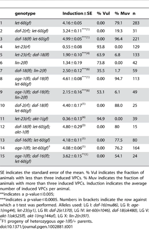
SE indicates the standard error of the mean. % Vul indicates the fraction of animals with less than three induced VPCs. % Muv indicates the fraction of animals with more than three induced VPCs. Induction indicates the average number of induced VPCs per animal. Tab. 2. RNAi against akt-2 and alternative PI3Ks. 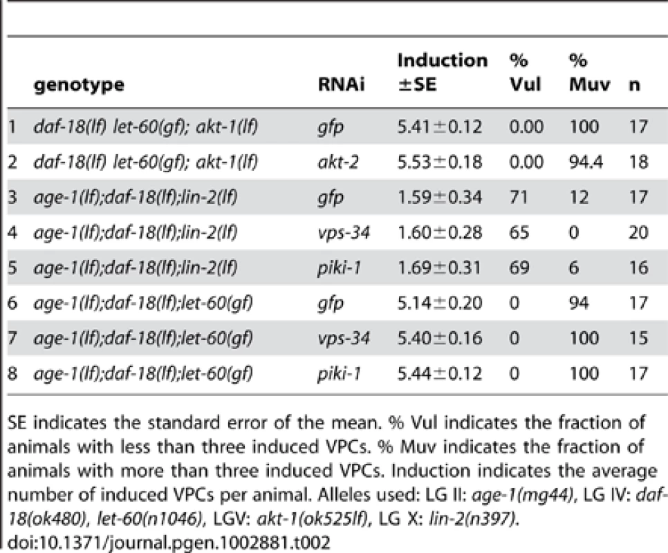
SE indicates the standard error of the mean. % Vul indicates the fraction of animals with less than three induced VPCs. % Muv indicates the fraction of animals with more than three induced VPCs. Induction indicates the average number of induced VPCs per animal. Alleles used: LG II: age-1(mg44), LG IV: daf-18(ok480), let-60(n1046), LGV: akt-1(ok525lf), LG X: lin-2(n397). We further tested the role of AGE-1 during vulval development. Since age-1(lf) leads to a constitutive dauer phenotype (DAF-c) that is maternally rescued, homozygous age-1(lf) worms could only be analyzed in the F1 progeny of heterozygous age-1(lf)/+ parents or when rescued by the daf-16(lf) mutation. Our analysis indicated that age-1 also exhibits a partial maternal rescue in vulval induction since the homozygous age-1(lf); let-60(gf) progeny obtained from heterozygous age-1(lf)/+ parents displayed similar levels of vulval induction as let-60(gf) single mutants (Table 1, row 14). In contrast, homozygous age-1(lf); let-60(gf) double mutants maintained in the daf-16(lf) background exhibited a partially suppressed Muv phenotype, though age-1(lf) suppressed the let-60(gf) Muv phenotype to a lesser extent than daf-2(rf) (Table 1, row 15, p-value≤0.05 compared to row 2).
Taken together, the genetic analysis indicates that the DAF-2 insulin receptor promotes and DAF-18 PTEN inhibits vulval induction. DAF-2 and DAF-18 both regulate vulval induction through AGE-1-dependent as well as AGE-1-independent pathways that do not utilize the canonical insulin pathway downstream of AGE-1.
DAF-18 inhibits vulval induction independently of other PI3Ks
AGE-1 is the only C. elegans member of the type I family of PI3Ks, which convert PI(4,5)P2 into PI(3,4,5)P3. To further investigate the AGE-1-independent effect of DAF-18 on vulval induction, we tested the roles of alternative PI3Ks that can phosphorylate PIs at position 3. vps-34 encodes a type III PI3K, which catalyzes the production of PI(3)P1, and piki-1 encodes a type II PI3K involved in the engulfment of apoptotic cell corpses [22]. In order to examine the role of these alternative PI3Ks during vulval induction, we performed RNAi against vps-34 and piki-1 in age-1(lf); daf-18(lf); lin-2(lf) animals and tested for a further reduction of vulval induction. RNAi to vps-34 and piki-1 has been previously shown to be effective in different tissues [23], [24]. Neither vps-34 nor piki-1 RNAi caused any significant reduction in the number of induced VPCs when compared to control (gfp) RNAi animals (Table 2, rows 3–5). Furthermore, vps-34 and piki-1 RNAi in an age-1(lf); daf-18(lf) let-60(gf) background did not cause a decrease in vulval induction (Table 2, rows 6–8). It thus seems unlikely that an alternative PI3K acts redundantly with AGE-1 during vulval induction, further supporting our observation that DAF-18 regulates vulval induction not only by regulating PIP3 levels but also via a lipid phosphatase-independent activity.
daf-18 inhibits 1° vulval cell fate specification
To characterize the nature of the cell fate transformation caused by daf-18(lf), we quantified the levels of the EGL-17::CFP reporter, whose expression is induced by RAS/MAPK signaling in the 1° vulval cell lineage [25] and of the LIP-1::GFP reporter whose expression is induced by LIN-12 NOTCH signaling in the 2° vulval cell lineage [26].
daf-18(lf) let-60(gf) double mutants showed an increased frequency of adjacent VPC descendants expressing high levels of EGL-17::CFP when compared to let-60(gf) single mutants (Figure 1A–1D). Specifically, in daf-18(lf); let-60(gf) double mutants 27% of adjacent VPC descendants showed strong EGL-17::CFP expression (i.e. at least 50% of the signal intensity seen in the P6.p lineage) versus 16% in let-60(gf) single mutants (Figure 1G). Furthermore, while the 2° P5.p and P7.p descendants in the wild-type displayed weak (i.e. less than 50% of the P6.px signal intensity) EGL-17::CFP expression in 22% of the cases, 52% of daf-18(lf) single mutants showed EGL-17::CFP expression in the 2° cells (Figure 1G).
Fig. 1. DAF-18 inhibits 1° vulval fate specification. 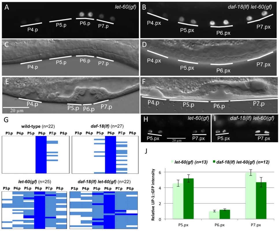
(A) Expression of the 1° cell fate marker EGL-17::CFP in let-60(gf) single and (B) daf-18(lf) let-60(gf) double mutants at the Pn.px stage. (C and D) show the corresponding Nomarski images of the animals shown in (A) and (B), respectively. (E) Morphology of the vulval invagination in a let-60(gf) mutant at the Pn.pxxx stage. Note that the descendants of P5.p and P7.p remain attached to the cuticle, which is characteristic of the 2° cell fate. (F) P5.p and P7.p often detach from the cuticle in daf-18(lf) let-60(gf) animals, resulting in a broad invagination with abnormal morphology. (G) Quantification of the 1° fate marker EGL-17::CFP expression at the Pn.px and Pn.pxx stages in the indicated genetic backgrounds. Dark blue represents cells with high EGL-17::CFP expression, corresponding to at least 50% of the intensity observed in the P6.px(x) cells, light blue represents cells with clearly visible but less than 50% of the P6.px(x) signal, and white spaces represent cells with undetectable levels of EGL-17::CFP. (H) Expression of the 2° fate marker LIP-1::GFP in let-60(gf) and (I) daf-18(lf) let-60(gf) animals. (J) Quantification of LIP-1::GFP expression in the P5.p through P7.p descendants. Signal intensities are indicated relative to P6.px(x) in let-60(gf). Besides the slight increase in EGL-17::CFP expression, the morphology of the vulval invagination at the L4 larval stage was changed in daf-18(lf) let-60(gf) double mutants. The vulval invagination formed by the P5.p to P7.p descendants of most let-60(gf) single mutants resembles the single invagination formed in the wild-type (Figure 1E). In daf-18(lf) let-60(gf) double mutants, on the other hand, the P5.p to P7.p descendants were often completely detached from the cuticle, resulting in an abnormal shape of the vulval invagination (Figure 1F) (37% detached P5.p and/or P7.p descendants in daf-18(lf) let-60(gf) versus 3% detached in let-60(gf), n = 54 and n = 35, respectively). A detachment of the P5.p and P7.p descendants from the cuticle is indicative of a 2° to 1° cell fate transformation as it has been observed in mutants exhibiting elevated MAPK activity in the 2° lineage [27].
In contrast to the 1° fate marker EGL-17::CFP, expression of the 2° fate marker LIP-1::GFP was not changed in daf-18(lf) mutants. In particular, LIP-1::GFP levels in the P5.p and P7.p descendants were unchanged in daf-18(lf) let-60(gf) double mutants compared to let-60(gf) single mutants (Figure 1H–1J).
Thus, daf-18(lf) enhances specification of the 1° cell fate and causes a partial 2° to 1° fate transformation in P5.p and P7.p without affecting the strength of the lateral LIN-12 NOTCH signal.
daf-18 negatively regulates vulval induction downstream of sos-1 and upstream or at the level of mpk-1
Since EGFR/RAS/MAPK signaling induces the 1° vulval cell fate and daf-18(lf) mutants exhibited an increased expression of the 1° cell fate marker EGL-17::CFP, we sought to determine at what level DAF-18 inhibits the activity of the EGFR/RAS/MAPK signaling pathway. For this purpose, we performed further epistasis analysis combining daf-18(lf) with mutations in different components of the RAS/MAPK pathway. Even though daf-18(lf) single mutants showed no obvious changes in the vulval fate pattern (Table 3, rows 1, 2), daf-18(lf) increased the levels of vulval induction in most of mutants in the RAS/MAPK pathway tested, confirming that DAF-18 negatively regulates the RAS/MAPK signaling during vulval induction. For example, when combined with mutations in different positive regulators of the RAS/MAPK pathway such as let-23(rf), lin-2(lf) or lin-45(rf), daf-18(lf) significantly suppressed the Vul phenotype of these mutants (Table 3, rows 5–8 and 11–12). In particular, daf-18(lf) suppressed a lf mutation in the RAS-GEF sos-1 when assayed in the let-60(gf) background to rescue the lethality caused by sos-1(lf), placing daf-18 function downstream of sos-1 (table 3, rows 9–10). However, since vulval induction in sos-1(lf); let-60(gf) animals is partly sensitive to the inductive anchor cell signal [28], we cannot exclude the possibility that DAF-18 might inhibit RAS/MAPK signaling through a SOS-1 independent branch of the pathway. As an exception, daf-18(lf) did not suppress the Vul phenotype of lin-3(rf) mutants (Table 3, rows 3–4), suggesting that daf-18(lf) alone is not sufficient to activate the RAS/MAPK pathway in the absence of the inductive AC signal. Furthermore, daf-18(lf) did not affect the completely penetrant Vul phenotype caused by mpk-1(lf) (Table 3, rows 13–14). Taken together, our epistasis analysis indicates that DAF-18 inhibits RAS/MAPK signaling downstream of or in parallel with the RAS-GEF SOS-1 and upstream or at the level of the MAP kinase MPK-1.
Tab. 3. Epistasis analysis of daf-18 with components of the RAS/MAPK pathway. 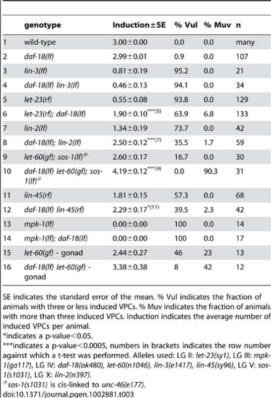
SE indicates the standard error of the mean. % Vul indicates the fraction of animals with three or less induced VPCs. % Muv indicates the fraction of animals with more than three induced VPCs. Induction indicates the average number of induced VPCs per animal. DAF-18 negatively regulates MPK-1 but not MEK-2 phosphorylation
Activation of the RAS/MAPK pathway results in an increased phosphorylation and activity of the downstream effectors RAF, MAPK kinase (MEK) and MAPK. We thus examined if daf-18(lf) mutants exhibit elevated levels of MEK and MAPK phosphorylation. Western blots of extracts from L4 larvae were probed with antibodies against mono-phosphorylated MEK (pMEK-2) and di-phosphorylated MAPK (dpMPK-1). Although the C. elegans genome encodes two MEK genes, MEK-1 and MEK-2, the phosphorylation site in human MEK to which the phospho-MEK antibody was raised (S217/S221) is only conserved in C. elegans MEK-2. Thus, we were able to specifically detect phosphorylated MEK-2 in whole animal extracts. Wild-type and daf-18(lf) L4 larvae contained only low levels of pMEK-2 and dpMPK-1 that could not be reliably quantified. As expected, let-60(gf) single mutants contained significantly higher levels of pMEK-2 and dpMPK-1 than wild-type larvae (Figure 2A and 2C). However, we observed no further increase in pMEK-2 levels in daf-18(lf) let-60(gf) double compared to let-60(gf) single mutants (Figure 2B). In contrast, dpMPK-1 levels were around two-fold increased daf-18(lf) let-60(gf) compared to let-60(gf) mutants (Figure 2D). Together with the genetic epistasis data presented above, the increase in MPK-1 phosphorylation in the absence of a significant change in MEK-2 phosphorylation indicates that DAF-18 most likely inhibits vulval induction at the level of MPK-1. Finally, the fact that we observed increased MPK-1 phosphorylation in total worm lysates suggests a global regulation of the RAS/MAPK pathway by DAF-18, probably including the germline.
Fig. 2. DAF-18 inhibits MPK-1 phosphorylation. 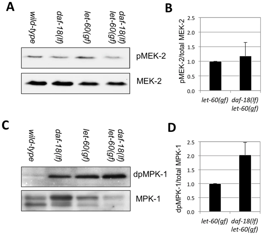
Total extracts of wild-type, daf-18(lf), let-60(gf) and daf-18(lf) let-60(gf) larvae at the L4 stage were analyzed on Western blots using antibodies against (A) phosphorylated and total MEK-2 and (C) against di-phosphorylated (dp) and total MPK-1. Signal intensities were quantified in four (for MEK-2) and three (for MPK-1) independent experiments, and the average ratios of (B) pMEK-2 to total MEK-2 and (D) dpMPK-1 to total MPK-1 intensities were calculated. daf-18 expression in the VPCs inhibits vulval induction
To further investigate the role of DAF-18 during vulval induction, we constructed a translational reporter by inserting a gfp cassette at the 3′ end of the ORF in a genomic daf-18 fragment (Figure 3). This DAF-18::GFP reporter rescued both the dauer defective (DAF-d) phenotype (data not shown) as well as the vulval phenotypes of daf-18(lf) with similar efficiency as a 6.5 kb genomic fragment spanning the entire daf-18 locus (Figure 4). DAF-18::GFP expression was observed in many tissues during all larval stages, including the developing vulva, the uterus, the ventral nerve cord and the distal tip cells (data not shown). In particular, equal levels of DAF-18::GFP expression were detected in the six VPCs of L2 larvae, and expression persisted in the descendants of the induced VPCs until the Pn.pxxx stage (Figure 3). Interestingly, the sub-cellular localization of DAF-18::GFP changed over the course of vulval development. In the VPCs of L2 larvae prior to and during induction (Pn.p stage), DAF-18::GFP was predominantly localized in the cytoplasm and the nucleus (Figure 3A, 3B and 3B′). However, at the subsequent stages (Pn.px to Pn.pxx stages), DAF-18::GFP became increasingly localized to the plasma membrane of the vulval cells (Figure 2C, 2D and 2D′). Plasma membrane staining peaked at the “Christmas tree” (Pn.pxxx) stage, when almost all the protein appeared to be localized to the membranes and nuclear staining was reduced to very low levels (Figure 3E and 3F). Since the DAF-18::GFP reporter was also expressed in tissues surrounding the vulval cells, we examined whether tissue-specific expression of DAF-18 in the VPCs reduces vulval induction. For this purpose, we expressed daf-18 cDNA fused to gfp under the control of the Pn.p cell-specific lin-31 promoter, which is active in the VPCs before and during vulval induction [29] (Plin-31::daf-18 cDNA::gfp::unc-54 3′ UTR). Indeed, introduction of the lin-31::daf-18::gfp transgene into daf-18(lf); let-23(rf) animals repressed vulval induction with similar efficiency as the daf-18::gfp reporter or a genomic daf-18 rescue construct (Figure 4).
Fig. 3. Expression pattern and sub-cellular localization of DAF-18::GFP. 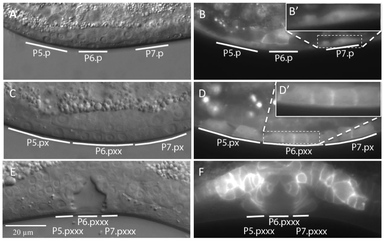
(A and B) Nomarski and fluorescence images of animals expressing the DAF-18::GFP translational reporter at the Pn.p cell stage, (C and D) the Pn.px(x) stage and (E and F) the Pn.pxxx “Christmas tree” stage. The insets in (B′) and (D′) show higher magnifications of the areas in (B) and (D) marked with dashed boxes. Fig. 4. DAF-18 expression in the Pn.p cells inhibits vulval induction. 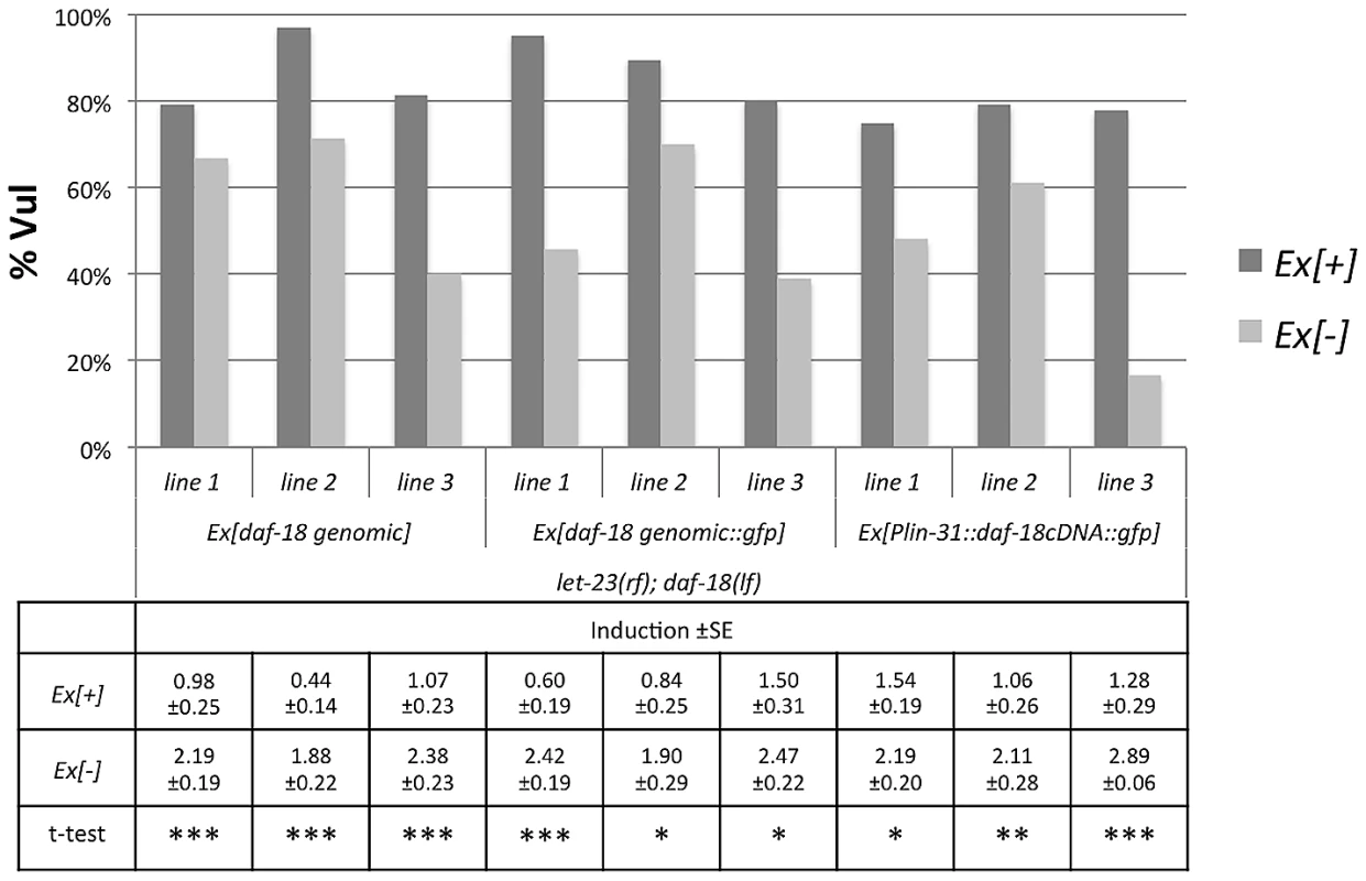
Vulval induction was scored at the L4 stage comparing let-23(rf); daf-18(lf) animals carrying the different extra-chromosomal arrays (Ex[+]) to their siblings lacking the array (Ex[−]). Three independent lines were scored per construct and at least 20 Ex[+] and Ex[−] animals were counted per line. For each line, a t-test was performed comparing induction in Ex[+] and Ex[−] animals on the same plates. * indicates a p-value≤0.05, ** a p-value≤0.005, and *** a p-value≤0.0005. Besides the vulval cells, the DAF-18::GFP reporter was also expressed at the L3 to L4 larval stages in several cells of the uterus, which is part of the somatic gonad (Figure 3). We thus tested if the daf-18-mediated repression of vulval induction requires the gonad by ablating the Z1 to Z4 somatic gonad and germline precursor cells at the L1 stage. In gonad-ablated daf-18(lf) let-60(gf) double mutants, vulval induction was higher than in gonad-ablated let-60(gf) single mutants, indicating that DAF-18 represses vulval induction independently of a signal from the gonad (Table 3, rows 15, 16). Thus, DAF-18 probably acts predominantly in the VPCs to inhibit MAPK signaling during vulval induction.
Both lipid and protein phosphatase activities of DAF-18 inhibit vulval induction
Mammalian PTEN acts as a lipid phosphatase as well as a dual-specificity protein phosphatase [5], [6]. Moreover, a recent report has shown that C. elegans DAF-18 can act as a protein phosphatase inhibiting signaling by the VAB-1 ephrin receptor during oocyte maturation [18]. The G129E mutation in the catalytic center of human PTEN eliminates the lipid phosphatase activity, while retaining the protein phosphatase activity [7]. The corresponding glycine 174 residue in C. elegans DAF-18 was therefore mutated to glutamic acid in the daf-18 genomic rescue construct. To quantify the rescuing activity of the daf-18 wild-type (daf-18 wt) and the G174E mutated lipid phosphatase mutant (daf-18 G174E), these constructs were expressed in the daf-18(lf) let-60(gf) and let-23(rf); daf-18(lf) backgrounds, and vulval induction was quantified. As expected, expression of daf-18 wt rescued both the DAF-d (data not shown) and vulval phenotypes of daf-18(lf) (Figure 5). In contrast, daf-18 G174E did not rescue the DAF-d phenotype ([15] and own observation), yet exhibited a weaker, though significant rescuing activity of the vulval induction phenotype (Figure 5). These results indicate that the DAF-18 protein and lipid phosphatase activities each play independent roles in negatively regulating the RAS/MAPK pathway and that both activities are required for the full inhibition of vulval induction by DAF-18.
Fig. 5. A lipid phosphatase deficient mutant of DAF-18 remains partially active. 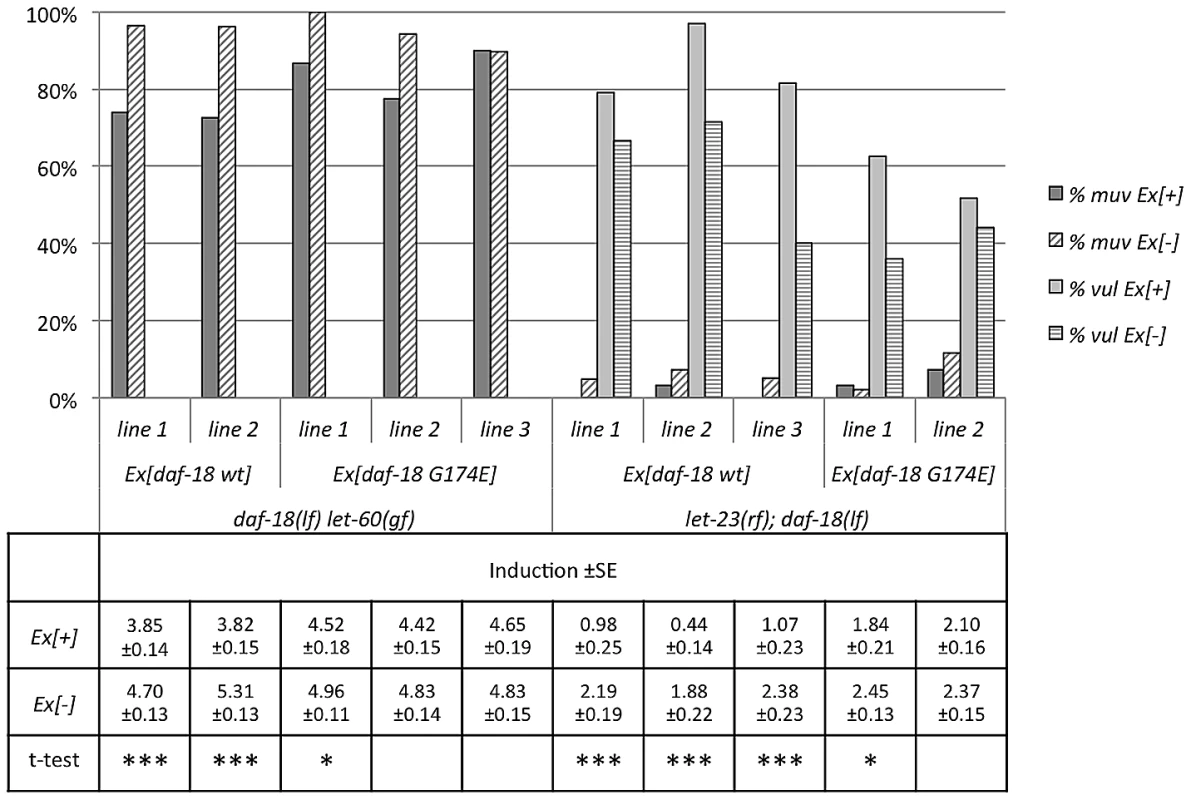
Animals carrying the daf-18 wt and daf-18 G174E extra-chromosomal arrays were scored at the L4 stage as described in the legend to Figure 4. Discussion
The Insulin pathway is a key regulator of development, reproduction, and life span in metazoans. In this study, we have discovered a new form of cross-talk between the Insulin and RAS/MAPK pathways during vulval development. Signaling by the Insulin receptor DAF-2 positively regulates MAPK activation. Surprisingly, the effect of DAF-2 on vulval development does not involve activation of the canonical Insulin pathway. DAF-2 signaling regulates vulval induction in at least two distinct manners, through AGE-1 dependent and independent pathways (Figure 6). One possible explanation for the AGE-1-independent branch of DAF-2 signaling is supported by mammalian data, which suggest that the Insulin receptor can directly stimulate RAS activation by recruiting GRB2 and the RAS-GEF SOS [30], [31]. Also in C. elegans, LET-60 RAS was found to act downstream of the DAF-2 Insulin receptor to modulate the effects of Insulin signaling during entry into the Dauer stage [32].
Fig. 6. Multiple modes of crosstalk between the RAS/MAPK and insulin pathways during vulval development. 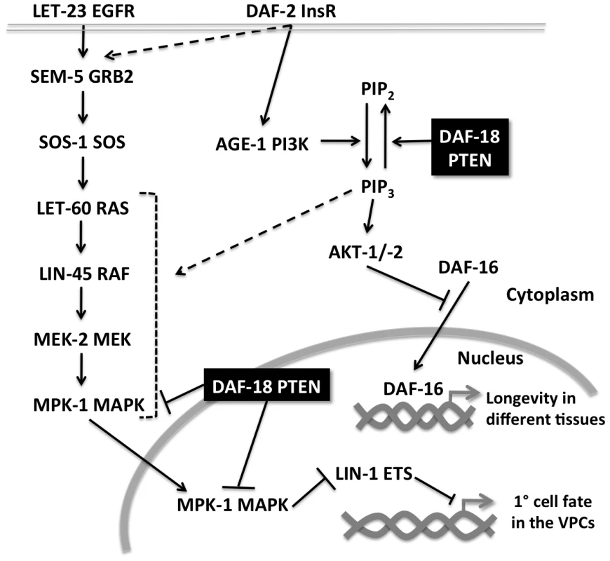
Activation of LET-60 RAS in the VPCs causes phosphorylated MPK-1 to enter the nucleus where it phosphorylates transcription factors such as LIN-1 ETS, which represses 1° fate-specific gene expression. The insulin signaling pathway interacts with the RAS/MAPK pathway at several levels. Firstly, DAF-2 InsR activation enhances RAS/MAPK signaling by stimulating the PI3K AGE-1, which positively regulates RAS/MAPK signaling via increased PIP3 production. In addition, DAF-2 signaling activates the RAS/MAPK signaling independently of AGE-1, possibly by recruiting SEM-5 to the plasma membrane. DAF-18 PTEN inhibits RAS/MAPK signaling in two distinct manners; by dephosphorylating PIP3 and by negatively regulating MAPK activation. Furthermore, we found that the PTEN ortholog DAF-18 strongly inhibits RAS/MAPK signaling. Vulval induction in daf-18(lf) let-60(gf) double mutants reaches levels similar to those seen in the strongest Muv mutants such as lin-15AB(lf) [33]. The increase in RAS/MAPK signaling in daf-18(lf) mutants could be partially reverted by loss of the PI3K activity, suggesting that elevated levels of PIP3 do stimulate RAS/MAPK signaling but cannot explain all the functions DAF-18 exerts during vulval induction. Accordingly, the inhibitory activity requires both the lipid and protein phosphatase activities of DAF-18. PIP3 acts as a second messenger that activates multiple downstream targets. One major PIP3 target in the Insulin pathway is the AKT kinases, which phosphorylate and thereby inhibits the FOXO transcription factor DAF-16. However neither akt-1, akt-2 nor daf-16 mutations had any detectable effect on vulval induction. Thus, PIP3 must act via other targets to stimulate RAS/MAPK signaling. Increased levels of PIP3 in the plasma membrane could, for example, enhance the recruitment of an alternative GEF that activates RAS signaling in parallel to the RAS-GEF SOS-1 [28] (Figure 6). However, we observed that prior to and at early stages of vulval induction, DAF-18::GFP was localized predominantly in the cytoplasm and nucleus of the VPCs, while membrane localization of DAF-18 only became apparent at later stages. Previous observations of mammalian PTEN localization suggested that PTEN performs different functions depending on its sub-cellular localization [34]. It has been proposed that the lipid phosphatase activity is important for the cytoplasmic and membrane functions of mammalian PTEN, while the protein phosphatase activity is rather required for its nuclear functions [34], [35]. The nuclear localization of PTEN in mammalian cells is often associated with cell-cycle arrest in G1 and accompanied by decreased levels of ERK phosphorylation. Prior to vulval induction, the VPCs are maintained in a long G1 arrest lasting the entire L2 stage [36]. It is therefore possible that initially DAF-18 acts predominantly in the nucleus as a protein phosphatase that negatively regulates vulval induction. Indeed, Western blot analysis revealed elevated levels of dpMPK-1 in daf-18(lf) mutants, supporting our model that DAF-18 –directly or indirectly - blocks MAPK activation (Figure 6).
In humans, PTEN is one of the most frequently mutated tumor suppressor genes. However, not all disease phenotypes associated with loss of PTEN can be explained by hyper-activation of the Insulin pathway alone. Thus, PTEN must have other functions that are independent of its inhibitory activity in the Insulin pathway. Accordingly, Suzuki and Han [19] observed many synthetic phenotypes in C. elegans daf-18(lf) mutants, including embryonic lethality and sterility, which are independent of DAF-16 FOXO and do not involve DAF-2 InsR signaling. Our work highlights the importance of C. elegans DAF-18 PTEN in regulating a range of biological processes and may serve as a basis to better understand the multiple roles human PTEN plays during cancer initiation and progression. Thus, single mutations in the PTEN tumor suppressor may result in the simultaneous hyper-activation of several oncogenic signaling pathways.
Materials and Methods
General worm methods
Standard methods were used for maintaining and manipulating Caenorhabditis elegans [37]. Animals were cultured at 20°C and the wild-type strain is the Bristol N2 strain. Information regarding the mutants used in this study can be found on WormBase (http://www.wormbase.org). Mutations used according to their linkage group:
LG I: daf-16(mu86), LG II: age-1(mg44), let-23(sy1), LG III: daf-2(e1370), mpk-1(ga117), LG IV: let-60(n1046), daf-18(ok480), lin-3(e1417), lin-45(sy96), LG V: akt-1(mg144gf), akt-1(ok525lf), LG X: lin-2(n397), sos-1(s1031), unc-46(e177) to cis link sos-1(s1031), LG X: lin-2(n397), gap-1(ga133). Transgenes used: syIs59[Pegl-17::cfp], zhIs4[Plip-1::gfp], zhEx382[daf-18 genomic] zhEx343[daf-18::gfp], zhEx358[Plin-31::daf-18::gfp], zhEx344[daf-18 G174E].
Plasmids and PCR fusion constructs
pIN05 (daf-18 genomic wt) was made by cloning the whole genomic fragment of daf-18 starting 1.3 kb upstream of the ATG and ending 0.5 kb downstream of the STOP and cloning to pGEM-T. pIN03 (daf-18 genomic G174E) was made by fusion PCR [38] of two overlapping fragments of the whole genomic daf-18 starting 1.3 kb upstream of the ATG and ending 0.5 kb downstream of the STOP using primers which contain the mutation G174E (GGC to GAA) in the overlapping region and cloning to pGEM-T. pIN17 (Plin-31::daf-18 cDNA::gfp::unc-54 3′UTR) was made by amplifying daf-18 cDNA::gfp from a previously cloned plasmid with primers containing NotI sites on both ends, digestion with NotI and cloning into the NotI site of the pB253 plasmid containing the lin-31 enhancer and promoter. The daf-18 genomic translational GFP reporter was made using fusion PCR of three parts by inserting a gfp cassette in frame between the last exon and the 3′ UTR into a genomic fragment encompassing 1.3 kb of 5′ regulatory sequences and the complete daf-18 coding sequences. Sequences of the primers used for the different constructs can be found in Table S1.
Transgenic lines
Worms expressing extra-chromosomal transgenic arrays were generated by microinjection of DNA into young adult worms [39]. pIN03 (zhEx344), pIN05 (zhEx382) and pIN17(zhEx358) were injected at a concentration of 50 ng/µl. The fusion PCR daf-18 genomic::gfp (zhEx343) translational reporter was injected at a concentration of 30 ng/ul. Co-markers used were either pCFJ90 (Pmyo-2::mCherry) at a concentration of 2 ng/ul or pTJ1157 (Plin-48::gfp) at a concentration of 50 ng/ul. Final concentration of injected DNA was filled up to 150 ng/ul using the empty plasmid pBluescript-KS.
Fluorescence microscopy
DAF-18::GFP, Plip-1::GFP and Pegl-17::CFP expression were observed under fluorescent light illumination with either a Leica DMRA microscope equipped with a cooled CCD camera (Hamamatsu ORCA-ER) or Olympus BX61 with Q Imaging Fast 1394 Retiga 2000R camera (Q Imaging Inc., Canada) controlled by the Openlab 5.x software (Improvision/PerkinElmer). Animals were mounted on 4% agarose pads in M9 solution with 20 mM tetramisole hydrochloride. Quantification of fluorescence levels was performed under the same picture acquisition settings for all conditions examined.
Vulval induction
Vulval induction was scored by examining worms at the L4 stage under Nomarski optics as described [40]. The number of VPCs that had adopted a 1° or 2° Vulval fate was counted for each animal and the induction index was calculated by dividing the number of 1° or 2° induced cells by the number of animals scored. Statistical analysis was performed using a t-test for independent samples.
RNA interference analysis
RNA interference analysis (RNAi) was performed by feeding animals dsRNA-producing bacteria as described previously [41]. Around 10 to 20 P0 animals at the L1 larval stage were transferred to plates containing RNAi bacteria grown on 3 mM IPTG. Vulval induction was scored in the F1 generation (or the P0 for akt-2 RNAi) at the L4 larval stage to count the number of induced VPCs. gfp, akt-2, piki-1 and vps-34 RNAi clones were all taken from the Ahringer RNAi library.
Western blot analysis
Forty-five animals at the L4 stage were placed into 15 µl of 1× SDS sample buffer, lysed at 95°C for 5 min, centrifuged at 14,000 rpm for 2 min and the supernatant was loaded on 10% acrylamide gels, which were analyzed by Western blotting. Anti-phospho-MEK1/2 (S217/S221) and Anti-MEK1/2 (D1A5) antibodies were purchased from Cell Signaling Technology (Beverly, MA). Anti-di-phosphorylated ERK-1&2 (M8159) antibody was purchased from Sigma-Aldrich (St. Louis, MO), and anti-ERK 2 (K-23) antibody was purchased from Santa Cruz Biotechnology (Santa Cruz, CA). Quantification of the bands was performed using the gel quantification plugin in ImageJ software [42]. The ratios between phosphorylated and total MEK-2 and MPK-1 levels, respectively, were calculated and normalized for each independent experiment to the ratios measured in the let-60(gf) single mutants.
Supporting Information
Zdroje
1. LiJ, YenC, LiawD, PodsypaninaK, BoseS, et al. (1997) PTEN, a putative protein tyrosine phosphatase gene mutated in human brain, breast, and prostate cancer. Science 275 : 1943–1947.
2. BlumenthalGM, DennisPA (2008) PTEN hamartoma tumor syndromes. Eur J Hum Genet 16 : 1289–1300 doi:10.1038/ejhg.2008.162.
3. LiawD, MarshDJ, LiJ, DahiaPL, WangSI, et al. (1997) Germline mutations of the PTEN gene in Cowden disease, an inherited breast and thyroid cancer syndrome. Nat Genet 16 : 64–67 doi:10.1038/ng0597-64.
4. MyersM, PassI, BattyI, Van der KaayJ, StolarovJ, et al. (1998) The lipid phosphatase activity of PTEN is critical for its tumor supressor function. Proc Natl Acad Sci USA 95 : 13513–13518.
5. DeyN, CrosswellHE, DeP, ParsonsR, PengQ, et al. (2008) The protein phosphatase activity of PTEN regulates SRC family kinases and controls glioma migration. Cancer Res 68 : 1862–1871 doi:10.1158/0008-5472.CAN-07-1182.
6. CaiX-M, TaoB-B, WangL-Y, LiangY-L, JinJ-W, et al. (2005) Protein phosphatase activity of PTEN inhibited the invasion of glioma cells with epidermal growth factor receptor mutation type III expression. Int J Cancer 117 : 905–912 doi:10.1002/ijc.21251.
7. MyersMP, StolarovJP, EngC, LiJ, WangSI, et al. (1997) P-TEN, the tumor suppressor from human chromosome 10q23, is a dual-specificity phosphatase. Proc Natl Acad Sci USA 94 : 9052–9057.
8. TamuraM, GuJ, MatsumotoK, AotaS, ParsonsR, et al. (1998) Inhibition of cell migration, spreading, and focal adhesions by tumor suppressor PTEN. Science 280 : 1614–1617.
9. GildeaJ, HerlevsenM, HardingM, GuldingK, MoskalukC, et al. (2004) PTEN can inhibit in vitro organotypic and in vivo orthotopic invasion of human bladder cancer cells even in the absence of its lipid phosphatase activity. Oncogene 23 : 6788–6797 doi:10.1038/sj.onc.1207599.
10. GuJ, TamuraM, PankovR, DanenEH, TakinoT, et al. (1999) Shc and FAK differentially regulate cell motility and directionality modulated by PTEN. J Cell Biol 146 : 389–403.
11. HuPJ (2007) Dauer. Worm Book 1–19 doi:10.1895/wormbook.1.144.1.
12. ChristensenR, la Torre-Ubieta deL, BonniA, Colón-RamosDA (2011) A conserved PTEN/FOXO pathway regulates neuronal morphology during C. elegans development. Development 138 : 5257–5267 doi:10.1242/dev.069062.
13. FukuyamaM, RougvieA, RothmanJ (2006) C. elegans DAF-18/PTEN mediates nutrient-dependent arrest of cell cycle and growth in the germline. Curr Biol 16 : 773–779 doi:10.1016/j.cub.2006.02.073.
14. MihaylovaV, BorlandC, ManjarrezL, SternM, SunH (1999) The PTEN tumor suppressor homolog in Caenorhabditis elegans regulates longevity and dauer formation in an insulin receptor-like signaling pathway. Proc Natl Acad Sci USA 96 : 7427–7432.
15. SolariF, Bourbon-PiffautA, MasseI, PayrastreB, ChanA, et al. (2005) The human tumour suppressor PTEN regulates longevity and dauer formation in Caenorhabditis elegans. Oncogene 24 : 20–27 doi:10.1038/sj.onc.1207978.
16. ParadisS, RuvkunG (1998) Caenorhabditis elegans Akt/PKB transduces insulin receptor-like signals from AGE-1 PI3 kinase to the DAF-16 transcription factor. Genes Dev 12 : 2488–2498.
17. CahillCM, TzivionG, NasrinN, OggS, DoreJ, et al. (2001) Phosphatidylinositol 3-kinase signaling inhibits DAF-16 DNA binding and function via 14-3-3-dependent and 14-3-3-independent pathways. J Biol Chem 276 : 13402–13410 doi:10.1074/jbc.M010042200.
18. BrisbinS, LiuJ, BoudreauJ, PengJ, EvangelistaM, et al. (2009) A role for C. elegans Eph RTK signaling in PTEN regulation. Dev Cell 17 : 459–469 doi:10.1016/j.devcel.2009.08.009.
19. SuzukiY, HanM (2006) Genetic redundancy masks diverse functions of the tumor suppressor gene PTEN during C. elegans development. Genes Dev 20 : 423–428 doi:10.1101/gad.1378906.
20. SternbergPW (2005) Vulval development. Worm Book 1–28 doi:10.1895/wormbook.1.6.1.
21. BattuG, HoierE, HajnalA (2003) The C. elegans G-protein-coupled receptor SRA-13 inhibits RAS/MAPK signalling during olfaction and vulval development. Development 130 : 2567–2577.
22. ZouW, LuQ, ZhaoD, LiW, MapesJ, et al. (2009) Caenorhabditis elegans myotubularin MTM-1 negatively regulates the engulfment of apoptotic cells. PLoS Genet 5: e1000679 doi:10.1371/journal.pgen.1000679.
23. AshrafiK, ChangFY, WattsJL, FraserAG, KamathRS, et al. (2003) Genome-wide RNAi analysis of Caenorhabditis elegans fat regulatory genes. Nature 421 : 268–272 doi:10.1038/nature01279.
24. KinchenJM, DoukoumetzidisK, AlmendingerJ, StergiouL, Tosello-TrampontA, et al. (2008) A pathway for phagosome maturation during engulfment of apoptotic cells. Nat Cell Biol 10 : 556–566 doi:10.1038/ncb1718.
25. BurdineRD, BrandaCS, SternMJ (1998) EGL-17(FGF) expression coordinates the attraction of the migrating sex myoblasts with vulval induction in C. elegans. Development 125 : 1083–1093.
26. BersetT, HoierE, BattuG, CanevasciniS, HajnalA (2001) Notch inhibition of RAS signaling through MAP kinase phosphatase LIP-1 during C. elegans vulval development. Science 291 : 1055–1058 doi:10.1126/science.1055642.
27. LacknerMR, KimSK (1998) Genetic analysis of the Caenorhabditis elegans MAP kinase gene mpk-1. Genetics 150 : 103–117.
28. ChangC, HopperN, SternbergP (2000) Caenorhabditis elegans SOS-1 is necessary for multiple RAS-mediated developmental signals. EMBO J 19 : 3283–3294 doi:10.1093/emboj/19.13.3283.
29. TanPB, LacknerMR, KimSK (1998) MAP kinase signaling specificity mediated by the LIN-1 Ets/LIN-31 WH transcription factor complex during C. elegans vulval induction. Cell 93 : 569–580.
30. SasaokaT, KobayashiM (2000) The functional significance of Shc in insulin signaling as a substrate of the insulin receptor. Endocr J 47 : 373–381.
31. SasaokaT, DrazninB, LeitnerJW, LangloisWJ, OlefskyJM (1994) Shc is the predominant signaling molecule coupling insulin receptors to activation of guanine nucleotide releasing factor and p21ras-GTP formation. J Biol Chem 269 : 10734–10738.
32. NanjiM, HopperN, GemsD (2005) LET-60 RAS modulates effects of insulin/IGF-1 signaling on development and aging in Caenorhabditis elegans. Aging Cell 4 : 235–245 doi:10.1111/j.1474-9726.2005.00166.x.
33. CuiM, ChenJ, MyersTR, HwangBJ, SternbergPW, et al. (2006) SynMuv Genes Redundantly Inhibit lin-3/EGF Expression to Prevent Inappropriate Vulval Induction in C. elegans. Dev Cell 10 : 667–672 doi:10.1016/j.devcel.2006.04.001.
34. PlanchonSM, WaiteKA, EngC (2008) The nuclear affairs of PTEN. J Cell Sci 121 : 249–253 doi:10.1242/jcs.022459.
35. WengLP, BrownJL, EngC (2001) PTEN coordinates G(1) arrest by down-regulating cyclin D1 via its protein phosphatase activity and up-regulating p27 via its lipid phosphatase activity in a breast cancer model. Hum Mol Genet 10 : 599–604.
36. HongY, RoyR, AmbrosV (1998) Developmental regulation of a cyclin-dependent kinase inhibitor controls postembryonic cell cycle progression in Caenorhabditis elegans. Development 125 : 3585–3597.
37. BrennerS (1974) The genetics of Caenorhabditis elegans. Genetics 77 : 71–94.
38. HobertO (2002) PCR fusion-based approach to create reporter gene constructs for expression analysis in transgenic C. elegans. Bio Techniques 32 : 728–730.
39. MelloCC, KramerJM, StinchcombD, AmbrosV (1991) Efficient gene transfer in C.elegans: extrachromosomal maintenance and integration of transforming sequences. EMBO J 10 : 3959–3970.
40. SternbergPW, HorvitzHR (1986) Pattern formation during vulval development in C. elegans. Cell 44 : 761–772.
41. KamathRS, FraserAG, DongY, PoulinG, DurbinR, et al. (2003) Systematic functional analysis of the Caenorhabditis elegans genome using RNAi. Nature 421 : 231–237 doi:10.1038/nature01278.
42. AbramoffMD, MagalhãesPJ, RamSJ (2004) Image processing with ImageJ. Biophotonics international 11 : 36–42.
Štítky
Genetika Reprodukční medicína
Článek Mutational Signatures of De-Differentiation in Functional Non-Coding Regions of Melanoma GenomesČlánek Rescuing Alu: Recovery of Inserts Shows LINE-1 Preserves Alu Activity through A-Tail ExpansionČlánek Genetics and Regulatory Impact of Alternative Polyadenylation in Human B-Lymphoblastoid CellsČlánek Retrovolution: HIV–Driven Evolution of Cellular Genes and Improvement of Anticancer Drug ActivationČlánek The Mi-2 Chromatin-Remodeling Factor Regulates Higher-Order Chromatin Structure and Cohesin DynamicsČlánek Identification of Human Proteins That Modify Misfolding and Proteotoxicity of Pathogenic Ataxin-1
Článek vyšel v časopisePLOS Genetics
Nejčtenější tento týden
2012 Číslo 8- Akutní intermitentní porfyrie
- Farmakogenetické testování pomáhá předcházet nežádoucím efektům léčiv
- Hypogonadotropní hypogonadismus u žen a vliv na výsledky reprodukce po IVF
- Molekulární vyšetření pro stanovení prognózy pacientů s chronickou lymfocytární leukémií
- Prof. Petr Urbánek: Potřebujeme najít pacienty s nediagnostikovanou akutní intermitentní porfyrií
-
Všechny články tohoto čísla
- Mutational Signatures of De-Differentiation in Functional Non-Coding Regions of Melanoma Genomes
- Rescuing Alu: Recovery of Inserts Shows LINE-1 Preserves Alu Activity through A-Tail Expansion
- Genetics and Regulatory Impact of Alternative Polyadenylation in Human B-Lymphoblastoid Cells
- Chromosome Territories Meet a Condensin
- It's All in the Timing: Too Much E2F Is a Bad Thing
- Fine-Mapping and Initial Characterization of QT Interval Loci in African Americans
- Genome Patterns of Selection and Introgression of Haplotypes in Natural Populations of the House Mouse ()
- A Combinatorial Amino Acid Code for RNA Recognition by Pentatricopeptide Repeat Proteins
- Advances in Quantitative Trait Analysis in Yeast
- Experimental Evolution of a Novel Sexually Antagonistic Allele
- Variation of Contributes to Dog Breed Skull Diversity
- , a Gene Involved in Axonal Pathfinding, Is Mutated in Patients with Kallmann Syndrome
- A Single Origin for Nymphalid Butterfly Eyespots Followed by Widespread Loss of Associated Gene Expression
- Cryptocephal, the ATF4, Is a Specific Coactivator for Ecdysone Receptor Isoform B2
- Retrovolution: HIV–Driven Evolution of Cellular Genes and Improvement of Anticancer Drug Activation
- The PARN Deadenylase Targets a Discrete Set of mRNAs for Decay and Regulates Cell Motility in Mouse Myoblasts
- A Sexual Ornament in Chickens Is Affected by Pleiotropic Alleles at and , Selected during Domestication
- Use of Allele-Specific FAIRE to Determine Functional Regulatory Polymorphism Using Large-Scale Genotyping Arrays
- Novel Loci for Metabolic Networks and Multi-Tissue Expression Studies Reveal Genes for Atherosclerosis
- The Genetic Basis of Pollinator Adaptation in a Sexually Deceptive Orchid
- Uncovering the Genome-Wide Transcriptional Responses of the Filamentous Fungus to Lignocellulose Using RNA Sequencing
- Inheritance Beyond Plain Heritability: Variance-Controlling Genes in
- The Metabochip, a Custom Genotyping Array for Genetic Studies of Metabolic, Cardiovascular, and Anthropometric Traits
- Reprogramming to Pluripotency Can Conceal Somatic Cell Chromosomal Instability
- Condensin II Promotes the Formation of Chromosome Territories by Inducing Axial Compaction of Polyploid Interphase Chromosomes
- PTEN Negatively Regulates MAPK Signaling during Vulval Development
- A Dynamic Response Regulator Protein Modulates G-Protein–Dependent Polarity in the Bacterium
- Population Genomics of the Facultatively Mutualistic Bacteria and
- Components of a Fanconi-Like Pathway Control Pso2-Independent DNA Interstrand Crosslink Repair in Yeast
- Polysome Profiling in Liver Identifies Dynamic Regulation of Endoplasmic Reticulum Translatome by Obesity and Fasting
- Stromal Liver Kinase B1 [STK11] Signaling Loss Induces Oviductal Adenomas and Endometrial Cancer by Activating Mammalian Target of Rapamycin Complex 1
- Reprogramming of H3K27me3 Is Critical for Acquisition of Pluripotency from Cultured Tissues
- Transgene Induced Co-Suppression during Vegetative Growth in
- Hox and Sex-Determination Genes Control Segment Elimination through EGFR and Activity
- A Quantitative Comparison of the Similarity between Genes and Geography in Worldwide Human Populations
- Minibrain/Dyrk1a Regulates Food Intake through the Sir2-FOXO-sNPF/NPY Pathway in and Mammals
- Comparative Analysis of Regulatory Elements between and by Genome-Wide Transcription Start Site Profiling
- Simple Methods for Generating and Detecting Locus-Specific Mutations Induced with TALENs in the Zebrafish Genome
- S Phase–Coupled E2f1 Destruction Ensures Homeostasis in Proliferating Tissues
- Cell-Nonautonomous Signaling of FOXO/DAF-16 to the Stem Cells of
- The Mi-2 Chromatin-Remodeling Factor Regulates Higher-Order Chromatin Structure and Cohesin Dynamics
- Comparative Analysis of the Genomes of Two Field Isolates of the Rice Blast Fungus
- Role of Mex67-Mtr2 in the Nuclear Export of 40S Pre-Ribosomes
- Genetic Modulation of Lipid Profiles following Lifestyle Modification or Metformin Treatment: The Diabetes Prevention Program
- HAL-2 Promotes Homologous Pairing during Meiosis by Antagonizing Inhibitory Effects of Synaptonemal Complex Precursors
- SLX-1 Is Required for Maintaining Genomic Integrity and Promoting Meiotic Noncrossovers in the Germline
- Phylogenetic and Transcriptomic Analysis of Chemosensory Receptors in a Pair of Divergent Ant Species Reveals Sex-Specific Signatures of Odor Coding
- Reduced Prostasin (CAP1/PRSS8) Activity Eliminates HAI-1 and HAI-2 Deficiency–Associated Developmental Defects by Preventing Matriptase Activation
- Dissecting the Gene Network of Dietary Restriction to Identify Evolutionarily Conserved Pathways and New Functional Genes
- Identification of Human Proteins That Modify Misfolding and Proteotoxicity of Pathogenic Ataxin-1
- and Link Transcription of Phospholipid Biosynthetic Genes to ER Stress and the UPR
- CDK9 and H2B Monoubiquitination: A Well-Choreographed Dance
- Rare Copy Number Variations in Adults with Tetralogy of Fallot Implicate Novel Risk Gene Pathways
- Ccdc94 Protects Cells from Ionizing Radiation by Inhibiting the Expression of
- NOL11, Implicated in the Pathogenesis of North American Indian Childhood Cirrhosis, Is Required for Pre-rRNA Transcription and Processing
- Human Developmental Enhancers Conserved between Deuterostomes and Protostomes
- A Luminal Glycoprotein Drives Dose-Dependent Diameter Expansion of the Hindgut Tube
- Melanophore Migration and Survival during Zebrafish Adult Pigment Stripe Development Require the Immunoglobulin Superfamily Adhesion Molecule Igsf11
- Dynamic Distribution of Linker Histone H1.5 in Cellular Differentiation
- Combining Comparative Proteomics and Molecular Genetics Uncovers Regulators of Synaptic and Axonal Stability and Degeneration
- Chemical Genetics Reveals a Specific Requirement for Cdk2 Activity in the DNA Damage Response and Identifies Nbs1 as a Cdk2 Substrate in Human Cells
- Experimental Relocation of the Mitochondrial Gene to the Nucleus Reveals Forces Underlying Mitochondrial Genome Evolution
- Rates of Gyrase Supercoiling and Transcription Elongation Control Supercoil Density in a Bacterial Chromosome
- Mutations in a P-Type ATPase Gene Cause Axonal Degeneration
- A General G1/S-Phase Cell-Cycle Control Module in the Flowering Plant
- Multiple Roles and Interactions of and in Development of the Respiratory System
- UNC-40/DCC, SAX-3/Robo, and VAB-1/Eph Polarize F-Actin during Embryonic Morphogenesis by Regulating the WAVE/SCAR Actin Nucleation Complex
- Epigenetic Remodeling of Meiotic Crossover Frequency in DNA Methyltransferase Mutants
- Modulating the Strength and Threshold of NOTCH Oncogenic Signals by
- Loss of Axonal Mitochondria Promotes Tau-Mediated Neurodegeneration and Alzheimer's Disease–Related Tau Phosphorylation Via PAR-1
- Acetyl-CoA-Carboxylase Sustains a Fatty Acid–Dependent Remote Signal to Waterproof the Respiratory System
- ATXN2-CAG42 Sequesters PABPC1 into Insolubility and Induces FBXW8 in Cerebellum of Old Ataxic Knock-In Mice
- Cohesin Rings Devoid of Scc3 and Pds5 Maintain Their Stable Association with the DNA
- The MicroRNA Inhibits Calcium Signaling by Targeting the TIR-1/Sarm1 Adaptor Protein to Control Stochastic L/R Neuronal Asymmetry in
- Rapid-Throughput Skeletal Phenotyping of 100 Knockout Mice Identifies 9 New Genes That Determine Bone Strength
- The Genes Define Unique Classes of Two-Partner Secretion and Contact Dependent Growth Inhibition Systems
- PLOS Genetics
- Archiv čísel
- Aktuální číslo
- Informace o časopisu
Nejčtenější v tomto čísle- Dissecting the Gene Network of Dietary Restriction to Identify Evolutionarily Conserved Pathways and New Functional Genes
- It's All in the Timing: Too Much E2F Is a Bad Thing
- Variation of Contributes to Dog Breed Skull Diversity
- The PARN Deadenylase Targets a Discrete Set of mRNAs for Decay and Regulates Cell Motility in Mouse Myoblasts
Kurzy
Zvyšte si kvalifikaci online z pohodlí domova
Autoři: prof. MUDr. Vladimír Palička, CSc., Dr.h.c., doc. MUDr. Václav Vyskočil, Ph.D., MUDr. Petr Kasalický, CSc., MUDr. Jan Rosa, Ing. Pavel Havlík, Ing. Jan Adam, Hana Hejnová, DiS., Jana Křenková
Autoři: MUDr. Irena Krčmová, CSc.
Autoři: MDDr. Eleonóra Ivančová, PhD., MHA
Autoři: prof. MUDr. Eva Kubala Havrdová, DrSc.
Všechny kurzyPřihlášení#ADS_BOTTOM_SCRIPTS#Zapomenuté hesloZadejte e-mailovou adresu, se kterou jste vytvářel(a) účet, budou Vám na ni zaslány informace k nastavení nového hesla.
- Vzdělávání


