-
Články
- Vzdělávání
- Časopisy
Top články
Nové číslo
- Témata
- Kongresy
- Videa
- Podcasty
Nové podcasty
Reklama- Kariéra
Doporučené pozice
Reklama- Praxe
Cell-Nonautonomous Signaling of FOXO/DAF-16 to the Stem Cells of
In Caenorhabditis elegans (C. elegans), the promotion of longevity by the transcription factor DAF-16 requires reduced insulin/IGF receptor (IIR) signaling or the ablation of the germline, although the reason for the negative impact of germ cells is unknown. FOXO/DAF-16 activity inhibits germline proliferation in both daf-2 mutants and gld-1 tumors. In contrast to its function as a germline tumor suppressor, we now provide evidence that somatic DAF-16 in the presence of IIR signaling can also result in tumorigenic activity, which counteracts robust lifespan extension. In contrast to the cell-autonomous IIR signaling, which is required for larval germline proliferation, activation of DAF-16 in the hypodermis results in hyperplasia of the germline and disruption of the surrounding basement membrane. SHC-1 adaptor protein and AKT-1 kinase antagonize, whereas AKT-2 and SGK-1 kinases promote, this cell-nonautonomous DAF-16 function. Our data suggest that a functional balance of DAF-16 activities in different tissues determines longevity and reveals a novel, cell-nonautonomous role of FOXO/DAF-16 to affect stem cells.
Published in the journal: . PLoS Genet 8(8): e32767. doi:10.1371/journal.pgen.1002836
Category: Research Article
doi: https://doi.org/10.1371/journal.pgen.1002836Summary
In Caenorhabditis elegans (C. elegans), the promotion of longevity by the transcription factor DAF-16 requires reduced insulin/IGF receptor (IIR) signaling or the ablation of the germline, although the reason for the negative impact of germ cells is unknown. FOXO/DAF-16 activity inhibits germline proliferation in both daf-2 mutants and gld-1 tumors. In contrast to its function as a germline tumor suppressor, we now provide evidence that somatic DAF-16 in the presence of IIR signaling can also result in tumorigenic activity, which counteracts robust lifespan extension. In contrast to the cell-autonomous IIR signaling, which is required for larval germline proliferation, activation of DAF-16 in the hypodermis results in hyperplasia of the germline and disruption of the surrounding basement membrane. SHC-1 adaptor protein and AKT-1 kinase antagonize, whereas AKT-2 and SGK-1 kinases promote, this cell-nonautonomous DAF-16 function. Our data suggest that a functional balance of DAF-16 activities in different tissues determines longevity and reveals a novel, cell-nonautonomous role of FOXO/DAF-16 to affect stem cells.
Introduction
The forkhead box O (FOXO) subfamily of Forkhead transcription factors is conserved from Caenorhabditis elegans (C. elegans) to mammals [1]. Mammalian FOXO transcription factors consist of four members: FOXO1, FOXO3, FOXO4 and FOXO6, whereas only one homologue, DAF-16, is encoded in the C. elegans genome. DAF-16/FOXO proteins are inactivated by the insulin/IGF-1 signaling (IIS) through PI3K and the AGC kinases Akt/SGK which promote its cytosolic localization [2]–[7]. Starvation reduces IIS, resulting in nuclear localization and activation of DAF-16. Stress stimuli also result in nuclear translocation and activate FOXO via JNK and MST1 even in the presence of Akt [8], [9].
The Akt/FOXO signaling network acts as a critical control mechanism at the intersection between cancer and stem cell biology. FOXO proteins have been considered as tumor suppressors, because of their ability to induce DNA damage repair, cell cycle arrest and apoptosis [10], [11]. Consistently, loss of functional FOXO is associated with tumorigenesis in various organs [11]–[13]. On the other hand, FOXO proteins are required for the long-term maintenance of both normal and cancer stem cells. Mice with FOXO1, FOXO3 and FOXO4 triple knockout display a marked reduction of hematopoietic stem cells due to increased physiological oxidative stress [14]. In the cancer stem cells of chronic myeloid leukemia, FOXO3 is enriched in the nucleus and essential for maintaining these cancer stem cells [15].
In C. elegans, DAF-16 is a key regulator of development, longevity and stress response. Active DAF-16 resulting from reduced IIS leads to dauer formation, enhanced stress response and lifespan extension [16]–[18]. Recent studies reveal that the cross-talk between the reproductive system and somatic tissues plays an important role in development and aging of C. elegans. During larval development DAF-16 inhibits robust proliferation of the germline through both cell-autonomous (the germline) and -nonautonomous (the musculature) mechanism [19]. In adult animals intestinal and muscular DAF-16 slow down reproductive aging via oocyte and germline quality maintenance [20]. Activation of DAF-16 upon reduced IIS also inhibits tumor growth in the germline of gld-1 mutants [21], indicating that the role of FOXO as tumor suppressor is evolutionarily conserved. On the other side, signals from the reproductive system regulate DAF-16 activity in the soma: Elimination of mitotic germ cells results in nuclear entry of intestinal DAF-16 and extends lifespan [22], [23]. Ablation of the somatic gonad precursors, however, abrogates the lifespan extension of the germline-ablated animals [23]. Even though several factors, such as KRI-1, TCER-1 and DAF-9, have been found to be involved in transduction of such signaling [24], [25], the details of the signaling mechanism are still not well known.
In a previous study we have shown that the C. elegans p52Shc homolog SHC-1 modulates DAF-16 activity through promoting its nuclear entry (Neumann-Haefelin et al., 2008). SHC-1 negatively regulates IIS by inhibition of the insulin/IGF receptor DAF-2. SHC-1 also associates with MEK-1, the mitogen-activated protein kinase kinase 7 (MAPKK7), to activate a C. elegans JNK homolog JNK-1, thus affecting stress response and longevity [9], [26]. SHC-1 and MEK-1 also mediate activation of an alternative C. elegans JNK homolog, KGB-1, upon heavy metal stress [27]. Shc-like proteins have been found in metazoan animals from nematodes to humans, suggesting their roles might also be conserved in evolution [28].
Here, we report a novel role of DAF-16 activity in epidermal cells affecting the reproductive system in a cell-nonautonomous manner, resulting in germline hyperplasia and disruption of the surrounding extracellular matrix of C. elegans. The adapter protein SHC-1 modulates both IIS and JNK pathways to antagonize this hypodermal function of DAF-16. Our finding reveals a new aspect of DAF-16 activity besides its well known roles in the regulation of longevity, stress response and dauer formation. Our data show that these two aspects affect longevity differently and indicate that the capability of DAF-16 to extend lifespan is dependent on the balance of these two opposing effects.
Results
Mitotic proliferation of the germline counteracts transgenic DAF-16–mediated longevity
SHC-1 positively regulates the activity of DAF-16 by both inhibiting DAF-2 and activating JNK signaling pathways [26]. shc-1 mutant animals are generally healthy, grow at a normal rate, and produce normal numbers of offspring [26], [27]. However, they live about 25% shorter than wild type animals and this reduced lifespan is accompanied by cytoplasmic retention of DAF-16 [26].
Since, according to this model, daf-16 acts downstream of shc-1, access of wild type DAF-16, in particular the proportion that escapes phosphorylation by active AKT-1/AKT-2/SGK-1, should compensate for the loss of shc-1 during the control of lifespan. We tested this hypothesis relying on the frequently used strain TJ356 which expresses the full length daf-16 isoform a fused to GFP in a wild type background [29]. TJ356 animals have increased DAF-16 activity, however, displayed lifespan comparable to wild type (Figure 1A and Table 1), consistent with a previous report [29]. To our surprise, the daf-16 transgene did not extend, but further reduced the already short lifespan of shc-1 (Figure 1A and Table 1). Remarkably, about 50% of the shc-1(ok198);Is[daf-16::gfp] adult animals died within the first five days of adulthood. In addition, about half of the animals were sterile and the remaining fertile animals showed a strongly reduced brood size (49 vs. 348, Table S2). In order to exclude that this phenotype is allele-specific or caused by a background mutation linked to the shc-1 locus, we crossed another allele of shc-1, tm1729, into the Is[daf-16::gfp] background and observed the same phenotype (Table 1 and Table S2). We conclude that both daf-16 expression and loss of shc-1 contribute to the observed phenotypes. This suggests that the combination of daf-16 transgene and shc-1 mutant results in a synergistic effect not previously reported in either TJ356 or shc-1(ok198).
Fig. 1. Lifespan of daf-16 transgenic animals. 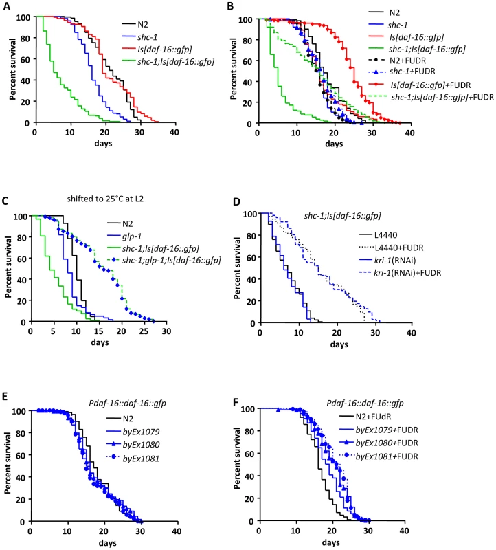
(A) shc-1(ok198);Is[daf-16::gfp] animals show reduced lifespan. (B) Treatment with FUDR extends the lifespan of both shc-1(ok198);Is[daf-16::gfp] and Is[daf-16::gfp] animals. (C) Knock-down of glp-1 extends lifespan of shc-1(ok198);Is[daf-16::gfp] animals (See also Figure S1, S2, S3 and Table S1). (D) The lifespan extension by FUDR does not require the adapter protein KRI-1. (E) In the absence of FUDR extrachromosomal daf-16::gfp transgene does not extend lifespan of wild type animals. (F) In the presence of FUDR extrachromosomal daf-16::gfp transgene extends lifespan of wild type animals. In the lifespan assay with FUDR treatment, animals were transferred onto the agar plates containing 0.1 mg/ml FUDR and E.coli 24 hours after the L4 larval stage. These were raised on the FUDR containing plates for four days and then transferred onto new plates without FUDR. The mean lifespan and statistical analyses in this figure are summarized in Table 1. Tab. 1. Lifespan of mutant strains. 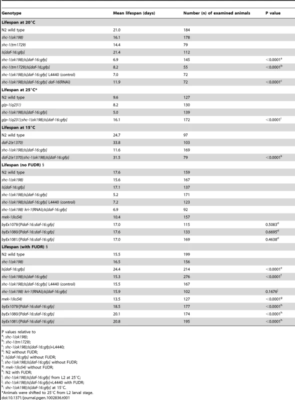
P values relative to Unexpectedly, raising the adult animals in the presence of 5-fluoro-2′-deoxyuridine (FUDR), an inhibitor of DNA synthesis which blocks cell proliferation, suppressed the early lethality and extended the lifespan of shc-1(ok198);Is[daf-16::gfp] animals up to two fold (Figure 1B and Table 1). This was surprising, since no lifespan extension was observed upon FUDR treatment of wild type. FUDR is one of the most commonly used drugs in the treatment of colorectal cancer and frequently used in C. elegans lifespan assays to facilitate strain handling due to its ability to block cell proliferation and generation of progeny. Based on this result, we suggest that the substantial lifespan reduction and lethality of shc-1(ok198);Is[daf-16::gfp] may be linked to proliferation and, possibly, abnormal mitosis, and that this detrimental effect is blocked by FUDR.
The germline is the only tissue that undergoes mitosis in adult animals, so we asked whether mitosis of the germline leads to the early lethality of shc-1(ok198);Is[daf-16::gfp] animals. The GLP-1 mediated Notch signaling represses meiosis of the germ cells and keeps them in mitotic proliferation from the L3 larval stage. In glp-1 mutants the germ cells stop mitotic cell division and enter meiosis [30]. If the lethality observed in shc-1(ok198);Is[daf-16::gfp] animals is indeed caused by proliferation of the germ cells, inactivation of glp-1 should result in a similar lifespan extension as seen upon FUDR treatment. To test this assumption, we used the temperature sensitive allele glp-1(q231), because glp-1(q231) animals at the restrictive temperature 25°C do not have a pronounced difference of mean lifespan compared to wild type probably due to a dysfunction in the atrophy intestine, in contrast to other long lived glp-1(lf) alleles, such as e2141ts or q158 (Figure S1) [22], [25]. Therefore, alterations of lifespan caused by detrimental germline proliferation should be easily detectable in this stain. shc-1(ok198);glp-1(q231);Is[daf-16::gfp] animals lived 240% longer than shc-1(ok198);Is[daf-16::gfp] when shifted at the L2 larval stage to the restrictive temperature (Figure 1C and Table 1). glp-1 mutation also extended lifespan of shc-1;Is[daf-16::gfp] animals when animals are re-shifted to 20°C after larval germline proliferation has ceased (Figure S2). In addition, the early lethality observed in shc-1;Is[daf-16::gfp] animals was completely abolished. Ablation of the mitotic germline or a glp-1(e2141) background extends lifespan of wild type animals, which require intestinal DAF-16 and the adaptor protein KRI-1. [23]. However, FUDR treatment did not extend lifespan of wild type animals (Figure 1B and Table 1), suggesting that FUDR uses a different mechanism as ablation of the germline to affect longevity of shc-1;Is[daf-16::gfp] animals. To validate this assumption, we further tested whether kri-1 knock-down could suppress the FUDR dependent lifespan extension. kri-1 RNAi treatment did not shorten the lifespan of shc-1;Is[daf-16::gfp] animals fed with FUDR (Figure 1D and Table 1). Based on these observations, we propose that early lethality of shc-1(ok198);Is[daf-16::gfp] animals is due to a negative input of germline proliferation rather than a general sickness of the strain.
We noticed in the strain TJ356 that transgenic daf-16 extended lifespan of wild type animals significantly only in the presence of FUDR (Figure 1B). To further exclude any effect of the transgene used, we generated three independent lines carrying extra-chromasomal daf-16::gfp transgenes in wild type background. In the absence of FUDR, these strains also had lifespan like wild type animals (Figure 1E), whereas they lived 19.3%, 20.6%, and 26.7%, respectively, longer than wild type animals in the presence of FUDR (Figure 1F). Thus, transgenic DAF-16 indeed extends lifespan if germline proliferation is inhibited. Recently, FUDR has been shown to extend lifespan of tub-1 mutant animals [31]. Together, this suggests that the use of FUDR in lifespan assays may be inconspicuous in some genetic backgrounds [32], but may strongly affect others.
Transgenic expression of daf-16 in shc-1 mutant animals causes pleiotropic germline and gonad development defects
Analysis of shc-1(ok198);Is[daf-16::gfp] animals using DIC microscopy revealed that the reproductive system exhibited multiple defects (Figure 2). The most prominent phenotype was the accumulation of cells in the pseudocoelom in almost all of the one day old adult animals (Figure 2B, 2J and Figure S3). These cells superficially looked like germ cells, yet were clearly localized outside the gonad. In the background of either glp-1(q231) or glp-1(e2141) these extra cells were absent, indicating that they may be germ cells (Figure S3). They showed substantial variations of shape and size. In some animals the boundary of the gonad was totally disrupted. It was, therefore, not possible to extract the gonad of these animals in an intact form (data not shown). In gonads with still recognizable morphology, a defect in the distal tip cell (DTC) migration was observed (Figure 2C and Figure S4). Analysis of shc-1(ok198);Is[daf-16::gfp] at different larval stages revealed that the gonad ruptured at the L3 larval stage at the proximal side next to the developing somatic gonad primordium. In animals in which germ cells leaked out into the pseudocoelom, a sharp gonad boundary indicative of an intact gonadal basement membrane, as seen in the wild type animals (Figure 2D), was absent in shc-1(ok198);Is[daf-16::gfp] animals (Figure 2E and Figure S5). The gonadal basement membrane can be visualized by staining with MitoTracker [33], and MitoTracker staining confirmed its partial disruption and leakage (Figure 2G and Figure S5).
Fig. 2. shc-1(ok198);Is[daf-16::gfp] animals display pleiotropic defects in the reproductive system. ![<i>shc-1(ok198);Is[daf-16::gfp]</i> animals display pleiotropic defects in the reproductive system.](https://www.prolekare.cz/media/cache/resolve/media_object_image_small/media/image/f5e575ad94caacabd1490747c9180182.png)
(A–C) DIC images of the gonad of one day adult animals. The white arrowhead denotes accumulated germ cells in the pseudocoelom (B) and a DTC migration defect (C). mito: mitotic region of the germline; meiot: meiotic region of the germline; o: oocyte; sp: sperm; em: embryo. (D and E) DIC images of the whole anterior gonad arm of L3 larvae. The white arrowhead denotes the germ cells leaking out from the gonad (E). (F and G) MitoTracker Red staining of the gonadal basement membrane. The white arrowhead points to the intact basement membrane in wild type (F) and disrupted basement membrane in shc-1(ok198);Is[daf-16::gfp] (G) animals. (H and I) DIC image (H) and anti-PGL-1 antibody staining (I) of shc-1(ok198);Is[daf-16::gfp] L3 larva. The white arrowhead indicates the anterior gonad with leakage (I). (J) Knock-down of shc-1 gene enhances the defect in the basement membrane of animals carrying daf-16 transgene. In this and the following figures data of defect in the basement membrane are presented as mean±SD; the mean values and statistic analysis are summarized in Table 2 and Table 3; the symbol * means P<0.01, ** means P<0.001 and *** means P<0.0001. (K) The gonad arms with disrupted basement membrane of shc-1(ok198);Is[daf-16::gfp] animals contain more germ cells than those with intact basement membrane at mid-L3 larval stage. For quantitative assessments PGL-1 antibody staining was performed to label the germ cells. Numbers of germ cells per gonad ±SD: wild type N2: 33±4 (n = 18); Is[daf-16::gfp]: 34±3 (n = 8); intact gonad of shc-1;Is[daf-16::gfp] animals: 32±5 (n = 24); disrupted gonad of shc-1;Is[daf-16::gfp] animals: 49±13 (n = 29) (P<0.0001 compared to N2, Is[daf-16::gfp] or intact gonad of shc-1;Is[daf-16::gfp] animals). (L and M) DAPI staining of wild type (L) and shc-1(ok198);Is[daf-16::gfp] (M) one day adult animals. The white arrowheads point to the chromosomes in the diakinetic oocytes of wild type animals and DNA in the endomitotic germ cells of shc-1(ok198);Is[daf-16::gfp] animals, respectively. Scale bar 10 µm. In order to determine the identity of the extra-gonadal cells, we stained shc-1(ok198);Is[daf-16::gfp] animals with an antibody against the germ cell specific protein PGL-1, a P-granule component. The released cells were PGL-1 positive, corroborating their identity as germ cells (Figure 2I, Movie S1). We noticed that at the mid-L3 stage the disrupted gonad arms of shc-1(ok198);Is[daf-16::gfp] animals contained significantly more germ cells than in wild type (46±14 vs. 35±4) (Figure 2K). In shc-1(ok198);Is[daf-16::gfp] animals in which one gonad arm was still intact, we counted more germ cells in the disrupted gonad arm than in the intact one. In adult animals the volume of some of these released cells increased so that they looked like oocytes. However, they lacked the condensed diakinetic chromosomes that are characteristic for oocytes (Figure 2L). Instead, some of them displayed endomitotic chromosomes (Figure 2M). Taking together, these data suggest that shc-1(ok198);Is[daf-16::gfp] animals show abnormal larval germline proliferation and disruption of the gonadal basement membrane. In the following results we focus mainly on the disruption of gonad phenotype.
Transgenic DAF-16 enhances a weakly penetrant phenotype present in shc-1(ok198)
Knock-down of daf-16 expression by RNAi significantly suppressed all phenotypic aspects (low brood size, sterility, gonad disruption and early adult lethality) of shc-1(ok198);Is[daf-16::gfp] animals (Figure 3A, Table 1 and Table S2). The suppression was only partial since knock-down of daf-16 by RNAi feeding was incomplete, indicated by a weak but persistent expression of DAF-16::GFP (Figure S6).
Fig. 3. Active DAF-16 derogates integrity of the gonadal basement membrane. 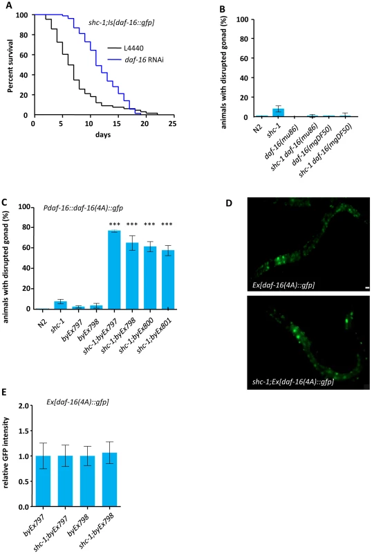
(A) Knock-down of daf-16 by RNAi suppressed the early lethality of shc-1(ok198);Is[daf-16::gfp] animals. Animals died within the first five days of adulthood: shc-1(ok198);Is[daf-16::gfp](L4440) as control: 45.7%, shc-1(ok198);Is[daf-16::gfp]+daf-16 RNAi: 1.4%. (B) daf-16 mutation suppresses the defects in gonad integrity of shc-1 mutant animals. (C) Transgenic expression of a constitutively nuclearly located daf-16(4A)::gfp results in disruption of the gonad in shc-1(−), but not in wild type animals (See also Figure S7 and Table 3). (D) SHC-1 does not affect the subcellular localization of DAF-16(4A)::GFP. In both wild type and shc-1(ok198) animals DAF-16(4A)::GFP is constitutively located in the nucleus. The mean lifespan and statistical analyses in this figure are summarized in Table 1. byEx represents extrachromosomal transgenic alleles which are summarized in Table 3. Mean values and statistic analysis of defect in gonadal integrity are summarized in Table 2 and Table 3. (E) SHC-1 does not affect expression level of transgenic daf-16(4A)::gfp. Columns represent pooled normalised values of three independent experiments plus standard deviation (SD). Mann Whitney test. Normalized GFP intensity: byEx797: 1.00±2.5 (n = 31); shc-1;byEx797: 1.00±2.1 (n = 33), P = 0.9670; byEx798: 1.00±0.19 (n = 37); shc-1;byEx798:1.06±0.22 (n = 48), P = 0.1591. In order to rule out an involvement of background mutations, we generated new transgenic animals carrying extra-chromosomal daf-16::gfp in shc-1 mutants. These strains displayed similar but less severe germline and gonad defects (Figure 2J and Table 3), which is probably due to the weaker DAF-16::GFP expression in extrachromosomal vs. integrated line (Figure S7) or due to the mosaic inheritance of extra-chromosomal transgenes.
The phenotype we observed in shc-1;Is[daf-16::gfp] animals was stronger than in Is[daf-16::gfp] animals, suggesting that reduced SHC-1 activity enhances DAF-16. This is in apparent contrast to our previous experiments in which we showed that DAF-16 activation is decreased in shc-1 mutant [26]. To understand this obvious discrepancy, we tested whether disruption of gonad phenotype could also be caused by inactivating DAF-16 in either wild type or shc-1(ok198) mutant background. No gonad disruption was seen in either daf-16(mu86) or daf-16(mgDF50) animals (Figure 3B and Table 2), suggesting that reduction of daf-16 activity was not the cause of this defect. However, about 7% of the shc-1(ok198) animals showed gonad disruption, yet with a delayed onset. Deletion of daf-16 almost fully suppressed this defect in shc-1(ok198) animals (Figure 3B and Table 2). This strongly suggests that the germline phenotype is not a synthetic or artificial phenotype seen only in shc-1;Is[daf-16::gfp] animals, and that DAF-16 and SHC-1 affect the reproductive system of C. elegans in opposite way.
Tab. 2. Defects in gonadal integrity. 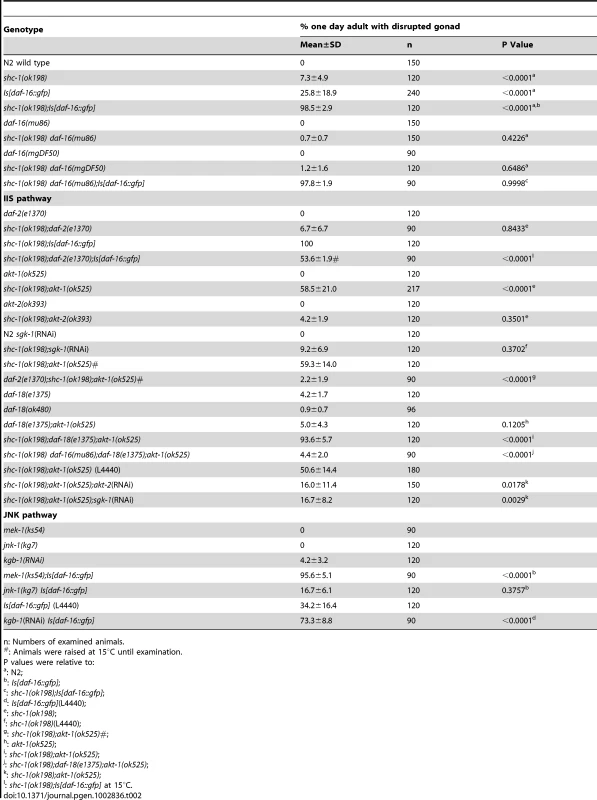
n: Numbers of examined animals. Nuclear DAF-16 signals to the germline
Since we have previously shown that SHC-1 promotes nuclear localization of DAF-16 by inhibiting DAF-2 and activating JNK-1, we expected an increase of cytosolic vs. nuclear DAF-16::GFP in shc-1(ok198);Is[daf-16::gfp] compared to Is[daf-16::gfp] animals [26]. If increased cytosolic vs. nuclear DAF-16::GFP would be the cause of the observed phenotype, then expression of a constitutively nuclear daf-16 mutant (daf-16(4A)::gfp) should not result in gonad disruption. Expressing daf-16(4A)::gfp in wild type animals caused only weak disrupted gonad phenotype (Figure 3C and Table 3). However, transgenic expression of daf-16(4A)::gfp in a shc-1(−) background caused severe defects. We observed that DAF-16(4A)::GFP was nuclearly localized and this nuclear localization was not affected by either presence or absence of SHC-1 (Figure 3D). In addition, shc-1 mutation did not affect the expression level of daf-16(4A)::gfp (Figure 3E). We also compared GFP intensities in the daf-16::gfp strains and correlated them to the severity of the phenotype. We found that, to result in a comparable phenotype, wild type daf-16::gfp required a higher expression level than daf-16(4A)::gfp (Figure S7). Given that wild type DAF-16 is mostly retained in the cytoplasm, this observation also indicates that nuclear instead of cytosolic DAF-16::GFP is the cause for the phenotype and SHC-1 antagonizes DAF-16 to ensure the gonadal integrity not simply via affecting its subcellular localization.
Tab. 3. Gonad basement membrane phenotype of animals with transgenic daf-16::gfp or daf-16(4A)::gfp. 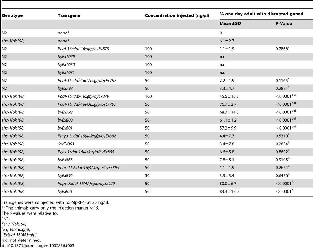
Transgenes were coinjected with rol-6(pRF4) at 20 ng/µl. AKT-1 ensures gonad integrity by inactivating DAF-16
AKT-1, AKT-2 and SGK-1 are known to phosphorylate DAF-16 directly and inhibit its nuclear entry [4], [7]. To test whether mutations in akt-1, akt-2 or sgk-1 that activate DAF-16 also influence the integrity of the gonadal basement membrane, shc-1(ok198);akt-1(ok525), shc-1(ok198);akt-2(ok393) and shc-1(ok198);sgk-1(RNAi) were analyzed (Figure 4A and Table 2). Neither akt-1(ok525), akt-2(ok393) single mutants nor sgk-1(RNAi) displayed gonad disruption. Inactivation of akt-2 or sgk-1 did not enhance the penetrance of the gonad defect of shc-1(ok198) animals, either. In contrast, 58.5±21.0% of first-day adult shc-1(ok198);akt-1(ok525) animals showed gonad disruption, providing further evidence that this phenotype is not an artificial effect of transgenic daf-16 since this strain does not harbor a daf-16 transgene. In addition, a loss-of-function allele of daf-16 suppressed this phenotype in shc-1;akt-1 animals, verifying its dependence on DAF-16 (Figure 4A). Taken together, these results strongly suggest that AKT-1 ensures gonadal integrity via inhibiting DAF-16.
Fig. 4. Active DAF-2, AKT-2 and SGK-1 antagonize AKT-1 mediated inhibition of DAF-16 to derogate gonad integrity. 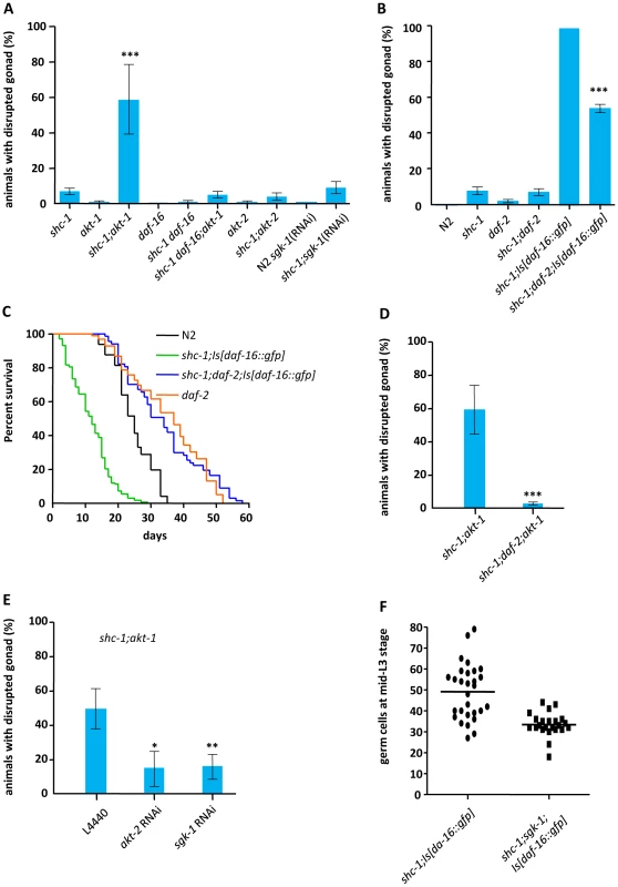
(A) DAF-16 activity affecting gonad integrity is inhibited by AKT-1. (B) Inactivation of IIR DAF-2 suppresses the defect in the gonadal basement membrane (See also Figure S8 and Table S2). (C) Inactivation of daf-2 extends lifespan of shc-1(ok198);Is[daf-16::gfp] animals. (D) daf-2 mutation suppresses gonad disruption in akt-1;shc-1 animals (See also Figure S9). (E) AKT-2 and SGK-1 antagonize AKT-1. (F) shc-1(ok198);sgk-1(ok538);Is[daf-16::gfp] L3 larvea have less germ cells than shc-1(ok198);Is[daf-16::gfp] L3 larvea. shc-1;Is[daf-16::gfp] animals: 49±13 (n = 29); shc-1;sgk-1;Is[daf-16::gfp] animals: 33±6 (n = 23) (P<0.0001). (See also Figure S10). The mean lifespan and statistical analyses in this figure are summarized in Table 1. Mean values and statistic analysis of gonad disruption are summarized in Table 2. Active IIR DAF-2 contributes to gonad disruption defect caused by DAF-16
Since akt-1 is downstream of daf-2, shc-1;daf-2 double mutant should therefore exhibit the same phenotype as akt-1;shc-1. To test this, we crossed the temperature sensitive mutant daf-2(e1370) into shc-1(ok198) background. At 25°C both daf-2(e1370) and shc-1(ok198);daf-2(e1370) mutants formed constitutive dauer larvae with a developmentally arrested germline that prevented the analysis of adult animals. At 20°C, at which daf-2 is partially inactivated, daf-2(e1370) did not alter the defect in the gonadal integrity in shc-1 mutant (Figure 4B and Table 2).
In order to explore whether DAF-2 inhibits a DAF-16 dependent effect on the germline, we crossed daf-2(e1370) into shc-1(ok198);Is[daf-16::gfp] animals and quantified the percentage of animals displaying disrupted gonad at 15°C, since at 20°C shc-1(ok198);daf-2(e1370);Is[daf-16::gfp] showed development arrest due to excessive daf-16 transgene expression (Figure S8). At 15°C, activity of DAF-2 in the daf-2(e1370) mutant is already reduced, indicated by an extended lifespan [34]. Surprisingly, daf-2(e1370) significantly reduced instead of increased the penetrance of animals with gonad disruption (Figure 4B and Table 2), indicating that this phenotype develops only in animals with intact IIS. In addition, the sterile phenotype of shc-1(ok198);Is[daf-16::gfp] animals was completely suppressed by daf-2 mutation and the brood size of the fertile animals was increased (Table S2). Consistent with the suppression of the defects in the reproductive system, the early lethality in adulthood was abolished and lifespan was extended from 11.6 days to 31.5 days (Figure 4C). These data suggest that the phenotypes in shc-1(ok198);Is[daf-16::gfp] animals require active DAF-2.
AKT-1 is known to act downstream of DAF-2 to inhibit DAF-16. Here, however, we observed that loss of akt-1 and loss of daf-2 have divergent consequences for DAF-16. To address whether the daf-2 mutation could still suppress the defect in the basement membrane in akt-1;shc-1 animals, we examined shc-1(ok198);daf-2(e1370);akt-1(ok525) triple mutant at 15°C, since only at this temperature the germline of the animals could proliferate after L3 stage. daf-2 mutation suppressed gonad disruption in shc-1(ok198);akt-1(ok525) animals completely (Figure 4D, Figure S9), indicating that active IIR DAF-2 contributes to gonad disruption caused by DAF-16.
AKT-2 and SGK-1 antagonize AKT-1 to affect gonadal integrity
As we observed antagonistic role of DAF-2 and AKT-1 affecting gonadal integrity, we further explored the downstream effectors of DAF-2 to antagonize AKT-1. In parallel to AKT-1, AKT-2 and SGK-1 also transmit input from DAF-2 to inhibit DAF-16. We asked whether AKT-2 or SGK-1 counteract AKT-1 to control gonadal integrity. Both akt-2 and sgk-1 RNAi clone strongly suppressed disruption of the gonad in shc-1(ok198);akt-1(ok525) mutant (Figure 4E and Table 2), indicating that AKT-2 and SGK-1 act downstream of DAF-2 to antagonize AKT-1.
SGK-1 promotes tumorous germline proliferation in shc-1;Is[daf-16::gfp] animals
Next we asked whether DAF-2, AKT-1/2 or SGK-1 also promote tumorous germline proliferation in shc-1;Is[daf-16::gfp] animals. We quantified number of the germ cells of shc-1;Is[daf-16::gfp] L3 larvae in daf-2, akt-1/akt-2 or sgk-1 mutant background. Knock-down of sgk-1 significantly reduced number of the germ cells (Figure 4F and Figure S10), suggesting SGK-1 contributes to proliferation of the germline tumor in shc-1;Is[daf-16::gfp] animals. Due to somatic missexpression of PGL-1 in daf-2 and akt-1/akt-2 animals [35], we were not able to quantify germ cells in these mutants (data not shown).
Both active PI3K signaling and inactive JNK signaling contribute to the DAF-16–dependent defect in the gonadal basement membrane
SHC-1 has been shown to inhibit IIS to activate the downstream PI3K signaling [26]. Therefore, one may speculate that active DAF-16 in the presence of hyperactive PI3K signaling is the cause for gonad disruption. The PTEN homolog DAF-18 antagonizes PI3 kinase AGE-1. Therefore daf-18 mutant should have enhanced PI3K signaling. To test whether active PI3K signaling contributes to the DAF-16 dependent phenotype, we analyzed daf-18(e1375);akt-1(ok525) and shc-1(ok198);daf-18(e1375);akt-1(ok525) animals. daf-18 mutation did not enhance gonad disruption of akt-1(ok525) one day adult animals (Figure 5A, Table 2), suggesting that active PI3K is not sufficient for DAF-16 to degenerate the basement membrane. However, daf-18 enhanced the defects in shc-1(ok198);akt-1(ok525) animals up to almost 100% and daf-16 mutation completely suppressed the defect in shc-1(ok198);daf-18(e1375);akt-1(ok525) animals (Figure 5A and Table 2), indicating that active IIR/PI3K signaling assists DAF-16 to degenerate the gonadal basement membrane. Taking together, DAF-2 mediated PI3K signaling is necessary but not sufficient for DAF-16 to cause disruption of the gonad.
Fig. 5. JNK signaling antagonizes hypodermal DAF-16 to ensure gonadal integrity. 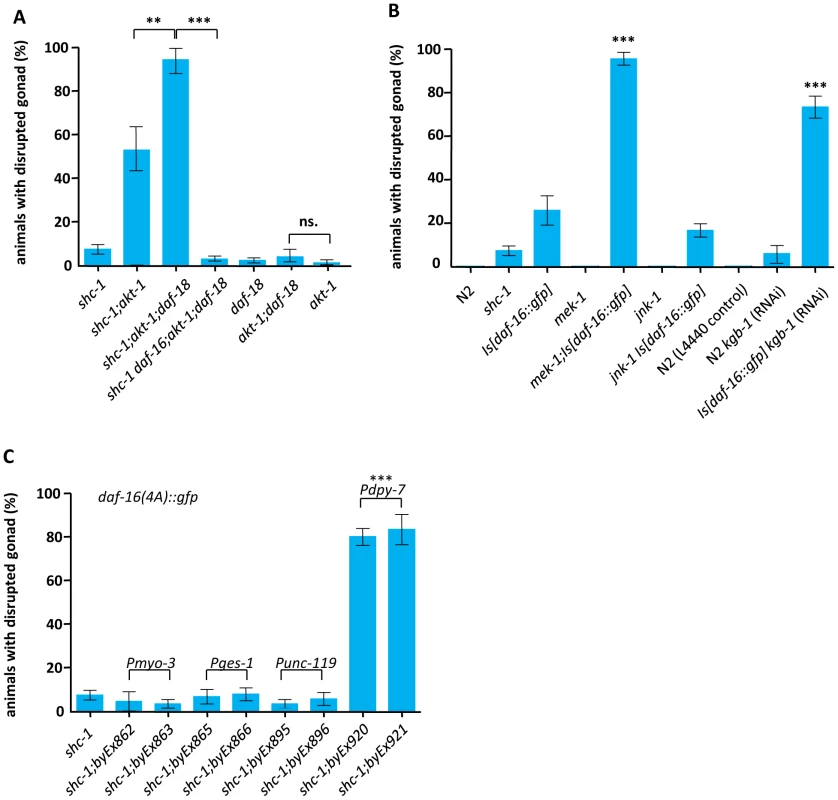
(A) daf-18 mutation enhances disruption of the gonad in shc-1(ok198);akt-1(ok525) but not in akt-1(ok525) one day adult animals. (B) Knock-down of mek-1 and kgb-1 enhances the defect in Is[daf-16::gfp] animals. The mean values and statistic analysis are summarized in Table 2. (C) Expression of hypodermal daf-16(4A)::gfp leads to disruption of the gonadal basement membrane. byEx represents extrachromosomal transgene alleles which are summarized in Table 3. Mean values and statistic analysis of defect in gonadal integrity are summarized in Table 3. Besides negatively modulating DAF-2, SHC-1 also activates MEK-1/JNK-1 to affect DAF-16 [26]. Upon heavy metal stress response, SHC-1 also mediates the activation of MEK-1, which in turn phosphorylates and activates another JNK homolog, KGB-1 [27]. Therefore, in the shc-1 mutant, hyperactive IIS is accomplished by inactive JNK signaling. Since hyperactive IIS is necessary but not sufficient for DAF-16 to derogate the gonadal basement membrane, we asked whether inactivated JNK signaling also plays a role. We generated mek-1(ks54);Is[daf-16::gfp], jnk-1(gk7) Is[daf-16::gfp] and kgb-1(um3) Is[daf-16::gfp] animals. Inactivation of JNK-1 did not further enhance disruption of the gonadal basement membrane in Is[daf-16::gfp] animals (Figure 5B, Table 2). 85% of mek-1(ks54);Is[daf-16::gfp] animals died at early larval stages. The remaining animals could develop to adulthood. However, 95.6% of these animals displayed germ cells outside of the gonad (Figure 5B and Table 2), phenocopying the shc-1;Is[daf-16::gfp] phenotype. Even though mek-1;Is[daf-16::gfp] animals showed more severe defects in early larval development than shc-1(ok198);Is[[daf-16::gfp] animals, fewer animals were sterile (29% vs. 43%; Table S2) and the fertile adult animals displayed a higher number of progeny (120 vs. 49; Table S2), suggesting that the early larval lethality is not due to a more severe defect in the basement membrane. All kgb-1(um3) Is[daf-16::gfp] animals died at larval stage. However, most of Is[daf-16::gfp] animals fed with kgb-1 RNAi could bypass the larval lethality and in 75% of these one day old adult animals germ cells in the body cavity were observed (Figure 5B and Table 2). These results suggest that the antagonistic function of SHC-1 to DAF-16 is mediated via MEK-1 and KGB-1.
Hypodermal expression of DAF-16 is sufficient to affect gonad integrity
The promoter used for the expression of daf-16 in TJ356 Is[daf-16::gfp] is a 6 kb genomic region upstream of the exon 1 of daf-16a isoform, which triggers expression in the hypodermis, intestine, neurons and body wall muscle cells [29], [36]. Due to a general germline silencing of transgenic promoters in C. elegans, this daf-16::gfp transgene is probably not expressed in the germline. Endogenous DAF-16 in the germline or somatic gonad is not required for this phenotype, since no phenotypic difference of shc-1;Is[daf-16::gfp] animals was observed in daf-16(+) and daf-16(mu86) null mutant backgrounds (Table 2). We conclude that DAF-16 outside of the reproductive system is the most likely cause of the phenotype we describe here.
In order to test how DAF-16 in different tissues promotes the phenotype and rule out an involvement of weak expression of the daf-16 transgene in the germline, we expressed the transgene in the specific somatic tissues. Since expressing the constitutively nuclear daf-16(4A)::gfp resulted in a stronger phenotype compared to wild type daf-16::gfp, we used daf-16(4A) for this analysis. Transgenic expression of daf-16(4A)::gfp in the neurons, intestine, or musculature did not increase the weak shc-1(−) phenotype (Figure 5C and Table 3). In contrast, expression of daf-16(4A)::gfp in the hypodermis was sufficient to cause disruption of the gonad in 80% of one day adult shc-1(−) animals, suggesting that hypodermal DAF-16 causes disruption of the gonadal basement membrane cell-nonautonomously.
Discussion
Two opposing qualities of DAF-16 signaling affect lifespan differently
In this manuscript we for the first time describe two qualities of DAF-16 function that affect longevity in opposite way. In contrast to the known lifespan extending effect, we discover that DAF-16 can shorten lifespan by inducing a tumor-like germline phenotype. It has probably affected the outcome of previously described experiments, however, to our knowledge this has not been reported before [17]–[36].
The starting point of our experiments was that we, as others before, failed to increase the lifespan of wild type (daf-2 +/+) animals by expressing transgenic copies of daf-16, unless the lifespan assays were performed in the presence of FUDR [37] (Figure 1B). In this respect daf-16 differs from other key regulators of stress response and lifespan such as skn-1, which typically shorten lifespan when inactivated, and increase lifespan when being overexpressed as a transgene. Although daf-16 transgene rescued the short life-span of daf-2;daf-16 double mutants, it did not or only modestly increases lifespan in wild type background [17]–[36]. It has been suggested that in a daf-2(+) background, AKT phosphorylation results in cytoplasmic retention of these extra copies of DAF-16, rendering them inactive [36]. However, phosphorylation was not sufficient to prevent the ability of transgenic SKN-1 to extend lifespan [38]. We found here that the lifespan extending effect of DAF-16 was balanced by its lifespan shortening effect which induced tumor-like growth in the germline. Consistently, blocking germline proliferation by using the cytostatic FUDR or a mutation inhibiting Notch signaling in the germline was sufficient to prevent negative aspects of DAF-16 signaling and increased lifespan of wild type animals containing a daf-16 transgene (Figure 1B and 1C).
DAF-16–mediated signaling results in a tumor-like germline phenotype
We found that not only a daf-16 transgene, but also a combination of mutants in the IIS pathway affecting DAF-16 activity caused a pleiotropic phenotype in the reproductive system. This phenotype occurred at low penetrance in wild type animals carrying the daf-16 transgene, which may be the reason why it had escaped detection in previous studies. The penetrance of this phenotype was strongly enhanced in shc-1 mutant background. Its most prominent phenotypic aspect was the disruption of the gonadal basement membrane at the proximal gonad adjacent to the developing somatic gonad primordium, so that eventually germline cells leaked into the body. We counted about 30% more germ cells in disrupted gonad arms of shc-1;Is[daf-16::gfp] L3 larvae compared to wild type animals (Figure 2K). A co-segregation of both phenotypic aspects was particularly obvious in animals in which only one gonad arm was disrupted, and typically contained significantly more germ cells than the other intact gonad arm. This phenotype is distinct from that of gld-1 mutants, in which the basement membrane ruptures in adult animals due to a massive excess of germ cells. gld-1 mutants at the equivalent L3 stage still have intact gonad arms despite their increased number of germ cells [39]. This indicates that in gld-1 mutants, but not in animals described in this study, the basement membrane of L3 animals is mechanically strong enough to withstand an increase in germline nuclei. It is therefore possible that the basement membrane of the gonad in shc-1;Is[daf-16] has a defect on its own, similar as e.g. seen in ten-1, dgn-1, ina-1 and epi-1 mutants, which also show disruption of gonadal basement membrane, but without an increase in germline proliferation [40]. An interesting hypothesis is that that the abnormally proliferating germ cells invade into the surrounding extracellular matrix. Although we observed germ cells that penetrate the basement membrane, and dissection and extraction of an intact gonad in those animals failed due to their leakage, we cannot prove at this point that this is an active process. Nevertheless, we discover for the first time that mutations in IIS result in both germline hyperplasia and disruption of the gonadal basement membrane in C. elegans, a phenotype that opposes the beneficial effects of DAF-16 on increasing healthspan.
DAF-16 activation in the hypodermis is sufficient to signal to the germline
The phenotype in the reproductive system was also observed in shc-1 mutants carrying extrachromosomal arrays with daf-16. Such arrays in C. elegans are typically not expressed in the germline [41]. In agreement with this generally accepted notion, we never observed germline expression of the GFP tagged daf-16 transgenes. One possibility is that overexpression of DAF-16 in the somatic tissues leads to activation of germline DAF-16, since it has been shown that active DAF-16 in one tissue elevated its activity in other tissues [42]. However, we observed no difference between shc-1;Is[daf-16::gfp] and shc-1 daf-16;Is[daf-16::gfp] animals, the latter lacking endogenous daf-16 in the germline [19] (Table 2). Therefore, we consider it more likely that daf-16 expression outside the reproductive system is the cause of the observed phenotype. The fact that expressing daf-16 in the hypodermis was sufficient to provoke this phenotype (Figure 5C) further excluded an involvement of very low levels of transgenic DAF-16 in the germline. These data suggest that DAF-16 mediates a cell-nonautonomous signaling to affect germline proliferation and basement membrane integrity.
SHC-1 regulates both IIS and MAPK signaling pathway to antagonize DAF-16 influence on the germline
Mutation in shc-1 strongly enhanced the germline and gonad phenotype of Is[daf-16::gfp] animals. In addition, we observed that DAF-16(4A) was nuclearly localized in both shc-1(+) and shc-1(−) animals (Figure 3D), while disruption of the gonad was only detectable in shc-1(−) background (Figure 3C). These data favor a model that SHC-1, in addition to its role in affecting the subcellular localization of DAF-16 upon stress response, can also antagonize nuclear DAF-16. Notably, SHC-1 is localized in both cytoplasm and nucleus [26]. Our data indicate that both active IIS and inactive JNK signaling contribute to the DAF-16 dependent phenotype in the reproductive system (Figure 4B, 4D, 5A and 5B). This may also explain why the mek-1;Is[daf-16::gfp] strain displayed a less severe basement membrane phenotype than shc-1;Is[daf-16::gfp] animals, as only SHC-1 affects both IIS and JNK signaling [26]. Our data also explain previous, contradictory data concerning the lifespan of mek-1 mutants. We reported previously that mek-1 mutants were short lived, while Oh and colleagues found mek-1 mutants to have a lifespan indistinguishable from wild type [9], [26]. We found that both mek-1 and shc-1 mutant animals lived as long as wild type worms in the presence of the drug FUDR that blocked germline proliferation (Table 2). However, in the absence of FUDR, mek-1 animals suffered from a pronounced reduction of lifespan that we can now attribute to defects in the reproductive system, rather than to the sensitivity for reactive oxygen species stress as proposed before [26].
AKT-2 and SGK-1 counteract AKT-1 to control DAF-16 signaling to the germline
We found that in a daf-2 loss-of-function mutant the DAF-16 mediated gonad dysintegrity phenotype of both shc-1;Is[daf-16::gfp] and shc-1;akt-1 animals was reduced (Figure 4B and 4D), thus abrogating lethality in early adulthood (Figure 4C). This was a surprising result, since our experiments so far had indicated that this gonad phenotype was a consequence of too much DAF-16 activity, and the daf-2 mutation should further increase DAF-16 activity. The PTEN homolog DAF-18 antagonizes the DAF-2 and PI3 kinase/AGE-1 pathway, thus a loss-of-function mutant is supposed to result in reduced DAF-16 activity via increased IIS. However, the daf-18 loss-of-function mutation further enhanced the phenotype of a shc-1;akt-1 mutant (Figure 5A). In summary, mutations in both IIS pathway genes daf-2 and daf-18 behave opposite to their known roles in the canonical IIS pathway. Similarly, while the increase of the phenotype in an akt-1 loss-of-function mutant correlated well with the increase of DAF-16 activity, akt-2 and sgk-1 mutations opposed akt-1, and behaved like daf-2 described above. We conclude that, while daf-2, akt-1, akt-2, and sgk-1 loss-of-function mutants all result in a longevity and stress resistance phenotype consistent with upregulation of DAF-16 nuclear activity, they differ in the way they regulate DAF-16 activity to cause the gonad phenotype.
One possible explanation for the antagonistic effect of distinct IIs pathway mutants is that daf-2, akt-2, and sgk-1 may provoke a distinct phosphorylation pattern of DAF-16 compared to akt-1, resulting in different outputs of DAF-16 transcriptional targets that affect germline proliferation/gonadal integrity compared to longevity and stress resistance. Whereas the akt-1(ok525) mutation might only prevent AKT-1 mediated DAF-16 phosphorylation, daf-2(e1370) supposedly reduces both AKT-1, AKT-2, and SGK-1 phosphorylation, and this may result in controlling distinct downstream genes for long lifespan and gonad integrity, respectively. We consider this model less likely, since there is currently no evidence for C. elegans AKT-1 phosphorylating distinct sites in DAF-16 compared to SGK-1 or AKT-2 [7]. However, we cannot exclude this possibility based on the existing data.
Another possible scenario is that mutation in daf-2, akt-2, and sgk-1 mediated activation of DAF-16 affects different tissues than akt-1 knock-down mediated activation of DAF-16, suggesting that the germline reads out and responds to a balance of DAF-16 activities in somatic tissues. This way, DAF-2 could prevent DAF-16 from counteracting hypodermal DAF-16 in some tissues, e.g. the intestine. It has been previously shown that DAF-2 promotes larval germline proliferation via inactivating germline DAF-16 [19]. Together with our results this suggests that DAF-16 activities in the hypodermis and the germline may antagonize each other. Moreover, previous studies suggested that AKT-1, AKT-2 and SGK-1 have different tissue specificity, expression levels, patterns, or activities to control DAF-16 [4], [7]. Notably, AKT-1 is expressed in the hypodermis, whereas AKT-2 and SGK-1 are probably not. Consistently, neither down-regulation of akt-2 nor sgk-1 caused disruption of the gonad in shc-1 mutant background (Figure 4A), but instead were both able to suppress the phenotype of akt-1;shc-1 mutants (Figure 4E). Based on our results presented here, we suggest that AKT-1 is active in the hypodermis to prevent DAF-16 mediated signal to the reproductive system, whereas AKT-2, SGK-1 and even AKT-1 in other tissues may inhibit DAF-16 activity that counteracts hypodermal DAF-16. Such antagonistic roles of SGK-1 and AKT-1 have been reported in regulation of lifespan [43]. Additional tissue specific studies of these three AGC family kinases will enable a further understanding of such interactions across tissue boundaries.
FOXO and tumor formation
It has been shown in different organisms that FOXO/DAF-16 functions as a tumor suppressor in a variety of cancers by promoting apoptosis or cell cycle arrest [44]. However, several recent data indicate that the function of FOXO/DAF-16 may be more complex than previously thought. An essential role of FOXO3a was proposed in the maintenance of cancer stem cells that are responsible for the reoccurrence of chronic myeloid leukemia [15]. In acute myeloid leukemia FOXO1/3/4 promote leukemic growth and maintenance by inhibiting myeloid maturation and apoptosis [45]. In C elegans, some DAF-16 target genes were identified which stimulate gld-1 germline tumor [46]. All these studies indicate that the function of FOXO/DAF-16 proteins is highly dependent on the cellular context. In mouse, FOXO3a promotes tumor cell invasion through the induction of matrix metalloproteases [47]. Our data show that FOXO/DAF-16 can also promote germline hyperplasia and disruption of the surrounding extracellular matrix through cell-nonautonomous signaling. It will be of great interest to further identify the signaling molecules to the germline activated by DAF-16, and to investigate whether the gonad dysintegrity phenotype we described involves active invasions of the germ cells into the extracellular matrix and, therefore, resembles the behavior of metastatic tumor cells.
Materials and Methods
Information about strains and constructs used are described in Text S1.
Transgenic strains
To generate transgenic animals carrying wild type daf-16::gfp or daf-16(4A)::gfp, the corresponding constructs were injected into wild type or shc-1(ok198) animals (for byEx numbers, see Table 3). 20 ng/µl pRF4 rol-6 (su1006) was used as co-injection marker. GFP expression of the transgenic animals was confirmed using fluorescent microscopy.
Antibody staining
A formaldehyde fixation procedure was used (Text S1) for the whole worm staining with 1∶200 anti-PGL-1 antibody (a gift from Dr. Susan Strome). Staining of dissected gonads was not possible because of the disruption of the gonad.
MitoTracker staining of the basement membrane
Basement membranes were stained with MitoTracker Red CMXRos (invitrogen) by placing worms in a solution of 10 µM MitoTracker Red at 25°C for 2 hours. The worms were then allowed to recover for 30 minutes on an NGM agar plate and analyzed.
Lifespan assay
Lifespan assays were initiated at the L4 larval stage. Synchronized animals were raised at 15°C prior to lifespan analysis. Then, L4 animals were transferred to the respective temperatures for the assays and examined every day. Animals that showed no response to touch were scored as dead. In assays without FUDR treatment animals were transferred every day onto new plates during the reproductive period. Animals died because of bagging of larvae were censored. In the lifespan assay with FUDR treatment, animals were transferred onto the agar plates containing 0.1 mg/ml FUDR and E.coli 24 hours after the L4 larval stage. These were raised on the FUDR containing plates for four days and then transferred onto new plates without FUDR. FUDR was administrated to adult instead of L4 animals in order to exclude a possible interference from seam cell development to the reproductive system, since the epidermal seam cells undergo a final mitotic division at the L4 to adult molt and DAF-16 mediated signaling is sent from the hypodermis. All the lifespan assays were performed at 20°C with exception of those with glp-1(q231) and daf-2(e1370) mutants, as glp-1(q231) required 25°C to inactivate GLP-1 and daf-2(e1370);shc-1(ok198);Is[daf-16::gfp] could develop to adulthood only at 15°C. All of the lifespan assays were performed at least twice. The short lifespan of shc-1(ok198);Is[daf-16::gfp] can only be observed, when no censoring for adult animals dying due to the development defect is done. As reported previously by us (Neumann-Haefelin et al., 2008), Is[daf-16::gfp] partially extends lifespan of shc-1(ok198) mutants, when adult animals dying due to the development defect are censored.
Scoring of gonad disruption
All animals in the tests except those with daf-2(e1370);shc-1(ok198);Is[daf-16::gfp] and daf-2(e1370);shc-1(ok198);akt-1(ok525), were raised at 20°C and analyzed via DIC microscopy 24 hours after L4 stage. Animals in which at least one germ cell was detected outside of the gonad were scored as positive. The percentage of positive one day old animals was calculated. Per test thirty animals were examined and each test was performed at least three times. shc-1(ok198);daf-2(e1370);Is[daf-16::gfp] and shc-1(ok198);daf-2(e1370);akt-1(ok525) animals were raised at 15°C and analyzed 24 hours after L4 larval stage.
Quantification of the germ cells
The numbers of anti-PGL-1 positive cells were quantified at the time point that vulva induction takes place.
Statistic analysis
GraphPad Prism 4.0 software (GraphPad Software Inc., San Diego, USA) was used to calculate mean value and to perform statistical analysis.
Supporting Information
Zdroje
1. FuZ, TindallDJ (2008) FOXOs, cancer and regulation of apoptosis. Oncogene 27 : 2312–2319.
2. BrunetA, BonniA, ZigmondMJ, LinMZ, JuoP, et al. (1999) Akt promotes cell survival by phosphorylating and inhibiting a Forkhead transcription factor. Cell 96 : 857–868.
3. ParadisS, AilionM, TokerA, ThomasJH, RuvkunG (1999) A PDK1 homolog is necessary and sufficient to transduce AGE-1 PI3 kinase signals that regulate diapause in Caenorhabditis elegans. Genes Dev 13 : 1438–1452.
4. ParadisS, RuvkunG (1998) Caenorhabditis elegans Akt/PKB transduces insulin receptor-like signals from AGE-1 PI3 kinase to the DAF-16 transcription factor. Genes Dev 12 : 2488–2498.
5. BerdichevskyA, ViswanathanM, HorvitzHR, GuarenteL (2006) C. elegans SIR-2.1 interacts with 14-3-3 proteins to activate DAF-16 and extend life span. Cell 125 : 1165–1177.
6. CalnanDR, BrunetA (2008) The FoxO code. Oncogene 27 : 2276–2288.
7. HertweckM, GobelC, BaumeisterR (2004) C. elegans SGK-1 is the critical component in the Akt/PKB kinase complex to control stress response and life span. Dev Cell 6 : 577–588.
8. LehtinenMK, YuanZ, BoagPR, YangY, VillenJ, et al. (2006) A conserved MST-FOXO signaling pathway mediates oxidative-stress responses and extends life span. Cell 125 : 987–1001.
9. OhSW, MukhopadhyayA, SvrzikapaN, JiangF, DavisRJ, et al. (2005) JNK regulates lifespan in Caenorhabditis elegans by modulating nuclear translocation of forkhead transcription factor/DAF-16. Proc Natl Acad Sci U S A 102 : 4494–4499.
10. RamaswamyS, NakamuraN, SansalI, BergeronL, SellersWR (2002) A novel mechanism of gene regulation and tumor suppression by the transcription factor FKHR. Cancer Cell 2 : 81–91.
11. ModurV, NagarajanR, EversBM, MilbrandtJ (2002) FOXO proteins regulate tumor necrosis factor-related apoptosis inducing ligand expression. Implications for PTEN mutation in prostate cancer. J Biol Chem 277 : 47928–47937.
12. PaikJH, KolliparaR, ChuG, JiH, XiaoY, et al. (2007) FoxOs are lineage-restricted redundant tumor suppressors and regulate endothelial cell homeostasis. Cell 128 : 309–323.
13. SuntersA, MadureiraPA, PomeranzKM, AubertM, BrosensJJ, et al. (2006) Paclitaxel-induced nuclear translocation of FOXO3a in breast cancer cells is mediated by c-Jun NH2-terminal kinase and Akt. Cancer Res 66 : 212–220.
14. TothovaZ, KolliparaR, HuntlyBJ, LeeBH, CastrillonDH, et al. (2007) FoxOs are critical mediators of hematopoietic stem cell resistance to physiologic oxidative stress. Cell 128 : 325–339.
15. NakaK, HoshiiT, MuraguchiT, TadokoroY, OoshioT, et al. (2010) TGF-beta-FOXO signalling maintains leukaemia-initiating cells in chronic myeloid leukaemia. Nature 463 : 676–680.
16. LinK, DormanJB, RodanA, KenyonC (1997) daf-16: An HNF-3/forkhead family member that can function to double the life-span of Caenorhabditis elegans. Science 278 : 1319–1322.
17. LinK, HsinH, LibinaN, KenyonC (2001) Regulation of the Caenorhabditis elegans longevity protein DAF-16 by insulin/IGF-1 and germline signaling. Nat Genet 28 : 139–145.
18. HondaY, HondaS (1999) The daf-2 gene network for longevity regulates oxidative stress resistance and Mn-superoxide dismutase gene expression in Caenorhabditis elegans. FASEB J 13 : 1385–1393.
19. MichaelsonD, KortaDZ, CapuaY, HubbardEJ (2010) Insulin signaling promotes germline proliferation in C. elegans. Development 137 : 671–680.
20. LuoS, KleemannGA, AshrafJM, ShawWM, MurphyCT (2010) TGF-beta and insulin signaling regulate reproductive aging via oocyte and germline quality maintenance. Cell 143 : 299–312.
21. PinkstonJM, GariganD, HansenM, KenyonC (2006) Mutations that increase the life span of C. elegans inhibit tumor growth. Science 313 : 971–975.
22. Arantes-OliveiraN, ApfeldJ, DillinA, KenyonC (2002) Regulation of life-span by germ-line stem cells in Caenorhabditis elegans. Science 295 : 502–505.
23. HsinH, KenyonC (1999) Signals from the reproductive system regulate the lifespan of C. elegans. Nature 399 : 362–366.
24. GhaziA, Henis-KorenblitS, KenyonC (2009) A transcription elongation factor that links signals from the reproductive system to lifespan extension in Caenorhabditis elegans. PLoS Genet 5: e1000639 doi:10.1371/journal.pgen.1000639..
25. BermanJR, KenyonC (2006) Germ-cell loss extends C. elegans life span through regulation of DAF-16 by kri-1 and lipophilic-hormone signaling. Cell 124 : 1055–1068.
26. Neumann-HaefelinE, QiW, FinkbeinerE, WalzG, BaumeisterR, et al. (2008) SHC-1/p52Shc targets the insulin/IGF-1 and JNK signaling pathways to modulate life span and stress response in C. elegans. Genes Dev 22 : 2721–2735.
27. MizunoT, FujikiK, SasakawaA, HisamotoN, MatsumotoK (2008) Role of the Caenorhabditis elegans Shc adaptor protein in the c-Jun N-terminal kinase signaling pathway. Mol Cell Biol 28 : 7041–7049.
28. LuziL, ConfalonieriS, Di FiorePP, PelicciPG (2000) Evolution of Shc functions from nematode to human. Curr Opin Genet Dev 10 : 668–674.
29. HendersonST, JohnsonTE (2001) daf-16 integrates developmental and environmental inputs to mediate aging in the nematode Caenorhabditis elegans. Curr Biol 11 : 1975–1980.
30. AustinJ, KimbleJ (1987) glp-1 is required in the germ line for regulation of the decision between mitosis and meiosis in C. elegans. Cell 51 : 589–599.
31. AitlhadjL, SturzenbaumSR (2010) The use of FUdR can cause prolonged longevity in mutant nematodes. Mech Ageing Dev 131 : 364–365.
32. MitchellDH, StilesJW, SantelliJ, SanadiDR (1979) Synchronous growth and aging of Caenorhabditis elegans in the presence of fluorodeoxyuridine. J Gerontol 34 : 28–36.
33. SherwoodDR, ButlerJA, KramerJM, SternbergPW (2005) FOS-1 promotes basement-membrane removal during anchor-cell invasion in C. elegans. Cell 121 : 951–962.
34. GemsD, SuttonAJ, SundermeyerML, AlbertPS, KingKV, et al. (1998) Two pleiotropic classes of daf-2 mutation affect larval arrest, adult behavior, reproduction and longevity in Caenorhabditis elegans. Genetics 150 : 129–155.
35. CurranSP, WuX, RiedelCG, RuvkunG (2009) A soma-to-germline transformation in long-lived Caenorhabditis elegans mutants. Nature 459 : 1079–1084.
36. LeeRY, HenchJ, RuvkunG (2001) Regulation of C. elegans DAF-16 and its human ortholog FKHRL1 by the daf-2 insulin-like signaling pathway. Curr Biol 11 : 1950–1957.
37. KwonES, NarasimhanSD, YenK, TissenbaumHA (2010) A new DAF-16 isoform regulates longevity. Nature 466 : 498–502.
38. TulletJM, HertweckM, AnJH, BakerJ, HwangJY, et al. (2008) Direct inhibition of the longevity-promoting factor SKN-1 by insulin-like signaling in C. elegans. Cell 132 : 1025–1038.
39. FrancisR, BartonMK, KimbleJ, SchedlT (1995) gld-1, a tumor suppressor gene required for oocyte development in Caenorhabditis elegans. Genetics 139 : 579–606.
40. TrzebiatowskaA, TopfU, SauderU, DrabikowskiK, Chiquet-EhrismannR (2008) Caenorhabditis elegans teneurin, ten-1, is required for gonadal and pharyngeal basement membrane integrity and acts redundantly with integrin ina-1 and dystroglycan dgn-1. Mol Biol Cell 19 : 3898–3908.
41. KellyWG, XuS, MontgomeryMK, FireA (1997) Distinct requirements for somatic and germline expression of a generally expressed Caernorhabditis elegans gene. Genetics 146 : 227–238.
42. LibinaN, BermanJR, KenyonC (2003) Tissue-specific activities of C. elegans DAF-16 in the regulation of lifespan. Cell 115 : 489–502.
43. AlamH, WilliamsTW, DumasKJ, GuoC, YoshinaS, et al. (2010) EAK-7 controls development and life span by regulating nuclear DAF-16/FoxO activity. Cell Metab 12 : 30–41.
44. DansenTB, BurgeringBM (2008) Unravelling the tumor-suppressive functions of FOXO proteins. Trends Cell Biol 18 : 421–429.
45. SykesSM, LaneSW, BullingerL, KalaitzidisD, YusufR, et al. (2011) AKT/FOXO Signaling Enforces Reversible Differentiation Blockade in Myeloid Leukemias. Cell 146 : 697–708.
46. Pinkston-GosseJ, KenyonC (2007) DAF-16/FOXO targets genes that regulate tumor growth in Caenorhabditis elegans. Nat Genet 39 : 1403–1409.
47. StorzP, DopplerH, CoplandJA, SimpsonKJ, TokerA (2009) FOXO3a promotes tumor cell invasion through the induction of matrix metalloproteinases. Mol Cell Biol 29 : 4906–4917.
Štítky
Genetika Reprodukční medicína
Článek Mutational Signatures of De-Differentiation in Functional Non-Coding Regions of Melanoma GenomesČlánek Rescuing Alu: Recovery of Inserts Shows LINE-1 Preserves Alu Activity through A-Tail ExpansionČlánek Genetics and Regulatory Impact of Alternative Polyadenylation in Human B-Lymphoblastoid CellsČlánek Retrovolution: HIV–Driven Evolution of Cellular Genes and Improvement of Anticancer Drug ActivationČlánek The Mi-2 Chromatin-Remodeling Factor Regulates Higher-Order Chromatin Structure and Cohesin DynamicsČlánek Identification of Human Proteins That Modify Misfolding and Proteotoxicity of Pathogenic Ataxin-1
Článek vyšel v časopisePLOS Genetics
Nejčtenější tento týden
2012 Číslo 8- Akutní intermitentní porfyrie
- Růst a vývoj dětí narozených pomocí IVF
- Vliv melatoninu a cirkadiálního rytmu na ženskou reprodukci
- Délka menstruačního cyklu jako marker ženské plodnosti
- Intrauterinní inseminace a její úspěšnost
-
Všechny články tohoto čísla
- Mutational Signatures of De-Differentiation in Functional Non-Coding Regions of Melanoma Genomes
- Rescuing Alu: Recovery of Inserts Shows LINE-1 Preserves Alu Activity through A-Tail Expansion
- Genetics and Regulatory Impact of Alternative Polyadenylation in Human B-Lymphoblastoid Cells
- Chromosome Territories Meet a Condensin
- It's All in the Timing: Too Much E2F Is a Bad Thing
- Fine-Mapping and Initial Characterization of QT Interval Loci in African Americans
- Genome Patterns of Selection and Introgression of Haplotypes in Natural Populations of the House Mouse ()
- A Combinatorial Amino Acid Code for RNA Recognition by Pentatricopeptide Repeat Proteins
- Advances in Quantitative Trait Analysis in Yeast
- Experimental Evolution of a Novel Sexually Antagonistic Allele
- Variation of Contributes to Dog Breed Skull Diversity
- , a Gene Involved in Axonal Pathfinding, Is Mutated in Patients with Kallmann Syndrome
- A Single Origin for Nymphalid Butterfly Eyespots Followed by Widespread Loss of Associated Gene Expression
- Cryptocephal, the ATF4, Is a Specific Coactivator for Ecdysone Receptor Isoform B2
- Retrovolution: HIV–Driven Evolution of Cellular Genes and Improvement of Anticancer Drug Activation
- The PARN Deadenylase Targets a Discrete Set of mRNAs for Decay and Regulates Cell Motility in Mouse Myoblasts
- A Sexual Ornament in Chickens Is Affected by Pleiotropic Alleles at and , Selected during Domestication
- Use of Allele-Specific FAIRE to Determine Functional Regulatory Polymorphism Using Large-Scale Genotyping Arrays
- Novel Loci for Metabolic Networks and Multi-Tissue Expression Studies Reveal Genes for Atherosclerosis
- The Genetic Basis of Pollinator Adaptation in a Sexually Deceptive Orchid
- Uncovering the Genome-Wide Transcriptional Responses of the Filamentous Fungus to Lignocellulose Using RNA Sequencing
- Inheritance Beyond Plain Heritability: Variance-Controlling Genes in
- The Metabochip, a Custom Genotyping Array for Genetic Studies of Metabolic, Cardiovascular, and Anthropometric Traits
- Reprogramming to Pluripotency Can Conceal Somatic Cell Chromosomal Instability
- Condensin II Promotes the Formation of Chromosome Territories by Inducing Axial Compaction of Polyploid Interphase Chromosomes
- PTEN Negatively Regulates MAPK Signaling during Vulval Development
- A Dynamic Response Regulator Protein Modulates G-Protein–Dependent Polarity in the Bacterium
- Population Genomics of the Facultatively Mutualistic Bacteria and
- Components of a Fanconi-Like Pathway Control Pso2-Independent DNA Interstrand Crosslink Repair in Yeast
- Polysome Profiling in Liver Identifies Dynamic Regulation of Endoplasmic Reticulum Translatome by Obesity and Fasting
- Stromal Liver Kinase B1 [STK11] Signaling Loss Induces Oviductal Adenomas and Endometrial Cancer by Activating Mammalian Target of Rapamycin Complex 1
- Reprogramming of H3K27me3 Is Critical for Acquisition of Pluripotency from Cultured Tissues
- Transgene Induced Co-Suppression during Vegetative Growth in
- Hox and Sex-Determination Genes Control Segment Elimination through EGFR and Activity
- A Quantitative Comparison of the Similarity between Genes and Geography in Worldwide Human Populations
- Minibrain/Dyrk1a Regulates Food Intake through the Sir2-FOXO-sNPF/NPY Pathway in and Mammals
- Comparative Analysis of Regulatory Elements between and by Genome-Wide Transcription Start Site Profiling
- Simple Methods for Generating and Detecting Locus-Specific Mutations Induced with TALENs in the Zebrafish Genome
- S Phase–Coupled E2f1 Destruction Ensures Homeostasis in Proliferating Tissues
- Cell-Nonautonomous Signaling of FOXO/DAF-16 to the Stem Cells of
- The Mi-2 Chromatin-Remodeling Factor Regulates Higher-Order Chromatin Structure and Cohesin Dynamics
- Comparative Analysis of the Genomes of Two Field Isolates of the Rice Blast Fungus
- Role of Mex67-Mtr2 in the Nuclear Export of 40S Pre-Ribosomes
- Genetic Modulation of Lipid Profiles following Lifestyle Modification or Metformin Treatment: The Diabetes Prevention Program
- HAL-2 Promotes Homologous Pairing during Meiosis by Antagonizing Inhibitory Effects of Synaptonemal Complex Precursors
- SLX-1 Is Required for Maintaining Genomic Integrity and Promoting Meiotic Noncrossovers in the Germline
- Phylogenetic and Transcriptomic Analysis of Chemosensory Receptors in a Pair of Divergent Ant Species Reveals Sex-Specific Signatures of Odor Coding
- Reduced Prostasin (CAP1/PRSS8) Activity Eliminates HAI-1 and HAI-2 Deficiency–Associated Developmental Defects by Preventing Matriptase Activation
- Dissecting the Gene Network of Dietary Restriction to Identify Evolutionarily Conserved Pathways and New Functional Genes
- Identification of Human Proteins That Modify Misfolding and Proteotoxicity of Pathogenic Ataxin-1
- and Link Transcription of Phospholipid Biosynthetic Genes to ER Stress and the UPR
- CDK9 and H2B Monoubiquitination: A Well-Choreographed Dance
- Rare Copy Number Variations in Adults with Tetralogy of Fallot Implicate Novel Risk Gene Pathways
- Ccdc94 Protects Cells from Ionizing Radiation by Inhibiting the Expression of
- NOL11, Implicated in the Pathogenesis of North American Indian Childhood Cirrhosis, Is Required for Pre-rRNA Transcription and Processing
- Human Developmental Enhancers Conserved between Deuterostomes and Protostomes
- A Luminal Glycoprotein Drives Dose-Dependent Diameter Expansion of the Hindgut Tube
- Melanophore Migration and Survival during Zebrafish Adult Pigment Stripe Development Require the Immunoglobulin Superfamily Adhesion Molecule Igsf11
- Dynamic Distribution of Linker Histone H1.5 in Cellular Differentiation
- Combining Comparative Proteomics and Molecular Genetics Uncovers Regulators of Synaptic and Axonal Stability and Degeneration
- Chemical Genetics Reveals a Specific Requirement for Cdk2 Activity in the DNA Damage Response and Identifies Nbs1 as a Cdk2 Substrate in Human Cells
- Experimental Relocation of the Mitochondrial Gene to the Nucleus Reveals Forces Underlying Mitochondrial Genome Evolution
- Rates of Gyrase Supercoiling and Transcription Elongation Control Supercoil Density in a Bacterial Chromosome
- Mutations in a P-Type ATPase Gene Cause Axonal Degeneration
- A General G1/S-Phase Cell-Cycle Control Module in the Flowering Plant
- Multiple Roles and Interactions of and in Development of the Respiratory System
- UNC-40/DCC, SAX-3/Robo, and VAB-1/Eph Polarize F-Actin during Embryonic Morphogenesis by Regulating the WAVE/SCAR Actin Nucleation Complex
- Epigenetic Remodeling of Meiotic Crossover Frequency in DNA Methyltransferase Mutants
- Modulating the Strength and Threshold of NOTCH Oncogenic Signals by
- Loss of Axonal Mitochondria Promotes Tau-Mediated Neurodegeneration and Alzheimer's Disease–Related Tau Phosphorylation Via PAR-1
- Acetyl-CoA-Carboxylase Sustains a Fatty Acid–Dependent Remote Signal to Waterproof the Respiratory System
- ATXN2-CAG42 Sequesters PABPC1 into Insolubility and Induces FBXW8 in Cerebellum of Old Ataxic Knock-In Mice
- Cohesin Rings Devoid of Scc3 and Pds5 Maintain Their Stable Association with the DNA
- The MicroRNA Inhibits Calcium Signaling by Targeting the TIR-1/Sarm1 Adaptor Protein to Control Stochastic L/R Neuronal Asymmetry in
- Rapid-Throughput Skeletal Phenotyping of 100 Knockout Mice Identifies 9 New Genes That Determine Bone Strength
- The Genes Define Unique Classes of Two-Partner Secretion and Contact Dependent Growth Inhibition Systems
- PLOS Genetics
- Archiv čísel
- Aktuální číslo
- Informace o časopisu
Nejčtenější v tomto čísle- Dissecting the Gene Network of Dietary Restriction to Identify Evolutionarily Conserved Pathways and New Functional Genes
- It's All in the Timing: Too Much E2F Is a Bad Thing
- Variation of Contributes to Dog Breed Skull Diversity
- The PARN Deadenylase Targets a Discrete Set of mRNAs for Decay and Regulates Cell Motility in Mouse Myoblasts
Kurzy
Zvyšte si kvalifikaci online z pohodlí domova
Autoři: prof. MUDr. Vladimír Palička, CSc., Dr.h.c., doc. MUDr. Václav Vyskočil, Ph.D., MUDr. Petr Kasalický, CSc., MUDr. Jan Rosa, Ing. Pavel Havlík, Ing. Jan Adam, Hana Hejnová, DiS., Jana Křenková
Autoři: MUDr. Irena Krčmová, CSc.
Autoři: MDDr. Eleonóra Ivančová, PhD., MHA
Autoři: prof. MUDr. Eva Kubala Havrdová, DrSc.
Všechny kurzyPřihlášení#ADS_BOTTOM_SCRIPTS#Zapomenuté hesloZadejte e-mailovou adresu, se kterou jste vytvářel(a) účet, budou Vám na ni zaslány informace k nastavení nového hesla.
- Vzdělávání


