-
Články
- Vzdělávání
- Časopisy
Top články
Nové číslo
- Témata
- Kongresy
- Videa
- Podcasty
Nové podcasty
Reklama- Kariéra
Doporučené pozice
Reklama- Praxe
High-Resolution Mapping of Spontaneous Mitotic Recombination Hotspots on the 1.1 Mb Arm of Yeast Chromosome IV
Although homologous recombination is an important pathway for the repair of double-stranded DNA breaks in mitotically dividing eukaryotic cells, these events can also have negative consequences, such as loss of heterozygosity (LOH) of deleterious mutations. We mapped about 140 spontaneous reciprocal crossovers on the right arm of the yeast chromosome IV using single-nucleotide-polymorphism (SNP) microarrays. Our mapping and subsequent experiments demonstrate that inverted repeats of Ty retrotransposable elements are mitotic recombination hotspots. We found that the mitotic recombination maps on the two homologs were substantially different and were unrelated to meiotic recombination maps. Additionally, about 70% of the DNA lesions that result in LOH are likely generated during G1 of the cell cycle and repaired during S or G2. We also show that different genetic elements are associated with reciprocal crossover conversion tracts depending on the cell cycle timing of the initiating DSB.
Published in the journal: . PLoS Genet 9(4): e32767. doi:10.1371/journal.pgen.1003434
Category: Research Article
doi: https://doi.org/10.1371/journal.pgen.1003434Summary
Although homologous recombination is an important pathway for the repair of double-stranded DNA breaks in mitotically dividing eukaryotic cells, these events can also have negative consequences, such as loss of heterozygosity (LOH) of deleterious mutations. We mapped about 140 spontaneous reciprocal crossovers on the right arm of the yeast chromosome IV using single-nucleotide-polymorphism (SNP) microarrays. Our mapping and subsequent experiments demonstrate that inverted repeats of Ty retrotransposable elements are mitotic recombination hotspots. We found that the mitotic recombination maps on the two homologs were substantially different and were unrelated to meiotic recombination maps. Additionally, about 70% of the DNA lesions that result in LOH are likely generated during G1 of the cell cycle and repaired during S or G2. We also show that different genetic elements are associated with reciprocal crossover conversion tracts depending on the cell cycle timing of the initiating DSB.
Introduction
A double-stranded break (DSB) is a potentially lethal DNA lesion that can lead to genomic instability and chromosome rearrangements if not promptly repaired. DSBs and other forms of DNA damage (for example, single-stranded nicks or base damage) can result from exogenous (for example, gamma - or UV-radiation) or endogenous sources [1].
Inverted repeats have been called “at-risk motifs” because of their ability to form recombinogenic secondary structures such as hairpins and cruciforms when intrastrand pairing takes place [2]. Hairpin structures can cause replication fork pausing and lead to DSBs [3]. Cruciform structures are thought to promote genome instability because they resemble Holliday junctions that can be cleaved by resolvases. The studies of the effects of inverted repeats on genomic stability have usually been done using repeats introduced into the genome by transformation [4], [5] or in strains with defective DNA replication [6], [7]. Below, we will show that naturally-occurring inverted repeats act as recombination hotspots in strains with unperturbed DNA replication.
In S. cerevisiae, DSBs can be repaired by non-homologous end joining (NHEJ) or by homologous repair (HR) [8]. In HR events, an intact donor DNA molecule (a sister chromatid or a homolog) is used to repair the broken molecule. HR can result in conversions (the non-reciprocal transfer of DNA sequence) and crossovers. Conversion events can be associated or unassociated with reciprocal crossovers (RCOs). Conversion events result from either correction of mismatches in heteroduplex DNA by the mismatch repair system [9], or by repair of a double-stranded DNA gap located near the site of the DSB [10]. Mitotic crossovers often result in LOH distal to the crossover (Figure 1).
Fig. 1. Patterns of gene conversion resulting from the repair of G1- or G2-generated DSBs. 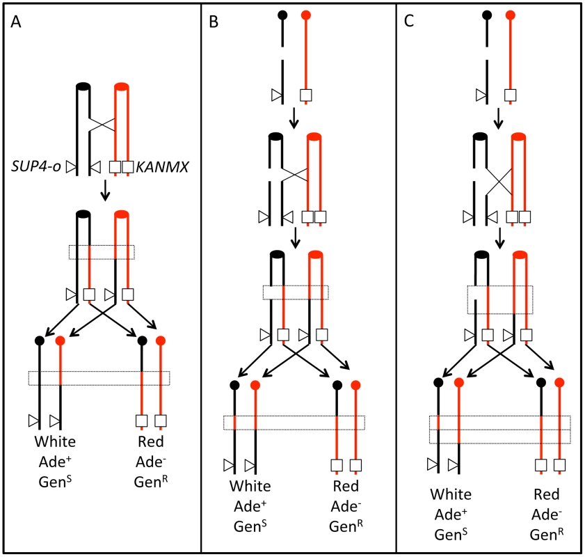
Chromatids that are depicted in black represent YJM789-derived chromatids, and those in red represent W303a-derived chromatids. The centromeres are shown as circles. A. 3∶1 conversion event. Repair of a G2-associated DSB results in a 3∶1 gene conversion tracts (enclosed in dotted lines) associated with the LOH event. B. 4∶0 conversion event. A chromosome in a G1 cell is replicated to form two sister chromatids that are broken at approximately the same position. Repair of these two broken chromatids in G2 can result in a region in which all four chromatids have identical SNPs, a 4∶0 conversion tract. In this example, both conversion tracts are of the same length. C. 4∶0/3∶1 hybrid tract. As in Figure 1B, a broken chromosome is replicated to form to two broken sister chromatids. The conversion tracts associated with the repair of the two DSBs, however, are of different lengths, resulting in a 4∶0/3∶1 hybrid tract. Previously, we showed several different types of gene conversion events that likely reflect differences in the timing of the recombination-initiating DSBs in the cell cycle [11], [12]. Figure 1 shows a comparison of conversion tracts resulting from repair of a DSB formed during S/G2 (Figure 1A) or G1 (Figure 1B and 1C); the red and black lines represent homologs with heterozygous SNPs. Figure 1A shows a DSB on a black chromatid during G2 of the cell cycle that is repaired by HR, resulting in a crossover and associated 3∶1 conversion tract (three chromatids with red SNPs and one with black SNPs). Figure 1B shows a DSB on the black homolog during G1 of the cell cycle that is replicated to generate two broken chromatids. Since both chromatids are broken in the same location, only the homolog is available as a template for HR. If one chromatid is repaired resulting in a crossover and associated conversion event, and the second chromatid is repaired as a conversion event unassociated with crossovers, there will be a 4∶0 conversion tract. Figure 1C also shows a DSB on the black homolog during G1 of the cell cycle. Following replication, repair occurs as in Figure 1B, but one broken chromatid has been processed to a greater extent than the other, resulting in unequal-sized conversion tracts. The resulting conversion tract is a 4∶0/3∶1 hybrid tract.
In this study, we map spontaneous crossovers and associated gene conversion events on the right arm of chromosome IV, an interval of about 1.1 Mb, using SNP microarrays to map events at approximately 500 bp resolution. We find that events occur throughout the chromosome arm, although their distribution is non-random. In particular, inverted pairs of transposons at two different locations result in homolog-specific hotspots for crossovers initiated by DSBs during G1. We also demonstrate that LOH events are more frequently a consequence of G1-initiated DSBs than G2-initiated DSBs, and that conversion tracts associated with the G1 DSBs are longer than those associated with G2-initiated DSBs. Finally, we show that different genetic elements are associated with crossovers initiated during G1 and G2. For example, regions of converging replication forks (TER sites; [13]) are associated with G2, but not G1, conversion events.
Results
Identification and mapping of crossovers on chromosome IV
We used a colony-color screening assay to identify reciprocal crossovers on chromosome IV, similar to the previously described system [11], [14] (Figure 1A). The diploid JSC25 was generated by mating two sequenced haploid strains, W303a and YJM789, that have 55,000 SNPs relative to each other [15]. We inserted the KANMX gene near the telomere on the right arm of the W303a chromosome IV homolog and SUP4-o (encoding an ochre-suppressing tRNA) allelically on the YJM789-derived homolog. The KANMX gene renders the cell resistant to geneticin (GenR), a drug related to the bacteria-specific antibiotic kanamycin. JSC25 is also homozygous for the ochre-suppressible ade2-1 mutation. Diploid strains of this genotype that lack SUP4-o form red colonies, strains with one copy of SUP4-o form pink colonies, and those with two copies form white colonies. If there is a crossover between the centromere and the SUP4-o locus, there is a 50% chance, following chromosome segregation that the two daughter cells will be homozygous for the region centromere-distal to the crossover. If the crossover occurs at the time that the diploid JSC25 is plated, a red/white sectored colony will be formed (Figure 1A), in which the white side of the sector is geneticin-sensitive due to loss of the KANMX gene, and the red side of the sector is Ade− due to loss of SUP4-O. The frequency of red/white sectored colonies (3.1×10⁻⁵) in JSC25 is reflective of the rate of crossovers between CEN4 and the KANMX/SUP4-o markers. Since only one half of reciprocal crossovers result in a sectored colony, the rate of crossovers on the right arm of chromosome IV is 6.2×10⁻⁵/division.
We mapped crossovers in about 140 sectored colonies of JSC25. We monitored about 2300 SNPs between CEN4 and the KANMX/SUP4-o loci within the 1.1 Mb interval. As in our previous study [16], for each SNP, we designed four 25-base oligonucleotides: one for each Watson and Crick strand of each homolog. The normalized ratio of experimental to reference hybridization of these samples to each oligonucleotide allowed us to determine whether the experimental strains were heterozygous or homozygous for a specific SNP.
Figure 2 shows an example of the analysis of genomic DNA from a sectored colony (JSC25 SP 129). The hybridization of genomic DNA to the W303a - and YJM789-derived SNPs is shown by red and blue lines, respectively. In the low-resolution analysis (Figure 2A), genomic DNA hybridizes equally efficiently to both W303a - and YJM789-derived SNPs (normalized ratio of 1) from SGD coordinates 445 kb (the location of CEN4) to 770 kb. Near coordinate 770 kb in Figure 2A, there is a transition from heterozygosity to homozygosity for the W303a and YJM789 forms of the SNPs in the red (top) and white (bottom) sectors, respectively. Based on the higher resolution depiction (Figure 2B), it is clear that the transition points between heterozygosity and homozygosity are different between the two sides of the sector, occurring near 762 kb and 772 kb in the red and white sectors, respectively. The boxed region in Figure 2B represents a 3∶1 gene conversion tract (Figure 1A). This pattern indicates that the gene conversion tract was initiated by a DNA lesion on the YJM789-derived chromosome, since the chromosome with the lesion acts as a recipient during the conversion event [17]. Microarray data illustrating a hybrid conversion tract are shown in Figure S1.
Fig. 2. Mapping of a crossover with an associated 3∶1 conversion event by SNP microarrays. 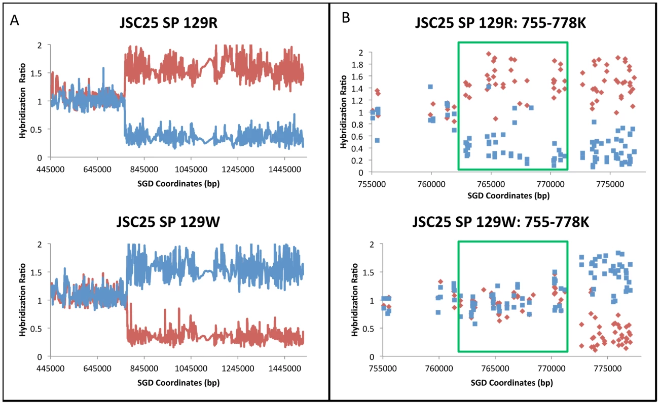
The values on the Y-axis show the experimental/reference hybridization ratio of genomic DNA to oligonucleotides that are specific to SNPs from the W303a and YJM789 backgrounds. The values on the X-axis indicate the SGD coordinates of the SNPs along chromosome IV. The red and blue lines or data points depict the hybridization levels of probes specific for the W303a and YJM789 homologs, respectively. Where both the red and blue lines have a value near 1, the diploid is heterozygous for the SNPs. If the strain is homozygous for the W303a variant, the red signal increases to about 1.6 and the blue signal diminishes to about 0.3. If the strain is homozygous for the YJM789 variant, the reverse pattern is observed. A. Low-resolution depiction of a reciprocal crossover analyzed by the SNP microarrays. The top and bottom plots represent the red and white sectors, respectively. In both the red and white sectors, there is a transition between heterozygosity and homozygosity at approximately SGD coordinate 770 kb. B. High-resolution depiction of a reciprocal crossover. Each data point indicates the hybridization level to a specific oligonucleotide. Each square and diamond indicates the hybridization level to a specific oligonucleotide. The genomic DNA from the red sector has a transition between heterozygosity and homozygosity at approximately coordinate 762.5 kb (top panel), whereas genomic DNA from the white sector has a transition at about coordinate 772 kb. The regions shown in green rectangles represent the 3∶1 gene conversion event. Of 139 crossovers analyzed, 121 (87%) were associated with a contiguous gene conversion event. Of the 121 conversion tracts, there were 29 simple 3∶1 conversions, 7 simple 4∶0 conversions, and 46 simple 4∶0/3∶1 or 3∶1/4∶0/3∶1 hybrid conversions; 39 tracts had more complicated patterns of conversion (Text S1). Summaries of various features of all conversion tracts observed in our study are presented in Tables S1, S2, S3. No aneuploidy of chromosome IV was observed for any of the sectored colonies. In addition, only one large deletion was detected. In the red sector of colony 42R/W (Class E34 in Table S1), we detected a deletion of about 100 kb located between pairs of inverted Ty elements at SGD coordinates 872 kb and 981 kb. We do not know whether this deletion occurred prior to the crossover or was associated with the crossover.
Location of crossovers and associated conversions on the right arm of chromosome IV
We collected and analyzed 138 sectors and 139 reciprocal crossovers in the strain JSC25; one sectored strain had a double crossover. The locations of crossovers and associated conversion tracts are shown in Figure 3. The crossovers associated with conversion tracts were separated based on which homolog had the initiating lesion. Events initiated in W303a - or YJM789-derived homologs are presented in the top or bottom of Figure 3, respectively. The conversion tracts are depicted as pairs of lines (discussed in the figure legend) with the lengths of the conversion tracts indicated on the Y-axis. Crossovers unassociated with conversions are not shown in Figure 3, because it is impossible to determine which homolog had the originating DSB. It is apparent that the distribution of conversion events along the chromosome arm is not even. In addition, in certain regions of the chromosome (between SGD coordinates 900 kb and 1000 kb), the conversion tracts appear longer than most of those in other regions of the chromosome.
Fig. 3. Crossovers and associated gene conversion events in JSC25 along the right arm of chromosome IV. 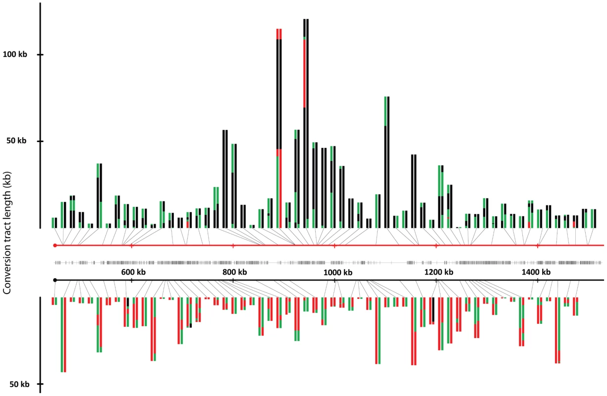
The X-axis represents the SGD coordinates (the centromere is at 449 kb), and the Y-axis represents the length of the individual conversion tract. The red and black horizontal lines in the center of the figure depict the W303a and YJM789 homologs, respectively. Individual conversion tracts are depicted as two parallel vertical lines that are attached to either homolog line by a black connector line which indicates the location of the centromere-proximal margin of the conversion tract. Conversion tracts depicted in the top portion of the figure and connected to the W303a homolog, resulted from DSBs initiated on the W303a homolog. Conversion tracts depicted in the lower half of the figure were initiated by DSBs on the YJM789 homolog. The vertical left and right members of the line pairs show the conversion tract of the red and white sides of the sector, respectively. Green, red, and black show heterozygosity, homozygosity for W303a SNPs, and homozygosity for YJM789 SNPs, respectively. Each segment of the depicted conversion tract is proportional to the size of that segment within the sector. The hash marks between the two homologs indicate the density of the SNPs represented on the microarray. Figure 4 summarizes the location of crossover-associated gene conversion events in a different way. In this figure, the X-axis shows the SGD coordinates and the Y-axis shows the number of conversion events that include the SNP located at that coordinate summed over all mapped events. The probability of a SNP being included within a conversion tract is a function of both the frequency of nearby DNA lesions and of the lengths of conversion tracts emanating from the initiating DNA lesions. In Figure 4, we label regions where multiple contiguous SNPs were involved multiple times in a gene conversion event as potential hotspots (HS1-HS7). In Figure 4B, we show separately those events initiated on the W303a - and YJM789-derived homologs. These distributions are strikingly different. It is also clear that the distributions of G1-initiated conversions (tracts with 4∶0 segments or other features that indicate a G1-associated DSB) and G2-associated conversions (3∶1 conversions without a 4∶0 segment) are different (Figure 4C). HS3 and HS4 are both G1-specific and specific for the W303a-derived homolog. The mechanism responsible for this specificity will be discussed below.
Fig. 4. Summary of conversion tracts associated with crossovers along the right arm of chromosome IV. 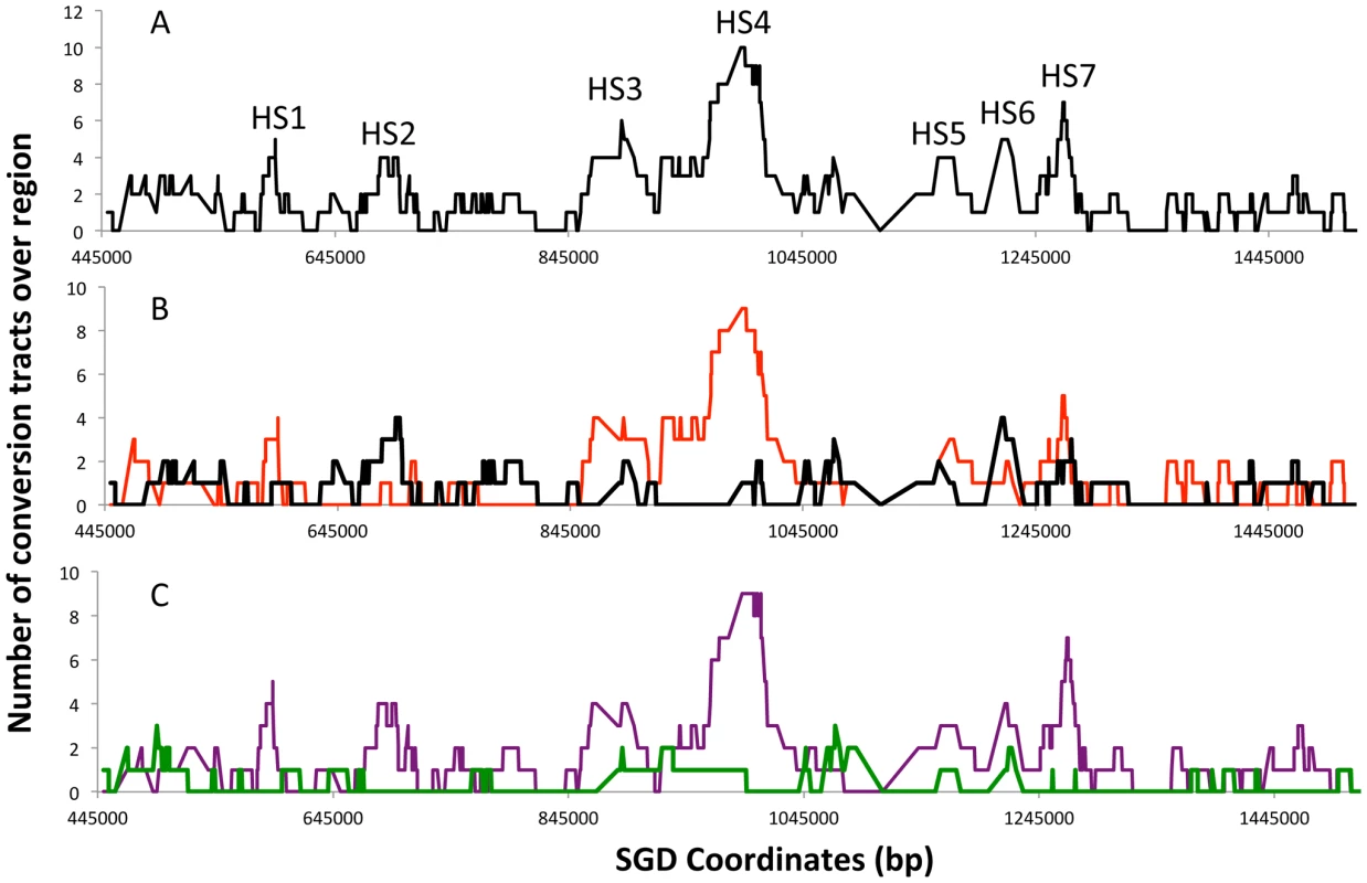
These plots represent the number of times a SNP represented on the microarray is involved in a conversion tract. A SNP was considered involved if it was between the start and end of the conversion tract. A. All conversion tracts. The labels HS1–HS7 represent potential hotspots for recombination. B. Conversion tract distribution of the events initiated on the W303a (red) and YJM789 (black) homolog. C. Conversion tract distribution of events initiated during G1 (purple) or G2 (green line) of the cell cycle. Statistical analysis of the distribution of crossovers and conversions
We used several different statistical methods to determine if the distribution of crossovers along the chromosome arm is non-random. In our first statistical analysis, we divided the right arm of chromosome IV into five bins of 200 kb (starting at CEN4) and a sixth bin of about 80 kb. We counted the number of crossovers that occurred within each region (additional details discussed in Text S1). If all events are examined, the number of crossovers per bin is approximately proportional to the size of each bin (p = 0.96 by chi-square Goodness-of-Fit). We also analyzed the events separated by the homolog and timing of the initiating lesion (depicted in Figure S2). Both the homolog-specific distributions of crossovers and cell cycle-specific distributions of events were significantly different with p values of 0.01 and 0.007, respectively. The bin from 845–1045 kb (containing HS3 and HS4) had a very significant enrichment of crossovers initiated on the W303a-derived homolog during G1 of the cell cycle compared to a random distribution of this class of event among all of the bins (p = 2.3×10⁻⁵).
Comparison of the physical and genetic maps of the right arm of chromosome IV
In Figure 5, we show a comparison between the physical map of the right arm of chromosome IV with the genetic maps based on recombination data from JSC25. In addition, we show separately the data from the W303a - and YJM789-derived homologs. This figure emphasizes the differences in recombination activity between the two homologs. For example, the region between 927 and 980 kb in the map of W303a is larger than expected from the physical map, but there are no events in this segment in the YJM789 map. Conversely, the segment between 768 and 821 kb has no events in the W303a map, but a greater-than-average number of events in the YJM789 map. Such differences are expected because of the differences in the distributions of hotspots on the two homologs. For example, the hotspot activity of HS4 is specific to the W303a chromosome. Thus, we expect an expansion of the genetic map on the W303a-derived homolog near HS4, but no expansion of the map on the YJM789-derived homolog.
Fig. 5. Comparison of physical and genetic mitotic recombination maps. 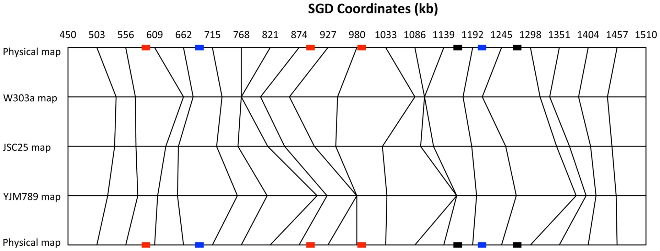
This figure shows a comparison of the physical map of the 1.1 Mb right arm of chromosome IV with a genetic map of mitotic crossovers. We show three genetic maps: all crossovers (JSC25 map), crossovers initiated from the W303a homolog (W303a map), and crossovers initiated from the YJM789 homolog (YJM789 map). The chromosome arm was divided into 20 regions of about 53 kb. The genetic distances are based on the proportion of crossovers within each 53 kb interval, using the “point-analysis” method employed for Figure S2 and described in Text S1. The top and bottom horizontal lines show the physical map. The vertical lines allow comparisons of the genetic maps with the physical maps, and of the different genetic maps with each other. The relative “thinness” and “thickness” of the same segment between the W303a and YJM789 maps indicates whether that region has more crossovers initiated on one homolog in comparison to the other. The recombination hotspots (labeled in Figure 4A) are indicated by rectangles on the physical map lines (top and bottom lines). The red, blue, and black colors indicate hotspots that are specific to the W303a-derived homolog, specific to the YJM789-derived homology, and hotspots that are found on both homologs, respectively. We compared the DNA sequences of W303a and YJM789 to determine if any features of the sequence would suggest possible mechanisms for the observed differences in hotspot activity on the two homologs. Although there are many SNPs that distinguish the two homologs, insertions/deletions (in/dels) greater than 50 bp are relatively rare [15], and most of these involve Ty elements or delta elements. These alterations and their approximate position by SGD coordinates are listed below; in this list, boldface indicates that the element is present in W303a and not in YJM789, and the regular typeface indicates that the element is present in YJM789 and not in W303a: 1) delta element at 489 kb, 2) delta element at 513 kb, 3) delta element at 520 kb, 4) Ty element at 645 kb, 5) sigma element at 668 kb, 6) partial Ty1 element at 805 kb, 7) inverted pair of Ty2/Ty1 elements at 872 kb, 8) sigma element at 946 kb, 9) inverted pair of Ty2/Ty1 elements at 981 kb, and 10) unannotated partial delta element at 1151 kb. In addition to retrotransposon-related sequences, there were a small number of other in/dels. The W303a-derived homology has three tandem ENA genes located near SGD coordinate 534 kb, whereas the YJM789-derived homolog has only two. The YJM789-derived homolog has a 153 bp insertion in YDR077W relative to the same gene on the W303a-derived homolog. The YJM789-derived homolog has a partial deletion of ARS420 relative to the W303a-derived homolog. Finally, the YJM789 allele of YDR150W has more copies of an internal 192 bp repeat than observed in the W303a allele. Except for these in/dels and numerous SNPs, the genomic sequences of the two homologs are well conserved. For example, no changes were observed for the positions of tRNA genes or G-quadruplex structures.
The significance of these genomic alterations on recombination hotspot activity is unclear with two exceptions. As discussed below, the inverted pairs of Ty elements located at positions 872 kb and 981 kb are likely to be important for the hotspot activities of HS3 and HS4 in the W303a-derived homolog. Other differences in hotspot activity between the two homologs could reflect multiple small changes or homolog-specific alterations in chromatin modifications. In addition, although it is difficult to determine what chromosome elements are associated with most of the hotspots, by analyzing the sequences within all of the conversion tracts, we were able to detect significant associations with certain chromosome elements; this analysis is described below.
Differences in hotspot activities on the two homologs demonstrate the difficulty and, perhaps, the futility of generating a universal mitotic recombination map for S. cerevisiae. As shown below, however, differences in the recombination activities of the two homologs can lead to mechanistic insights into mitotic recombination.
The HS4 hotspot is associated with a pair of inverted Ty elements
The HS4 hotspot, located between coordinates 970 and 1,000 kb, has the highest level of recombination as measured by the number of conversion events (Figure 4A). This hotspot is specific to the W303a-derived homolog (Figure 4B) and is associated with G1-initiated gene conversion events (Figure 4C). Analysis of the DNA sequences in the hotspot region shows that the peak of recombination activity of HS4 overlaps with a closely-spaced inverted pair of Ty elements (YDRWTy2-3 and YDRCTy1-3) located between coordinates 981 and 992 kb. Ty elements are 6 kb retrotransposons present in about 40 copies per haploid genome [18].
To determine whether the hotspot activity of HS4 was related to the inverted pair of Ty elements, we first examined whether both of the haploid parental strains contained the inverted pair of elements. PCR analysis demonstrated that the inverted repeat is in the W303a homolog but not the YJM789 homolog (Figure S3). The observation that the HS4 hotspot is specific to the W303a-derived homolog that contains the inverted pair of Ty elements implicates this structure in the hotspot activity. The spacer between the Ty elements is about 25–66 bp, depending on the degree of mismatches allowed between the repeats (Figure S4).
To examine some of the structural features of the HS4 hotspot, we measured hotspot activity in four different isogenic strains (Figure 6) with the wild-type HS4 hotspot (JSC71-1), with a deletion of the HS4-associated Ty2 element (JSC73-2), with an expansion of the spacer between Ty2 and Ty1 (JSC74-1), and with a deletion of one of the delta elements associated with Ty2. For all four strains, we inserted URA3 about 23 kb centromere-distal to HS4 (1013 kb) and the hygromycin-resistance gene HYG about 20 kb centromere-proximal to HS4 (957 kb). These strains are depicted in Figure 6A and 6B.
Fig. 6. Deletion analysis of the HS4 hotspot. 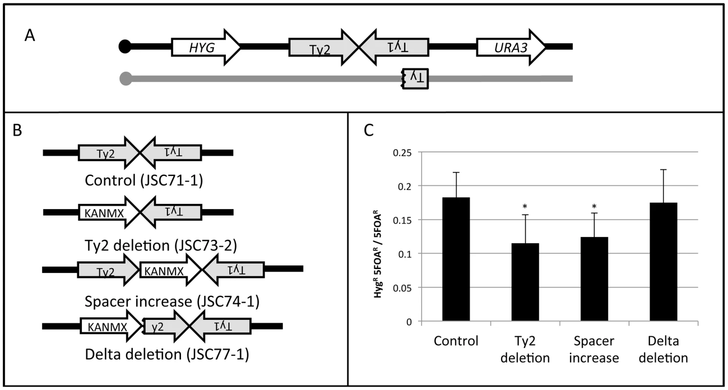
A. Experimental system used to monitor the activity of the wild-type HS4 hotspot and various deletion derivatives. The URA3 and HYG genes were inserted centromere-distal and -proximal to the inverted Ty elements, respectively, on the W303a-derived homolog (black). The YJM789-derived homolog (grey) was unaltered. B. Modifications of the inverted repeat structure. In the control strain JSC71-1, the unmodified hotspot consists of a Ty2 element on the Watson strand, 25 bp of non-Ty sequence, and a Ty1 element on the Crick strand. In the JSC73-2 strain, the Ty2 element is replaced with KANMX. In the JSC74-1 strain, the space between the two Ty elements, is increased by about 2 kb by insertion of the KANMX marker. In the strain JSC77-1, the centromere-proximal delta element of Ty2 was replaced by the KANMX gene. C. Frequency of recombination between HYG and URA3 in the strain with wild-type HS4 activity and in various deletion derivatives. Asterisks indicate a significant (p<0.05) reduction in recombination activity compared to the control strain. A crossover initiated at HS4 will result in a daughter cell that is HygR and 5-FOAR half of the time. A crossover initiated between CEN4 and HYG will result in a daughter cell that is HygS and 5-FOAR half of the time. By monitoring the ratio of HygR 5-FOAR recombinants to all 5-FOAR recombinants, we measured the recombination rate between the HYG and URA3 markers. The distance between CEN4 and URA3 is about 568 kb and the distance between HYG and URA3 is about 56 kb. Thus, if the numbers of crossovers are proportional to the size of the physical intervals, 10% of the 5-FOAR strains should be HygR. The observed frequency of derivatives of this class in the strain with the wild-type hotspot (JSC71-1) was 0.17 (Figure 6C). The difference in the number of observed and expected crossovers is statistically significant (p<0.001), as expected since this region contains the HS4 hotspot. In a strain with the deletion of the Ty2 element (JSC73-2) or a strain with an expanded spacer (JSC74-1), the hotspot activity is significantly reduced (Figure 6C). In contrast, in a strain in which the centromere-proximal δ element of Ty2 is deleted (JSC77-1), no significant loss of hotspot activity is observed.
Deletion of the Ty2 element eliminates the possibility of hairpin formation at HS4 and an increase in the spacer between the repeated elements would be expected to substantially reduce the probability of hairpin formation [4], [7]. The 330 bp δ elements flank Ty elements [18] and the 5′ δ element acts as a transcriptional promoter [19]. In the strain containing the deletion of the 5′ δ, Ty2 should no longer be transcriptionally active. The results summarized in Figure 6C, therefore, indicate that the formation of a secondary structure between the two Ty elements is necessary, but the transcriptional activities of both of the Ty elements are not required for hotspot activity at HS4.
The HS3 hotspot (Figure 4) also maps at the positions of an inverted pair of Ty elements (YDRWTy2-2 and YDRCTy1-2) separated by a spacer of about 100 bp. As with HS4, this hotspot is specific to the W303a-derived homolog and G1-specific. Since the YJM789-derived homolog does not have this inverted pair of Ty elements [15], we assume that the hotspot activity of HS3 is a consequence of formation of a secondary structure.
Types of gene conversion events: Inferences about the timing of the recombinogenic DSBs during the cell cycle
Most (121/139; 87%) of the crossovers examined in our study were associated with gene conversion. Of these conversion tracts, about two-thirds (82/121) are simple 3∶1 conversions, simple 4∶0 conversions, or hybrid 3∶1/4∶0 or 3∶1/4/0/3∶1 tracts as diagrammed in Figure 1. As discussed previously, the simple 3∶1 tracts are most simply interpreted as events initiated in S or G2, and the other two classes are likely to reflect events initiated in G1. Thus, if we count only the simple conversions as illustrated in Figure 1, two-thirds of the spontaneous crossovers have gene conversions indicative of a G1-initiated event, supporting previous conclusions based on a more limited dataset [11]. The numbers of conversion events of all types, as well as their schematic depictions, are given Table S1. About 1/3 of the conversion tracts were more complicated than those predicted by Figure 1. These complex tracts are discussed further in Text S1.
We also examined the conversion tract lengths for the G2 - and G1-associated conversions. The median tract lengths of G2 - and G1-associated crossovers (Table S1 and S2; Text S1) were 4.7 kb (95% confidence limits of 2.6–9.5 kb) and 14.8 kb (11.7–17.5 kb), respectively. By the Mann-Whitney test, the G1 and G2 conversion tract lengths are significantly different (p<0.0001). The median tract length for all conversion events was 10.6 kb (8.2–13.6 kb).
Association of conversion tracts with elements of chromosome sequence or structure
From many genetic studies in yeast, it has been shown that the site at which recombination initiates is within or adjacent to the conversion tract [17]. There are a number of elements of chromosome structure in yeast that have been identified as potential chromosome fragile sites or potential hotspots of recombination. We compared our mapping of gene conversion events with the 18 possible recombination-inducing elements listed in Table S4. The locations of some of these elements (G-quadruplexes, replication termination regions, Ty retrotransposons, and long terminal repeats) are depicted in relation to the crossover map in Figure S5.
The methods used for examining potential element enrichments are explained in detail in Text S1. In brief, we determined whether there was a statistically significant excess of elements within the gene conversion tracts relative to that expected based on the known number of elements on the right arm of chromosome IV. In addition to examining all events collectively, we performed data analysis on more exclusive categories: conversion events initiated on the W303a-derived chromosome, conversion events initiated on the YJM789-derived chromosome, G1 conversion events, and G2 conversion events. The statistically significant associations from these analyses are in Table S5.
In the analysis in which all conversions are included, the only significant enrichments were with Ty elements, long terminal repeats (δ elements) that flank Ty elements, and tRNA genes. These same associations were also significant for G1 events and events initiated on the W303a-derived homolog. These associations are expected since the HS3 and HS4 hotspots are G1 - and W303a-specific (Figure 4B and 4C). No significant associations were observed between conversion tracts and δ elements unassociated with Ty elements. The non-random linkage between Ty elements and tRNA genes in the genome [18] may explain the tRNA enrichment.
Replication forks do not proceed at a constant rate in the genome and replication-pause sites have been associated with a number of chromosome elements including the ribosomal RNA fork barrier, centromeres, tRNA genes, convergent replication forks, G-quadruplex motifs, and highly-transcribed RNA polymerase II genes (reviewed in [20]). We found several similar motifs associated with conversion events. G2 conversion events were significantly associated with replication-termination regions. We also found a significant association between G-quadruplex motifs [21] and G1 conversion events, as well as an association between G-quadruplex motifs and conversion events initiated on the YJM789-derived chromosome; G-quadruplex structures slow replication fork progression [22]. Finally, the conversion events that are initiated on the YJM789-derived chromosome have an over-representation of sites of stalling of the Rrm3p helicase; this helicase is thought to aid replication through genomic regions at which DNA polymerase is paused [20]. We also found some elements of chromosome structure that were negatively associated with the conversion tracts (Table S6).
In addition to looking for associations of various elements within the conversion tracts, we looked for associations of these elements with the termini of the tracts. The details of this analysis are described in Text S1. In brief, we examined windows defined by transitions between heterozygous and homozygous markers at the ends of the conversion tracts for over - or under-representation of the chromosome elements described in Table S4. One significant positive association was observed. We found that the LTR elements associated with Ty elements were significantly over-represented at the termini of conversion events. Although it is possible that these regions are preferred sites for the termination of conversion events, there are several alternative interpretations. First, since the YJM789-derived chromosome IV is missing five of the eight Ty elements that are present on the right arm of the W303a-derived chromosome IV [15], several of these LTRs border large regions of heterology. It is possible, therefore, that the conversion tracts are terminated as a consequence of this heterology rather than as a consequence of the sequence/structure of the LTR. Second, HS3 and HS4, the two strongest hotspots, are flanked by Ty-associated LTR events. Thus, these preferred sites for initiating DSBs may lead to preferred sites of tract termination indirectly. A choice among these alteratives will require additional experiments. Finally, it should be emphasized that, despite an enrichment for Ty-associated LTRs at the ends of conversion tracts, the termini of most tracts are not associated with these elements.
Discussion
In this study, we developed a high-resolution map of spontaneous crossovers on the right arm of chromosome IV, a region that includes about 10% of the yeast genome. Below, we will discuss: 1) the frequency of mitotic crossovers, 2) the distribution of crossover events, 3) the properties of mitotic gene conversion tracts, and 4) the timing of recombination events during the cell cycle.
Frequency of reciprocal crossovers
The right arm of chromosome IV has about 6.2×10⁻⁵ crossovers/division or about 6.2×10⁻⁸ crossovers per kb. As a convenient unit of mitotic crossovers, we suggest that 10−6 crossovers/division be defined as one micro Stern (µS), named after Curt Stern, the discoverer of mitotic recombination [23]. We emphasize that this unit is restricted to crossovers that occur between homologs; sister-chromatid exchanges are undetectable by our methods since they do not result in loss of heterozygosity. We calculate that the right arm of chromosome IV has a genetic length of about 62 µS. Extrapolating the data from chromosome IV, we calculate that the yeast genome has a genomic crossover rate of about 6.2×10⁻⁴/cell division and a genetic map length of about 620 µS. This value is only a rough estimate since the frequency of mitotic recombination hotspots may vary from strain-to-strain, chromosome-to-chromosome, and homolog-to-homolog. However, since mitotic recombination hotspots appear weaker than meiotic recombination hotspots (as described below), some of these variables may have only small effects. For example, although the W303a-derived chromosome IV has more hotspots than the YJM789-derived chromosome IV, the numbers of events initiated on each homolog (determined by which homolog is the donor in the conversion event) are almost identical: 61 events initiated on the W303a-derived homolog and 59 events initiated on the YJM789-derived homolog.
Most previous studies of the rates of spontaneous crossovers utilize a diploid strain that is heterozygous for a can1 mutation on chromosome V [24]. The heterozygous diploid is sensitive to canavanine, and mitotic crossovers resulting in LOH for the can1 locus produce cananvanine-resistant derivatives. In several representative studies performed in different genetic backgrounds, the rates of crossovers per division in the 120 kb interval between CEN5 and CAN1 were measured as 6×10⁻⁶ [24], 2×10⁻⁶ [25], and 2×10⁻⁶ [11]. These rate estimates are in reasonable agreement with our rate measurement of 6.2×10⁻⁵ on chromosome IV, since the interval measured in our study is about nine-fold greater than the interval on chromosome V.
Measurements of the rate of mitotic gene conversion events are usually done using diploids that have different non-complementing mutant alleles (heteroalleles) of genes affecting the biosynthesis of an amino acid or nucleotide. The rates of gene conversion, therefore, are estimated by the rate that prototrophic derivatives are produced from the auxotrophic diploid. The rates of heteroallelic gene conversion vary considerably in different studies. Representative rates per division (the heteroallelic gene indicated in parentheses) are: 2.5×10⁻⁷ (arg4) [26], 2.6×10⁻⁶ (trp5) [27] and 3×10⁻⁷ (leu2) [28]. Since gene conversion events are initiated by DSBs, the frequency of these events will be affected by the hotspot distribution. In addition, the distance between the mutant substitutions would be expected to influence the rate of gene conversion with this assay. It is important to note that gene conversion requires a recombinogenic DSB near the heteroalleles, whereas the crossover assays detect DSBs distributed throughout the region between the reporter gene and the centromere. In the present study, our analysis of conversion events was restricted to those associated with crossovers on chromosome IV. In a previous study, using microarrays that covered the entire yeast genome, no unselected spontaneous gene conversion events were detected in thirteen strains examined [16].
The yeast genome has a genetic length of about 4100 cM (SGD), representing about 81 meiotic crossovers/division. Thus, the rate of crossovers in meiosis is about 105-fold higher than the mitotic rate. Although the rate of mitotic crossovers/division is low relative to the meiotic rate, since the number of mitotic divisions is likely to greatly exceed the number of meiotic divisions, mitotic crossovers are likely to be a potent mechanism for generating novel combinations of alleles.
General considerations concerning the distribution of crossovers
The distributions of mitotic crossovers and their associated gene conversion tracts are shown in Figure 3 and Figure 4. Two points concerning the distribution should be emphasized. First, by our statistical analysis, not all of the peaks labeled as hotspots have significantly elevated levels of recombination. Second, for conversion-associated crossovers, the position of the initiating DNA lesion can be anywhere within the conversion tract. Previously, we showed that 230 repeats of the GAA trinucleotide was a hotspot for DSB formation [29]. In individual conversion events associated with this hotspot, the conversion tracts were propagated either toward the centromere, away from the centromere or bidirectionally from the tract. Our mapping of DNA lesions and the resolution of the mitotic recombination map, therefore, are limited by the size of the conversion tracts rather than the distribution of SNPs.
An important conclusion from our analysis is that two homologs can differ significantly in their distribution of recombination events (Figure 4B and Figure 5). From the data shown in Figure 4B, it appears that HS1, HS3, and HS4 are hotspots on the W303a-derived chromosome, HS2 and HS6 are hotspots on the YJM789-derived chromosomes, and HS5 and HS7 are hotspots on both homologs. Thus, construction of a universal genetic map of mitotic events is difficult.
Meiotic recombination events have been mapped throughout the genome using various types of microarrays [30]–[35]. The patterns of meiotic and mitotic recombination events on the right arm of chromosome IV show no evident similarity (Figure S6). Since meiotic recombination are initiated by meiosis-specific cleavage of the genome by Spo11p and the DNA lesions that initiate mitotic recombination are likely to have a variety of sources (discussed further below), this difference in meiotic and mitotic patterns of recombination is expected. The levels of meiosis-specific DSBs at different places in the genome vary at least 400-fold [34]. We can estimate the relative “heat” of a chromosome region by determining the number of times that SNPs within that region are included within conversion tracts. For example, the peak SNP in HS4 (the strongest hotspot) was included in eleven recombination events (Figure 4). Since the average number of events for all SNPs throughout the right arm of chromosome IV is two, HS4 is only about five times more active than an average region. Thus, our data suggest that hotspots and coldspots are more pronounced for meiotic than for mitotic recombination.
Inverted repeats as hotspots for mitotic recombination
Both HS3 and HS4 co-localize with inverted Ty elements. Inverted repeats have been previously shown to be hotspots for certain types of mitotic recombination [5], [36]. Most of these studies have involved non-yeast DNA sequences inserted into the genome by transformation. In our previous study [7], a hotspot for chromosome rearrangements on chromosome III in cells that have low levels of DNA polymerase alpha was an inverted pair of Ty elements separated by about 280 bp. In contrast, HS3 and HS4 function as hotspots in cells with wild-type levels of DNA polymerases.
The conversion tracts associated with inverted repeats at HS3 and HS4 are primarily 4∶0 or hybrid events, arguing that the recombinogenic lesion is formed prior to replication. Two types of secondary structures have been associated with inverted repeats: cruciforms and hairpins. Evidence for cruciform processing (presumably by a Holliday junction-like resolvase) resulting in two hairpin-capped ends has been obtained [5]. However, spacers greater than 20 bases substantially inhibit cruciform formation in E. coli [37]–[39], and reduce the recombinogenic properties of palindromes in yeast [40]. An alternative model is that a single-stranded nick near the junction of the inverted repeats is processed into a single-stranded DNA gap, allowing formation of a hairpin on the intact DNA strand. The loop formed by the spacer in the hairpin could then be nicked by a nuclease such as the Mre11p complex [5]. We do not know the source of the nick that initiates hairpin formation. It is possible that DNA supercoils produced by Ty transcription are nicked by topoisomerases [41]. Our deletion analysis, however, demonstrates that convergent transcription of both Ty elements is not necessary for HS4 activity. It is also possible that the nick that initiates hairpin formation could represent a lesion generated by mismatch repair. Since the hotspot activity of HS3 and HS4 appears G1-specific, it is likely that the initiating nick occurs in G1.
Mitotic recombination events that are initiated independently of inverted repeats
In addition to inverted repeats, several other factors have been associated with mitotic recombination or DSB formation. Using several different assays, researchers have found that elevated levels of transcription stimulate recombination (reviewed in [1]). Since mutations in many enzymes involved in DNA replication result in a hyper-Rec phenotype, it is likely that some spontaneous recombination events result from DNA lesions generated during DNA replication [42]. It has been shown that some DNA sequences or motifs are associated with slow or stalled replication forks [42]. Consequently, we analyzed our conversion tracts to determine if there was an over-representation of these structure/sequence motifs. We found that G4 quadruplex motifs were significantly associated with G1-initiated DSBs (Table S5). In yeast cells, G4 motifs have been shown previously to be enriched in areas of the genome that have high γ-H2AX binding, a signal associated with DNA damage [21]. In our study, the events initiated during S or G2 of the cell cycle were significantly associated with replication-termination (TER) regions. This association is especially intriguing because sister chromatids are intercalated following replication, and need to be resolved by Top2p [43], [44]. Since Top2p induces DSBs to allow decatenation of sister-chromatids, it is possible that some of these Top2p induced cleavages are misrepaired, leading to homologous recombination.
Conversion tract lengths
The median conversion tract length for all analyzed crossovers on the right arm of chromosome IV was 10.6 kb (95% confidence limits: 8.2–13.6 kb). As discussed in Results, we found that conversion tract lengths associated with G1-initiated breaks were significantly longer than G2-initiated breaks. Since the crossovers of both G1 - and G2-initiated events occur in G2, the broken ends generated by a G1-initiated lesion have a longer time for 5′-to-3′ resection prior to repair of the DSBs. In addition, the G1-initiated events involve the repair of two broken chromatids. If the conversion tracts associated with these two repair events are propagated in different directions, we would expect that the G1-initiated conversions would be longer than those initiated in G2. As noted previously [11], the mitotic conversion tracts characterized in our system are considerably longer than the average meiotic conversion tract of about 2 kb [45].
One of the unique characteristics of recombination events associated with HS4 is that their conversion tracts are substantially longer than those that occur at other locations along chromosome IV. The median conversion tract length associated with HS4 is 48.4 kb (95% confidence limits: 17.1–118.3 kb), much longer than the median tract length for all conversions (10.6 kb) and longer than the median length of G1-induced conversions (14 kb). One factor that would be expected to affect tract length is that HS3 has two intact Ty elements on the W303a-derived homolog whereas the YJM789-derived homolog has only a 2 kb fragment of a Ty (Figure 6, Figure S3). Thus, a DSB formed in this hemizygous insertion on the W303a-derived chromosome would need to be processed about 10 kb before exposing homology on the YJM789-derived chromosome. In addition, since the homology would be located internally on the resected strand, a recombination intermediate with single-stranded branches would be formed [17]. Removal of these single-stranded branches likely requires the nucleotide excision repair proteins Rad1p and Rad10p [46]–[47], as well as the mismatch repair proteins Msh2p and Msh3p [48]–[51]. Thus, the hotspot activity of HS4 would also be dependent on these proteins. It is possible that the more extensive processing of the DSBs occurring in HS4 delays completion of the crossover, resulting in a longer crossover-associated gene conversion tract.
DSBs initiated during G1 versus G2 of the cell cycle
As described above, about two-thirds of the observed conversion events involve the repair of two sister chromatids broken at approximately the same position. We infer these events are a consequence of a G1-induced DSB on one of the homologs, followed by replication of the broken chromosome. The repair of two broken chromatids subsequently occurs in G2 (Figure 1B and 1C). This model is supported by the observation that γ-irradiation of G1-synchronized yeast cells results in 4∶0 and 4∶0/3∶1 hybrid tracts, whereas irradiation of G2-synchronized cells results in primarily 3∶1 tracts [12]. Although we prefer the interpretation that these events are initiated by a G1 DSB, we cannot rule out the possibility that a DNA lesion produces two broken chromatids during DNA synthesis. Conversion tracts more complicated than those depicted in Figure 1 were observed. These are discussed in Text S1, and a mechanism to explain a class E2 (Table S1) conversion tract is presented in Figure S7.
Although about two-thirds of the LOH-inducing events were caused by G1-DSBs in our study, it is likely that more spontaneous DSBs occur in S than in G1. Rad52 foci, indicative of HR, are more frequent in the S - and G2-phases than in G1 [52]. A related observation is that the extent of resection of broken ends is substantially greater in G2 than in G1 [53], [54]. One simple model that reconciles these observations is shown in Figure 7. We suggest that most spontaneous DSBs are generated in S as a consequence of broken replication forks. These DSBs are preferentially repaired by sister chromatid recombination with only a small fraction involving an interaction with the homolog; this preference is likely to be ensured by sister-chromatid cohesion [55]. In yeast, DSBs generated by ionizing radiation during G2 of the cell cycle are usually repaired using the sister chromatid as the template [56]; such events are undetectable by our analysis. In contrast, the DSBs generated in G1, even though less frequent than DSBs initiated in S, are preferentially repaired by recombination between the homologs, because both sister chromatids have a break at the same location. Thus, the model accounts for the preferential the use of the homolog in a G1-initiated event.
Fig. 7. Model for homologous recombination repair choices depending on the timing of the initiating DSB lesion. 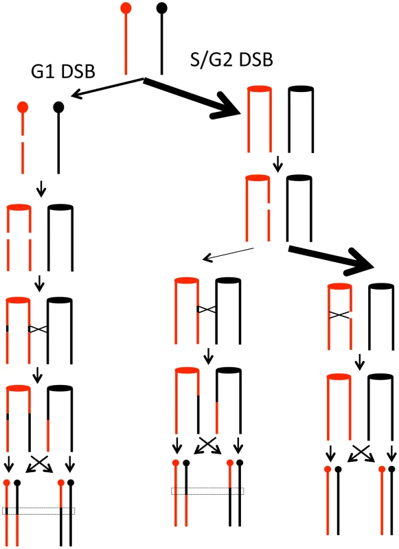
We suggest that most spontaneous DSBs occur during S- or G2 of the cell cycle as indicated by the relative widths of the arrows. The DSBs that occur in G1, however, are repaired preferentially by recombination with the homolog, whereas the DSBs that occur in G2 are repaired preferentially by recombination with the sister chromatids. The rationale for these preferences is given in the text. In summary, our analysis shows that mitotic recombination events are distributed broadly along the chromosome. One well-defined class of hotspots is a consequence of secondary structure formation between inverted repeats. We suggest that most of the recombination events leading to loss of heterozygosity are initiated by a DSB in G1 since DSBs that occur in S or G2 are usually repaired by sister-chromatid exchanges.
Materials and Methods
Strains and construction
A list of all strains used in this study is in Table S7, and a list of all primers used is in Table S8, unless otherwise specified. Mapping of crossovers was performed with the strain JSC25 (MATa/MATα::HYG leu2-3,112/LEU2 his3-11,15/HIS3 ura3-1/ura3 GAL2/gal2 ade2-1/ade2-1 trp1-1/TRP can1-100::NAT/CAN1::NAT RAD5/RAD5 IV1510386::KANMX-can1-100/IVI1510386::SUP4-o), a diploid generated by mating the haploids W303a and YJM789. Details of strain constructions are in Text S1.
Media
All media were prepared using standard recipes [57], except that the SD-Arg plates contained only 10 ìg/mL of adenine, as previously described [14].
Assay for crossovers
To detect crossovers, we struck JSC25 to generate single colonies on rich solid medium (YPD plates). The cells were grown for three days at 30°. A single colony was then resuspended in water and plated onto SD-Arg plates at a density of about 3000 cells per plate. Plates were incubated at 25° for three days and then at 4° for an additional day to let the red pigmentation develop. Red/white sectors were detected using a dissecting microscope. In colonies in which the red sector was at least one-eighth of the colony, we purified colonies from both the red and white sides of the sector. We confirmed that the red colonies were Ade− (due to loss of SUP4-o) and that the white colonies were GenS (due to loss of the KANMX gene). Single red and single white colonies from each sectored colony were subsequently analyzed.
Microarray methods and analysis
Microarray samples were prepared, hybridized, and analyzed as previously described [16]. Briefly, agarose plugs containing genomic DNA were prepared from strains derived from the sectored colonies and from the control diploid reference strain. DNA from both the experimental and reference strains was sonicated to yield DNA fragments of about 200–400 bp; these samples were labeled with Cy5-dUTP and Cy3-dUTP, respectively. DNA samples from the experimental and control strains were mixed and competitively hybridized to SNP microarrays. Following washing and scanning of the arrays, probe signals were analyzed as ratios of hybridization of the experimental and control samples for each SNP-specific oligonucleotide represented on the arrays.
Microarray design
The chromosome IV SNP microarray was designed and optimized following the principles described previously [16]. For the majority of the probes, the SNP is located in the center of the oligonucleotide, and the melting temperature for the oligonucleotide/genomic DNA hybrid is between 55 and 59°C. Minor deviations from these principles are described in Text S1. The location and sequence of each SNP on the microarray are listed in Table S9.
Assay of recombination activity of HS4 containing inverted repeats
Recombination between the URA3 and HYG markers was assayed by monitoring the HYG gene in 5-FOAR strains. Each strain was struck for single colonies on YPD plates. After incubating the cultures at 30° for 30 hours, we made patches of individual colonies on plates containing medium with 5-FOA. The plates were then incubated for two days at 30°. A single 5-FOAR colony from each patch was then transferred as a patch to YPD plates. After one day of growth at 30°, the samples were replica-plated to YPD plates containing hygromycin. About 400 5-FOAR colonies were analyzed for each strain.
Statistical analysis
We used chi-square goodness-of-fit tests to compare expected values with observed values. VassarStats (http://vassarstats.net/) and Microsoft Excel were used for this statistical test. We used Table B11 of [58] to calculate 95% confidence intervals on median estimates of conversion tract length. The Mann-Whitney test from the VassarStats website was used to compare conversion tract lengths. Data were corrected for multiple comparisons in the element enrichment analysis [59].
Supporting Information
Zdroje
1. AguileraA (2002) The connection between transcription and genomic instability. EMBO J 21 : 195–201.
2. GordeninDA, ResnickMA (1998) Yeast ARMs (DNA at-risk motifs) can reveal sources of genome instability. Mutat Res 400 : 45–58.
3. VoineaguI, NarayananV, LobachevKS, MirkinSM (2008) Replication stalling at unstable inverted repeats: interplay between DNA hairpins and fork stabilizing proteins. Proc Natl Acad Sci U S A105 : 9936–41.
4. GordeninDA, LobachevKS, DegtyarevaNP, MalkovaAL, et al. (1993) Inverted DNA repeats: a source of eukaryotic genomic instability. Mol Cell Biol 13 : 5315–22.
5. LobachevKS, GordeninDA, ResnickMA (2002) The Mre11 complex is required for repair of hairpin-capped double-strand breaks and prevention of chromosome rearrangements. Cell 108 : 183–93.
6. LemoineFJ, DegtyarevaNP, KokoskaRJ, PetesTD (2008) Reduced levels of DNA polymerase delta induce chromosome fragile site instability in yeast. Mol Cell Biol 28 : 5359–68.
7. LemoineFJ, DegtyarevaNP, LobachevK, PetesTD (2005) Chromosomal translocations in yeast induced by low levels of DNA polymerase a model for chromosome fragile sites. Cell 120 : 587–98.
8. SymingtonLS, GautierJ (2011) Double-strand break end resection and repair pathway choice. Annu Rev Genet 45 : 247–71.
9. ModrichP, LahueR (1996) Mismatch repair in replication fidelity, genetic recombination, and cancer biology. Annu Rev Biochem 65 : 101–33.
10. Orr-WeaverTL, SzostakJW (1983) Yeast recombination: the association between double-strand gap repair and crossing-over. Proc Natl Acad Sci U S A 80 : 4417–21.
11. LeePS, GreenwellPW, DominskaM, GawelM, HamiltonM, et al. (2009) A fine-structure map of spontaneous mitotic crossovers in the yeast Saccharomyces cerevisiae. PLoS Genet 5: e1000410 doi: 10.1371/journal.pgen.1000410.
12. LeePS, PetesTD (2010) Mitotic gene conversion events induced in G1-synchronized yeast cells by gamma rays are similar to spontaneous conversion events. Proc Natl Acad Sci U S A 107 : 7383–8.
13. FachinettiD, BermejoR, CocitoA, MinardiS, KatouY, et al. (2010) Replication termination at eukaryotic chromosomes is mediated by Top2 and occurs at genomic loci containing pausing elements. Mol Cell 39 : 595–605.
14. BarberaMA, PetesTD (2006) Selection and analysis of spontaneous reciprocal mitotic cross-overs in Saccharomyces cerevisiae. Proc Natl Acad Sci U S A 103 : 12819–24.
15. WeiW, McCuskerJ, HymanR, JonesT, NingY, et al. (2007) Genome sequencing and comparative analysis of Saccharomyces cerevisiae strain YJM789. Proc Natl Acad Sci U S A 104 : 12825–30.
16. St CharlesJ, Hazkani-CovoE, YinY, AndersenSL, DietrichFS, et al. (2012) High-resolution genome-wide analysis of irradiated (UV and gamma-rays) diploid yeast cells reveals a high frequency of genomic loss of heterozygosity (LOH) events. Genetics 190 : 1267–84.
17. PaquesF, HaberJE (1999) Multiple pathways of recombination induced by double-strand breaks in Saccharomyces cerevisiae. Microbiol Mol Biol Rev 63 : 349–404.
18. KimJM, VanguriS, BoekeJD, GabrielA, VoytasDF (1998) Transposable elements and genome organization: a comprehensive survey of retrotransposons revealed by the complete Saccharomyces cerevisiae genome sequence. Genome Res 8 : 464–78.
19. MorillonA, BenardL, SpringerM, LesageP (2002) Differential effects of chromatin and Gcn4 on the 50-fold range of expression among individual yeast Ty1 retrotransposons. Mol Cell Biol 22 : 2078–88.
20. AzvolinskyA, GiresiPG, LiebJD, ZakianVA (2009) Highly transcribed RNA polymerase II genes are impediments to replication fork progression in Saccharomyces cerevisiae. Mol Cell 34 : 722–34.
21. CapraJA, PaeschkeK, SinghM, ZakianVA (2010) G-quadruplex DNA sequences are evolutionarily conserved and associated with distinct genomic features in Saccharomyces cerevisiae. PLoS Comput Biol 6: e1000861 doi:10.1371/journal.pcbi.1000861.
22. PaeschkeK, CapraJA, ZakianVA (2011) DNA replication through G-quadruplex motifs is promoted by the Saccharomyces cerevisiae Pif1 DNA helicase. Cell 145 : 678–91.
23. SternC (1936) Somatic Crossing over and Segregation in Drosophila Melanogaster. Genetics 21 : 625–730.
24. HartwellLH, SmithD (1985) Altered fidelity of mitotic chromosome transmission in cell cyle mutants of S. cerevisiae. Genetics 110 : 381–395.
25. KleinHL (2001) Spontaneous chromosome loss in Saccharomyces cerevisiae is suppressed by DNA damage checkpoint functions. Genetics 159 : 1501–1509.
26. MaloneyDH, FogelS (1980) Mitotic recombination in yeast: isolation and characterization of mutants with enhanced spontaneous mitotic gene conversion rates. Genetics 94 : 825–839.
27. GolinJE, EspositoMS (1984) Coincident gene conversion during mitosis in Saccharomyces. Genetics 107 : 355–365.
28. SteeleDF, MorrisME, Jinks-RobertsonS (1991) Allelic and ectopic interactions in recombination-defective yeast strains. Genetics 127 : 53–60.
29. TangW, DominskaM, GreenwellPW, HarvanekZ, LobachevKS, et al. (2011) Friedreich's ataxia (GAA)n*(TTC)n repeats strongly stimulate mitotic crossovers in Saccharomyces cerevisae. PLoS Genet 7: e1001270 doi:10.1371/journal.pgen.1001270.
30. BlitzblauHG, BellGW, RodriguezJ, BellSP, HochwagenA (2007) Mapping of meiotic single-stranded DNA reveals double-stranded-break hotspots near centromeres and telomeres. Curr Biol 17 : 2003–12.
31. BordeV, LinW, NovikovE, PetriniJH, LichtenM, et al. (2004) Association of Mre11p with double-strand break sites during yeast meiosis. Mol Cell 13 : 389–401.
32. BuhlerC, BordeV, LichtenM (2007) Mapping meiotic single-strand DNA reveals a new landscape of DNA double-strand breaks in Saccharomyces cerevisiae. PLoS Biol 5: e324 doi:10.1371/journal.pbio.0050324.
33. GertonJL, DeRisiJ, ShroffR, LichtenM, BrownPO, et al. (2000) Global mapping of meiotic recombination hotspots and coldspots in the yeast Saccharomyces cerevisiae. Proc Natl Acad Sci U S A 97 : 11383–90.
34. PanJ, SasakiM, KniewelR, MurakamiH, BlitzblauHG, et al. (2011) A hierarchical combination of factors shapes the genome-wide topography of yeast meiotic recombination initiation. Cell 144 : 719–31.
35. PetesTD (2001) Meiotic recombination hot spots and cold spots. Nat Rev Genet 2 : 360–9.
36. LobachevKS, ShorBM, TranHT, TaylorW, KeenJD, et al. (1998) Factors affecting inverted repeat stimulation of recombination and deletion in Saccharomyces cerevisiae. Genetics 148 : 1507–24.
37. AllersT, LeachDR (1995) DNA palindromes adopt a methylation-resistant conformation that is consistent with DNA cruciform or hairpin formation in vivo. J Mol Biol 252 : 70–85.
38. KogoH, InagakiH, OhyeT, KatoT, EmanuelBS, et al. (2007) Cruciform extrusion propensity of human translocation-mediating palindromic AT-rich repeats. Nucleic Acids Res 35 : 1198–208.
39. SindenRR, ZhengGX, BrankampRG, AllenKN (1991) On the deletion of inverted repeated DNA in Escherichia coli: effects of length, thermal stability, and cruciform formation in vivo. Genetics 129 : 991–1005.
40. LobachevKS, StengerJE, KozyrevaOG, JurkaJ, GordeninDA, et al. (2000) Inverted Alu repeats unstable in yeast are excluded from the human genome. EMBO J 19 : 3822–3830.
41. WangJC (2002) Cellular roles of DNA topoisomerases: a molecular perspective. Nat Rev Mol Cell Biol 3 : 430–40.
42. BranzeiD, FoianiM (2010) Maintaining genome stability at the replication fork. Nat Rev Mol Cell Bio 11 : 208–19.
43. BermejoR, DoksaniY, CapraT, KatouYM, TanakaH, et al. (2007) Top1 - and Top2-mediated topological transitions at replication forks ensure fork progression and stability and prevent DNA damage checkpoint activation. Gene Dev 21 : 1921–36.
44. DiNardoS, VoelkelK, SternglanzR (1984) DNA topoisomerase II mutant of Saccharomyces cerevisiae: topoisomerase II is required for segregation of daughter molecules at the termination of DNA replication. Proc Natl Acad Sci U S A 81 : 2616–20.
45. ManceraE, BourgonR, BrozziA, HuberW, SteinmetzLM (2008) High-resolution mapping of meiotic crossovers and non-crossovers in yeast. Nature 454 : 479–85.
46. Fishman-LobellJ, HaberJE (1992) Removal of nonhomologous DNA ends in double-strand break recombination: the role of the yeast ultraviolet repair gene RAD1. Science 258 : 480–4.
47. IvanovEL, HaberJE (1992) RAD1 and RAD10, but not other excision repair genes, are required for double-strand break-induced recombination in Saccharomyces cerevisiae. Mol Cell Biol 15 : 2245–51.
48. InbarO, KupiecM (1999) Homology search and choice of homologous partner during mitotic recombination. Mol Cell Biol 19 : 4134–42.
49. PaquesF, HaberJE (1997) Two pathways for removal of nonhomologous DNA ends during double-strand break repair in Saccharomyces cerevisiae. Mol Cel Biol 17 : 6765–71.
50. SaparbaevM, PrakashL, PrakashS (1996) Requirement of mismatch repair genes MSH2 and MSH3 in the RAD1-RAD10 pathway of mitotic recombination in Saccharomyces cerevisiae. Genetics 142 : 727–36.
51. SugawaraN, PâquesF, ColaiacovoM, HaberJE (1997) Role of Saccharomyces cerevisiae Msh2 and Msh3 repair proteins in double-strand break-induced recombination. P Natl Acad Sci USA 94 : 9214–9.
52. LisbyM, RothsteinR, MortensenUH (2001) Rad52 forms DNA repair and recombination centers during S phase. Proc Natl Acad Sci U S A 98 : 8276–82.
53. AylonY, LiefshitzB, KupiecM (2004) The CDK regulates repair of double-strand breaks by homologous recombination during the cell cycle. EMBO J 23 : 4868–75.
54. IraG, PellicioliA, BalijjaA, WangX, FioraniS (2004) DNA end resection, homologous recombination and DNA damage checkpoint activation require CDK1. Nature 431 : 1011–7.
55. CovoS, WestmorelandJW, GordeninDA, ResnickMA (2010) Cohesin is limiting for the suppression of DNA damage-induced recombination between homologous chromosomes. PLoS Genet 7: e1001006 doi:10.1371/journal.pgen.1001006.
56. KadykLC, HartwellLH (1992) Sister chromatids are preferred over homologs as substrates for recombinational repair in Saccharomyces cerevisiae. Genetics 132 : 387–402.
57. Guthrie C, Fink GR (1991) Guide to yeast genetics and molecular biology. San Diego: Academic Press.
58. Altman DG (1991) Practical statistics for medical research. London: Chapman & Hall.
59. HochbergY, BenjaminiY (1990) More powerful procedures for multiple significance testing. Stat Med 9 : 811–8.
Štítky
Genetika Reprodukční medicína
Článek The G4 GenomeČlánek Mondo/ChREBP-Mlx-Regulated Transcriptional Network Is Essential for Dietary Sugar Tolerance inČlánek RpoS Plays a Central Role in the SOS Induction by Sub-Lethal Aminoglycoside Concentrations inČlánek Tissue Homeostasis in the Wing Disc of : Immediate Response to Massive Damage during DevelopmentČlánek Disruption of TTDA Results in Complete Nucleotide Excision Repair Deficiency and Embryonic LethalityČlánek DJ-1 Decreases Neural Sensitivity to Stress by Negatively Regulating Daxx-Like Protein through dFOXO
Článek vyšel v časopisePLOS Genetics
Nejčtenější tento týden
2013 Číslo 4- Růst a vývoj dětí narozených pomocí IVF
- Délka menstruačního cyklu jako marker ženské plodnosti
- Vztah užívání alkoholu a mužské fertility
- Intrauterinní inseminace a její úspěšnost
- Šanci na úspěšný průběh těhotenství snižují nevhodné hladiny progesteronu vznikající při umělém oplodnění
-
Všechny články tohoto čísla
- Epigenetic Upregulation of lncRNAs at 13q14.3 in Leukemia Is Linked to the Downregulation of a Gene Cluster That Targets NF-kB
- A Big Catch for Germ Cell Tumour Research
- The Quest for the Identification of Genetic Variants in Unexplained Cardiac Arrest and Idiopathic Ventricular Fibrillation
- A Nonsynonymous Polymorphism in as a Risk Factor for Human Unexplained Cardiac Arrest with Documented Ventricular Fibrillation
- The Hourglass and the Early Conservation Models—Co-Existing Patterns of Developmental Constraints in Vertebrates
- Smaug/SAMD4A Restores Translational Activity of CUGBP1 and Suppresses CUG-Induced Myopathy
- Balancing Selection on a Regulatory Region Exhibiting Ancient Variation That Predates Human–Neandertal Divergence
- The G4 Genome
- Extensive Natural Epigenetic Variation at a Originated Gene
- Mouse Oocyte Methylomes at Base Resolution Reveal Genome-Wide Accumulation of Non-CpG Methylation and Role of DNA Methyltransferases
- The Environment Affects Epistatic Interactions to Alter the Topology of an Empirical Fitness Landscape
- TIP48/Reptin and H2A.Z Requirement for Initiating Chromatin Remodeling in Estrogen-Activated Transcription
- Aconitase Causes Iron Toxicity in Mutants
- Tbx2 Terminates Shh/Fgf Signaling in the Developing Mouse Limb Bud by Direct Repression of
- Mondo/ChREBP-Mlx-Regulated Transcriptional Network Is Essential for Dietary Sugar Tolerance in
- Sex-Differential Selection and the Evolution of X Inactivation Strategies
- Identification of a Tissue-Selective Heat Shock Response Regulatory Network
- Phosphorylation-Coupled Proteolysis of the Transcription Factor MYC2 Is Important for Jasmonate-Signaled Plant Immunity
- RpoS Plays a Central Role in the SOS Induction by Sub-Lethal Aminoglycoside Concentrations in
- Six Homeoproteins Directly Activate Expression in the Gene Regulatory Networks That Control Early Myogenesis
- Rtt109 Prevents Hyper-Amplification of Ribosomal RNA Genes through Histone Modification in Budding Yeast
- ATP-Dependent Chromatin Remodeling by Cockayne Syndrome Protein B and NAP1-Like Histone Chaperones Is Required for Efficient Transcription-Coupled DNA Repair
- Iron-Responsive miR-485-3p Regulates Cellular Iron Homeostasis by Targeting Ferroportin
- Mutations in Predispose Zebrafish and Humans to Seminomas
- Cytotoxic Chromosomal Targeting by CRISPR/Cas Systems Can Reshape Bacterial Genomes and Expel or Remodel Pathogenicity Islands
- Tissue Homeostasis in the Wing Disc of : Immediate Response to Massive Damage during Development
- All SNPs Are Not Created Equal: Genome-Wide Association Studies Reveal a Consistent Pattern of Enrichment among Functionally Annotated SNPs
- Functional 358Ala Allele Impairs Classical IL-6 Receptor Signaling and Influences Risk of Diverse Inflammatory Diseases
- The Tissue-Specific RNA Binding Protein T-STAR Controls Regional Splicing Patterns of Pre-mRNAs in the Brain
- Neutral Genomic Microevolution of a Recently Emerged Pathogen, Serovar Agona
- Genetic Requirements for Signaling from an Autoactive Plant NB-LRR Intracellular Innate Immune Receptor
- SNF5 Is an Essential Executor of Epigenetic Regulation during Differentiation
- Dialects of the DNA Uptake Sequence in
- Reference-Free Population Genomics from Next-Generation Transcriptome Data and the Vertebrate–Invertebrate Gap
- Senataxin Plays an Essential Role with DNA Damage Response Proteins in Meiotic Recombination and Gene Silencing
- High-Resolution Mapping of Spontaneous Mitotic Recombination Hotspots on the 1.1 Mb Arm of Yeast Chromosome IV
- Rod Monochromacy and the Coevolution of Cetacean Retinal Opsins
- Evolution after Introduction of a Novel Metabolic Pathway Consistently Leads to Restoration of Wild-Type Physiology
- Disruption of TTDA Results in Complete Nucleotide Excision Repair Deficiency and Embryonic Lethality
- Insulators Target Active Genes to Transcription Factories and Polycomb-Repressed Genes to Polycomb Bodies
- Signatures of Diversifying Selection in European Pig Breeds
- The Chromosomal Passenger Protein Birc5b Organizes Microfilaments and Germ Plasm in the Zebrafish Embryo
- The Histone Demethylase Jarid1b Ensures Faithful Mouse Development by Protecting Developmental Genes from Aberrant H3K4me3
- Regulates Synaptic Development and Endocytosis by Suppressing Filamentous Actin Assembly
- Sensory Neuron-Derived Eph Regulates Glomerular Arbors and Modulatory Function of a Central Serotonergic Neuron
- Analysis of Rare, Exonic Variation amongst Subjects with Autism Spectrum Disorders and Population Controls
- Scavenger Receptors Mediate the Role of SUMO and Ftz-f1 in Steroidogenesis
- DNA Double-Strand Breaks Coupled with PARP1 and HNRNPA2B1 Binding Sites Flank Coordinately Expressed Domains in Human Chromosomes
- High-Resolution Mapping of H1 Linker Histone Variants in Embryonic Stem Cells
- Comparative Genomics of and the Bacterial Species Concept
- Genetic and Biochemical Assays Reveal a Key Role for Replication Restart Proteins in Group II Intron Retrohoming
- Genome-Wide Association Studies Identify Two Novel Mutations Responsible for an Atypical Hyperprolificacy Phenotype in Sheep
- The Genetic Correlation between Height and IQ: Shared Genes or Assortative Mating?
- Comprehensive Assignment of Roles for Typhimurium Genes in Intestinal Colonization of Food-Producing Animals
- An Essential Role for Zygotic Expression in the Pre-Cellular Drosophila Embryo
- The Genome Organization of Reflects Its Lifestyle
- Coordinated Cell Type–Specific Epigenetic Remodeling in Prefrontal Cortex Begins before Birth and Continues into Early Adulthood
- Improved Detection of Common Variants Associated with Schizophrenia and Bipolar Disorder Using Pleiotropy-Informed Conditional False Discovery Rate
- Site-Specific Phosphorylation of the DNA Damage Response Mediator Rad9 by Cyclin-Dependent Kinases Regulates Activation of Checkpoint Kinase 1
- Npc1 Acting in Neurons and Glia Is Essential for the Formation and Maintenance of CNS Myelin
- Identification of , a Retrotransposon-Derived Imprinted Gene, as a Novel Driver of Hepatocarcinogenesis
- Aag DNA Glycosylase Promotes Alkylation-Induced Tissue Damage Mediated by Parp1
- DJ-1 Decreases Neural Sensitivity to Stress by Negatively Regulating Daxx-Like Protein through dFOXO
- Asynchronous Replication, Mono-Allelic Expression, and Long Range -Effects of
- Differential Association of the Conserved SUMO Ligase Zip3 with Meiotic Double-Strand Break Sites Reveals Regional Variations in the Outcome of Meiotic Recombination
- Focusing In on the Complex Genetics of Myopia
- Continent-Wide Decoupling of Y-Chromosomal Genetic Variation from Language and Geography in Native South Americans
- Breakpoint Analysis of Transcriptional and Genomic Profiles Uncovers Novel Gene Fusions Spanning Multiple Human Cancer Types
- Intrinsic Epigenetic Regulation of the D4Z4 Macrosatellite Repeat in a Transgenic Mouse Model for FSHD
- Bisphenol A Exposure Disrupts Genomic Imprinting in the Mouse
- Genetic and Genomic Architecture of the Evolution of Resistance to Antifungal Drug Combinations
- Transposable Elements Are Major Contributors to the Origin, Diversification, and Regulation of Vertebrate Long Noncoding RNAs
- Functional Dissection of the Condensin Subunit Cap-G Reveals Its Exclusive Association with Condensin I
- PLOS Genetics
- Archiv čísel
- Aktuální číslo
- Informace o časopisu
Nejčtenější v tomto čísle- The G4 Genome
- Neutral Genomic Microevolution of a Recently Emerged Pathogen, Serovar Agona
- The Histone Demethylase Jarid1b Ensures Faithful Mouse Development by Protecting Developmental Genes from Aberrant H3K4me3
- The Tissue-Specific RNA Binding Protein T-STAR Controls Regional Splicing Patterns of Pre-mRNAs in the Brain
Kurzy
Zvyšte si kvalifikaci online z pohodlí domova
Autoři: prof. MUDr. Vladimír Palička, CSc., Dr.h.c., doc. MUDr. Václav Vyskočil, Ph.D., MUDr. Petr Kasalický, CSc., MUDr. Jan Rosa, Ing. Pavel Havlík, Ing. Jan Adam, Hana Hejnová, DiS., Jana Křenková
Autoři: MUDr. Irena Krčmová, CSc.
Autoři: MDDr. Eleonóra Ivančová, PhD., MHA
Autoři: prof. MUDr. Eva Kubala Havrdová, DrSc.
Všechny kurzyPřihlášení#ADS_BOTTOM_SCRIPTS#Zapomenuté hesloZadejte e-mailovou adresu, se kterou jste vytvářel(a) účet, budou Vám na ni zaslány informace k nastavení nového hesla.
- Vzdělávání


