-
Články
- Vzdělávání
- Časopisy
Top články
Nové číslo
- Témata
- Kongresy
- Videa
- Podcasty
Nové podcasty
Reklama- Kariéra
Doporučené pozice
Reklama- Praxe
Collaborative Action of Brca1 and CtIP in Elimination of Covalent Modifications from Double-Strand Breaks to Facilitate Subsequent Break Repair
Topoisomerase inhibitors such as camptothecin and etoposide are used as anti-cancer drugs and induce double-strand breaks (DSBs) in genomic DNA in cycling cells. These DSBs are often covalently bound with polypeptides at the 3′ and 5′ ends. Such modifications must be eliminated before DSB repair can take place, but it remains elusive which nucleases are involved in this process. Previous studies show that CtIP plays a critical role in the generation of 3′ single-strand overhang at “clean” DSBs, thus initiating homologous recombination (HR)–dependent DSB repair. To analyze the function of CtIP in detail, we conditionally disrupted the CtIP gene in the chicken DT40 cell line. We found that CtIP is essential for cellular proliferation as well as for the formation of 3′ single-strand overhang, similar to what is observed in DT40 cells deficient in the Mre11/Rad50/Nbs1 complex. We also generated DT40 cell line harboring CtIP with an alanine substitution at residue Ser332, which is required for interaction with BRCA1. Although the resulting CtIPS332A/−/− cells exhibited accumulation of RPA and Rad51 upon DNA damage, and were proficient in HR, they showed a marked hypersensitivity to camptothecin and etoposide in comparison with CtIP+/−/− cells. Finally, CtIPS332A/−/−BRCA1−/− and CtIP+/−/−BRCA1−/− showed similar sensitivities to these reagents. Taken together, our data indicate that, in addition to its function in HR, CtIP plays a role in cellular tolerance to topoisomerase inhibitors. We propose that the BRCA1-CtIP complex plays a role in the nuclease-mediated elimination of oligonucleotides covalently bound to polypeptides from DSBs, thereby facilitating subsequent DSB repair.
Published in the journal: . PLoS Genet 6(1): e32767. doi:10.1371/journal.pgen.1000828
Category: Research Article
doi: https://doi.org/10.1371/journal.pgen.1000828Summary
Topoisomerase inhibitors such as camptothecin and etoposide are used as anti-cancer drugs and induce double-strand breaks (DSBs) in genomic DNA in cycling cells. These DSBs are often covalently bound with polypeptides at the 3′ and 5′ ends. Such modifications must be eliminated before DSB repair can take place, but it remains elusive which nucleases are involved in this process. Previous studies show that CtIP plays a critical role in the generation of 3′ single-strand overhang at “clean” DSBs, thus initiating homologous recombination (HR)–dependent DSB repair. To analyze the function of CtIP in detail, we conditionally disrupted the CtIP gene in the chicken DT40 cell line. We found that CtIP is essential for cellular proliferation as well as for the formation of 3′ single-strand overhang, similar to what is observed in DT40 cells deficient in the Mre11/Rad50/Nbs1 complex. We also generated DT40 cell line harboring CtIP with an alanine substitution at residue Ser332, which is required for interaction with BRCA1. Although the resulting CtIPS332A/−/− cells exhibited accumulation of RPA and Rad51 upon DNA damage, and were proficient in HR, they showed a marked hypersensitivity to camptothecin and etoposide in comparison with CtIP+/−/− cells. Finally, CtIPS332A/−/−BRCA1−/− and CtIP+/−/−BRCA1−/− showed similar sensitivities to these reagents. Taken together, our data indicate that, in addition to its function in HR, CtIP plays a role in cellular tolerance to topoisomerase inhibitors. We propose that the BRCA1-CtIP complex plays a role in the nuclease-mediated elimination of oligonucleotides covalently bound to polypeptides from DSBs, thereby facilitating subsequent DSB repair.
Introduction
CtIP was isolated as a binding partner of CtBP (C-terminal binding protein), and has subsequently been shown to interact with a number of molecules, including BRCA1 (Breast Cancer Susceptibility Gene 1) [1]. CtIP is a functional homolog of yeast Sae2 (Sporulation in the Absence of Spo Eleven), and acts at the initial step of homologous recombination (HR)-dependent double-strand break (DSB) repair [2],[3]. HR is initiated by forming 3′ single-strand (ss) overhangs at DSBs. In this resection step, Sae2/CtIP works together with a complex composed of Mre11/Rad50/Xrs2 in budding yeast, or with Mre11/Rad50/Nbs1 in mammals [4]–[7]. The Rad51 recombinase protein polymerizes on the ss DNA overhang, and the resulting ssDNA-Rad51 complex undergoes homology search. Resection activity is upregulated by phosphorylation of a conserved residue in Sae2 by the cyclin-dependent kinase (CDK) [8]. This phosphorylation site is conserved in human CtIP (Thr847), and is also phosphorylated by CDK [7].
BRCA1 was originally identified as a tumor suppressor gene associated with familial breast and ovarian cancer [9]. BRCA1 contains an N-terminal RING-finger domain, and is associated with structurally related BARD1 to form an E3-ubiquitin ligase. BRCA1/BARD1 forms three distinct complexes with Abraxas, Bach1 and CtIP, and plays a role in DNA repair [10]. BRCA1 binds to CtIP in a manner that is dependent on the phosphorylation of CtIP at Ser327 [11],[12]. Following DNA damage, the ubiquitylation of CtIP by BRCA1 causes the migration of CtIP towards a chromatin fraction [12]. However, the biological significance of the complex formed between BRCA1 and CtIP has not yet been clarified.
Topoisomerases 1 and 2 (Topo1 and Topo2) have been drawing increasing attention as important targets for cancer therapy, since the inhibition of these enzymes causes DSBs during DNA replication [13]. Topo1 and Topo2 induce single-strand breaks (SSBs) and DSBs, respectively. Covalent bonds are transiently formed between Topo1 and the 3′ end of the SSB and between Topo2 and the 5′ end of the DSB [14]. The anti-cancer agent camptothecin (CPT) inhibits Topo1 by stabilizing the Topo1-cleavage complex, which interferes with replication, and thereby induces DSBs in one of the sister chromatids [15]. Topo2 inhibitors such as etoposide (VP16) and ICRF-193 also kill cycling cells and are used in cancer therapy. VP16 stabilizes the Topo2-cleavage complex, while ICRF-193 stabilizes the closed clamp which forms after the strand passage [16],[17]. Topo1-mediated DNA damage caused by CPT is repaired primarily by homologous recombination (HR), while Topo2-mediated DNA damage caused by VP16 or ICRF-193 is mainly repaired by nonhomologous end joining (NHEJ) [18],[19]. It should be noted that the repair of CPT - and VP16-induced DSBs requires an additional step: the elimination of covalently bound polypeptides from the DNA ends. Hartsuiker et al. demonstrated that Topo2 is removed from DNA by the collaborative action of the MRX complex and ctp1 (the ortholog of CtIP) in fission yeast [20]. It remains to be seen whether vertebrate CtIP shares the same function as in yeast, which does not have BRCA1 ortholog.
To understand the role of the BRCA1-CtIP interaction, we substituted the Ser332 residue (equivalent to human Ser327) of CtIP with alanine in the chicken DT40 B lymphocyte line [21],[22]. In addition, to analyze the function of CtIP, we conditionally depleted CtIP in DT40 cells. We here show that the depletion of CtIP is lethal to cells as is the inactivation of Mre11, Rad50, and Nbs1 [23],[24], indicating the critical role played by CtIP in HR. Remarkably, although the CtIP S332A mutation had no significant impact on HR, it made cells hypersensitive to CPT and VP16 but not to ICRF193. These observations unmasked an unexpected function of the BRCA1-CtIP interaction: cellular tolerance to the DSBs that are covalently associated with the polypeptides. Our data therefore support two distinct functions of CtIP: the resection of DSBs in HR and the elimination of polypeptides from the cleavage complex.
Results
CtIP is required for the assembly of Rad51 at DNA damage sites
In order to determine the function of CtIP, we conditionally disrupted the CtIP gene in chicken DT40 cells, using a chicken CtIP transgene under the control of a tetracycline-repressible promoter (tetCtIP transgene, Figure S1A). We designed CtIP gene-disruption constructs, so that the amino acid sequences from 96 to 335 would be replaced by selection-marker genes. Since the gene is encoded on chromosome 2, which is in trisomy in DT40, we disrupted three CtIP alleles (Figure S1B and S1C). The resulting CtIP−/−/−tetCtIP cells tended to grow more slowly than did wild-type cells, presumably due to overexpression of the tetCtIP transgene (Figure 1A and 1B). To deplete the CtIP in the CtIP−/−/−tetCtIP cells, we added doxycycline (modified tetracycline) to the culture medium. One day after the addition of doxycycline, the amount of CtIP was reduced to around 20% of wild-type cells (Figure 1B), and the cells started dying as evidenced by an increase in the sub-G1 fraction (Figure 1C). This lethality can be attributed to abolished HR, because the cells showed a significant increase in the number of spontaneous chromosomal breaks (Table 1), as do Mre11 - and Rad51-depleted cells [23],[25]. By day 3, the vast majority of the CtIP−/−/−tetCtIP cells had stopped growing and died (Figure 1A and 1C). We therefore conclude that CtIP is essential for maintenance of chromosomal DNA and cellular proliferation.
Fig. 1. CtIP is essential for cell survival. 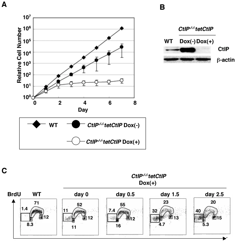
(A) Defective proliferation of CtIP−/−/−tetCtIP cells following addition of doxycycline (2 µg/ml) to deplete CtIP, from time zero. Data shown are the average results from three separate clones. Points indicate mean (n = 3); bars indicate S.D. (B) Western blot analysis of chicken CtIP expression. Whole cell lysates were prepared from CtIP−/−/−tetCtIP cells 24 h after addition of doxycycline. β-actin was used as a loading control. (C) Cell cycle distribution of CtIP−/−/−tetCtIP cells at the indicated time after addition of doxycycline. Cells were pulse-labeled with BrdU for 10 min and subsequently stained with FITC-conjugated anti-BrdU antibody (Y axis, log scale) and propidium iodide (PI) (X axis, linear scale). Each square on the left-hand side represents apoptic cells (sub-G1 fraction). The small rectangle at the bottom of the tubular arch, the tubular arch, and the small square to the right represent cells in the G1, S, and G2/M phases, respectively. Tab. 1. Chromosomal aberrations in CtIP−/−/−tetCtIP mutants. 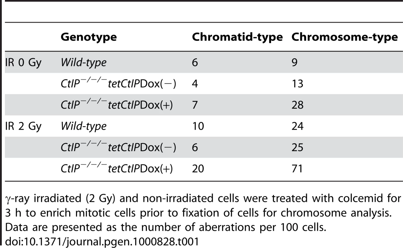
γ-ray irradiated (2 Gy) and non-irradiated cells were treated with colcemid for 3 h to enrich mitotic cells prior to fixation of cells for chromosome analysis. Data are presented as the number of aberrations per 100 cells. To assess the HR capability of CtIP−/−/−tetCtIP cells, we monitored the recruitment of Rad51 and RPA to DNA damage sites one day after addition of doxycycline. Clear Rad51 foci appeared in wild-type cells one hour after ionizing radiation (IR), whereas Rad51 foci were hardly detectable in the CtIP-depleted cells (Figure 2A). Likewise, the depletion of CtIP abolished the accumulation of RPA on DNA lesions induced by microlaser treatment (Figure 2B). This is consistent with a phenotype shown in the previous report [3]. Thus, CtIP plays an essential role in the resection of DSBs during HR in DT40 cells as well as in mammalian cells.
Fig. 2. Impaired γ-ray–induced Rad51 focus formation in CtIP-depleted cells. 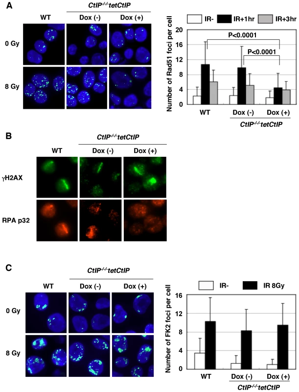
(A) CtIP−/−/−tetCtIP cells, with or without 24 h doxycycline treatment, were exposed to 8 Gy ionizing radiation (IR). One hour after irradiation, cells were stained with anti-Rad51 antibody. The graph shows the quantification of Rad51 foci per cell. More than 100 cells were analyzed. (B) Accumulation of RPA protein in the cells exposed to 405 nm pulse laser. Cells were fixed with 4% PFA 1 h post laser stimulation and stained with anti-γH2AX and anti-RPAp32 antibodies. (C) Conjugated-ubiquitin (FK2) focus formation in wild-type (WT) or CtIP−/−/−tetCtIP DT40 cells (with or without doxycycline) at 1 h post IR (8Gy). Quantification of FK2 foci per cell is shown in the graph. More than 100 cells were analyzed. We next investigated whether or not CtIP facilitates the activation of BRCA1 at DSBs. To this end, we measured the formation of conjugated-ubiquitin foci at DSBs, since Brca1 promotes extensive ubiquitylation at IR-induced DSBs [26]. Previous studies showed that BRCA1−/− DT40 cells exhibit a prominent defect in the formation of conjugated-ubiquitin foci [27]. In contrast, CtIP depletion did not reduce the ubiquitylation of DNA damage sites (Figure 2C), suggesting that CtIP is not required for the activation of Brca1.
Proficient HR in CtIPS332A/−/− DT40 clones
To functionally analyze the interaction of CtIP with BRCA1, we generated CtIPS332A/−/− cells, in which the critical amino acid in the binding interface has been mutated (Figure S2A, S2B, S2C). The CtIPS332A/−/− DT40 clones were capable of proliferating at a rate similar to the CtIP+/−/− cells without a prominent change in the cell-cycle profile (Figure 3A and Figure S2D). Western blot analysis showed that the S332A CtIP proteins were expressed at the similar level to the wild-type protein, indicating that amino acid substitutions do not affect the stability of the CtIP protein (Figure S2E). As expected, given the results of a previous study [12], the S332A mutation of CtIP indeed inhibited its interaction with BRCA1 (Figure S2F).
Fig. 3. Proficient homologous recombination in CtIPS332A/−/− clones. 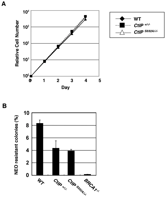
(A) Growth kinetics of the indicated CtIP mutant clones. (B) I-Sce1–induced DSBs stimulate homologous recombination (HR) in an artificial substrate, SCneo. The recombination frequency was calculated by dividing the number of neomycin-resistant colonies by the number of the total colonies. Error bars indicate S.D. To evaluate the capability of HR in CtIPS332A/−/− cells, we integrated an artificial substrate, SCneo, in the Ovalbumin locus [28], and measured the efficiency of I-SceI-induced gene conversion. The CtIPS332A/−/− clones showed no significant decrease in the appearance of neomycin-resistant colonies compared to CtIP+/−/− cells (Figure 3B). The proficient HR in CtIPS332A/−/− DT40 clones is in marked contrast to the severe phenotype of the Nbs1p70 hypomorphic mutant, which exhibited a 10-fold reduction of the gene-targeting frequency and a 103-fold decrease in the efficiency of HR in the SCneo substrate [29]. Next, we measured the frequency of gene targeting at the CENP-H and Ovalbumin loci. In contrast to I-SceI-induced gene conversion, the gene-targeting frequency of the CtIPS332A/−/− clones decreased moderately in comparison with CtIP+/−/− cells (Table 2). We speculate that this is because unknown recombination intermediates that require processing by CtIP/BRCA1 may arise during gene targeting event (see Discussion).
Tab. 2. Targeted integration frequencies of CtIPS332A/−/− clones. 
Gene-targeting efficiencies were determined by flow cytometry for CENP-H locus and by Southern blot analysis for Ovalbumin locus. N.D. indicates not determined. Fluorescent immunostaining revealed that the kinetics of Rad51 focus formation after γ-irradiation was indistinguishable between CtIPS332A/−/− cells and the CtIP+/−/− control cells, while BRCA1−/− cells showed the significant reduction in the Rad51 focus formation at 1–6 h after irradiation (Figure 4A). Furthermore, the CtIPS332A/−/− mutants displayed laser-induced RPA accumulation as did the CtIP+/−/−cells (Figure 4B). Laser-generated RPA accumulation following BrdU incorporation largely arises from the resection rather than other routes of single strand formation such as the damage caused by laser itself or replication-associated single strand formation, because RPA accumulation is abolished specifically in Ubc13 deficient cells [27]. This suggests that CtIPS332A/−/− cells are proficient in resection at DSB sites. Taken together, we conclude that the S332A mutation of CtIP does not significantly compromise HR.
Fig. 4. Accumulation of Rad51 and RPA at DNA damage sites is indistinguishable between CtIP+/−/− and CtIPS332A/−/− clones. 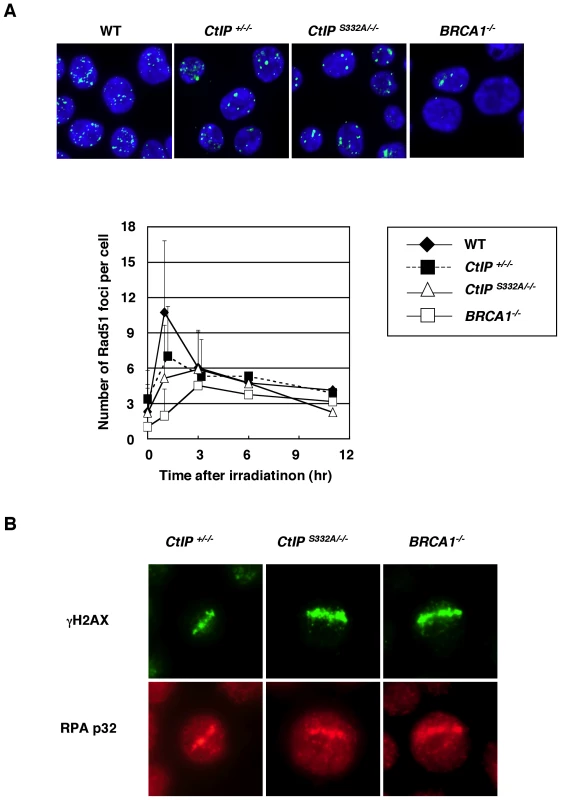
(A) Rad51 focus formation in CtIP mutant cells. The cells of indicated genotype were irradiated with 8Gy of γ-ray, and were immuno-stained with anti-Rad51 antibodies 1 h after irradiation. The lower graph shows the quantification of Rad51 foci per cell. More than 100 cells were analyzed. Error bars indicate S.D. (B) The subnuclear accumulation of RPA in the cells exposed to 405 nm pulse laser. The cells were fixed with 4% PFA 1 h after exposure, and were stained with anti-γH2AX and anti-RPAp32 antibodies. CtIPS332A/−/− cells display a marked hypersensitivity to both CPT and VP16, which stabilize the Topo-cleavage complexes
To determine the role of CtIP in the cellular response to DNA damage, we measured the sensitivity of the CtIP mutant cells to various genotoxic agents using a colony survival assay. CtIP+/−/− cells exhibited the slightly elevated sensitivity toward CPT and VP16 (Figure 5A and 5B), though they expressed the similar level of CtIP protein to the wild-type cells (Figure S2E). It is possible that the difference in the amount of CtIP protein between CtIP+/−/− and wild-type cells is too subtle to detect, and that even the suboptimal level of CtIP protein renders the cells sensitive to genotoxic stimuli. A compensatory post-translational regulation may be present because CtIP+/−/− cells exhibited about 80% reductions in CtIP mRNA level compared to the wild-type level (Figure S2G). In contrast to CtIP+/−/− cells, CtIPS332A/−/− mutants showed a significantly increased sensitivity to VP16 and MMS (Figure 5B and 5D), but not to γ-rays (data not shown). Furthermore, the sensitivity to CPT was dramatically elevated in the CtIPS332A/−/− mutants, in comparison with the CtIP+/−/− cells (Figure 5A).
Fig. 5. Colony survival assay of CtIP mutant cells to various genotoxic agents. 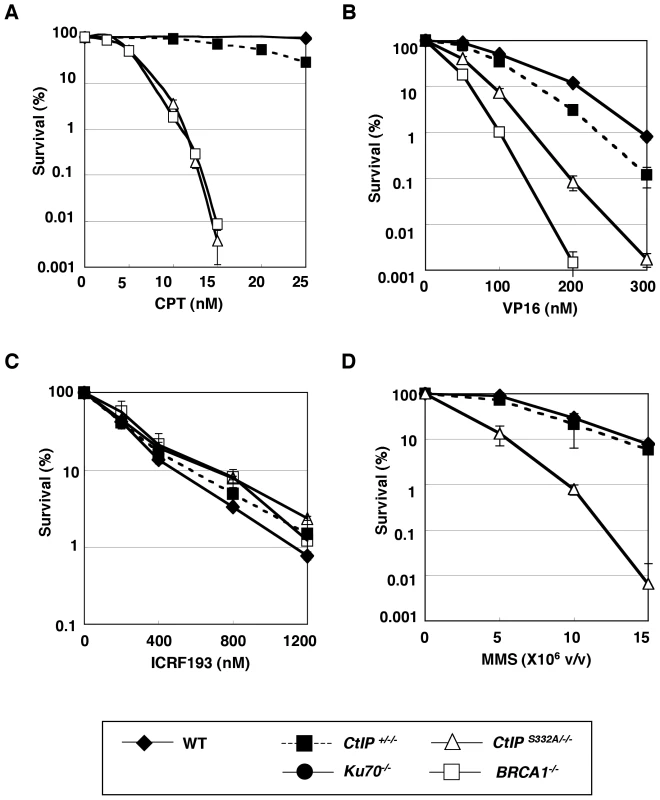
Colony survival assay of asynchronous populations of cells exposed to (A) camptothecin (CPT), (B) etoposide (VP16), (C) ICRF-193, and (D) methyl methanesulfonate (MMS). The dose is displayed on the X axis on a linear scale, while the percent fraction of surviving colonies is displayed on the Y axis on a logarithmic scale. To count the surviving colonies, cells were grown for 8–10 days in methylcellulose medium containing various concentrations of DNA–damaging agents. At least three independent experiments were performed with triplicate samples, and the representative results are shown. The error bars indicate S.D. The contribution of CtIP to the cellular tolerance to VP16 indicated that CtIP might play a role in NHEJ [18]. To test this hypothesis, we evaluated NHEJ by measuring the sensitivity of CtIP mutant cells to ICRF-193, because ICRF193-induced DNA lesions are repaired exclusively by NHEJ, whereas a fraction of the VP16-induced DSBs are repaired by HR [18]. The CtIPS332A/−/− clones exhibited no increased ICRF193 sensitivity (Figure 5C). NHEJ can also be evaluated by measuring the IR sensitivity of the cell population at the G1 phase, where NHEJ plays a dominant role in DSB repair [30]. The CtIP hypomorphic mutants synchronized at the G1 phase did not show significant IR hypersensitivity (Figure S3). These observations indicate that NHEJ is not impaired in CtIPS332A/−/− clones. In summary, in comparison with CtIP+/−/− cells, CtIPS332A/−/− clones exhibited a significantly higher sensitivity to CPT and VP16, although these clones exhibited no decrease in the efficiency of HR or NHEJ. We conclude that CtIP can therefore contribute to cellular tolerance to CPT and VP16, independently of HR or NHEJ, most likely by eliminating covalently bound polypeptides from the DSBs.
Epistatic relationship of CtIP to BRCA1 in cellular tolerance to CPT and VP16
CtIP physically interacts with BRCA1 in a manner dependent on phosphorylation of Ser332 [12]. In order to assess the functional relationship between Ser332 phosphorylation of CtIP and BRCA1, we disrupted the BRCA1 gene in the CtIPS332A/−/− and CtIP+/−/− clones (Figure S4A and S4B), as was done previously [31]. Both the CtIPS332A/−/−BRCA1−/− and the CtIP+/−/−BRCA1−/− clones proliferated with similar rates at significantly reduced growth rates, in comparison with BRCA1−/−cells (doubling time ± SD: 8.3±0.2 h for wild-type, 9.3±0.3 h for BRCA1−/−, 11±0.3 h for CtIP+/−/−BRCA1−/−, 12.6±0.9 h for CtIPS332A/−/−BRCA1−/−). The viability of CtIPS332A/−/−BRCA1−/− cells is in marked contrast with the lethality of CtIP-null cells, supporting the idea that the CtIP-BRCA1 interaction works independently from the function of CtIP in resection.
We next examined the sensitivity of double mutant cells to CPT and VP16. To this end, we measured the number of live cells after 48-hour continuous exposure to the DNA-damaging agents [32], during which the double mutant cells are able to divide four to five times. We did not use a conventional colony formation assay for this purpose, because CtIP+/−/−BRCA1−/− and CtIPS332A/−/−BRCA1−/− clones grew very badly from a single cell in semi-solid methylcellulose medium. The number of viable cells cultured in the presence of CPT was significantly decreased for CtIPS332A/−/− and BRCA1−/− cells compared to the wild-type cells, whereas CtIP+/−/− cells grew to the similar extent to the wild-type cells in the presence of CPT (Figure 6A). The sensitivity of CtIP+/−/−BRCA1−/− cells to CPT was greater than that of BRCA1−/− clones. This observation is in agreement with the idea that BRCA1 and CtIP can independently contribute to HR, where CtIP promotes the resection of DSBs, while BRCA1 subsequently loads Rad51 at resected ssDNA overhang. Importantly, although the CtIPS332A mutation significantly increased cellular sensitivity to CPT in the presence of BRCA1, the CtIPS332A/−/−BRCA1−/− and CtIP+/−/−BRCA1−/− clones exhibited a very similar sensitivity to CPT (Figure 6A). Likewise, the CtIPS332A/−/−BRCA1−/− and CtIP+/−/−BRCA1−/− clones exhibited indistinguishable cellular sensitivities to VP16 (Figure 6B). These observations suggest that CtIP and BRCA1 can act in collaboration to repair DSBs that are chemically modified by topoisomerases.
Fig. 6. Epistasis analysis of BRCA1 and CtIP in cellular tolerance to topoisomerase inhibitors. 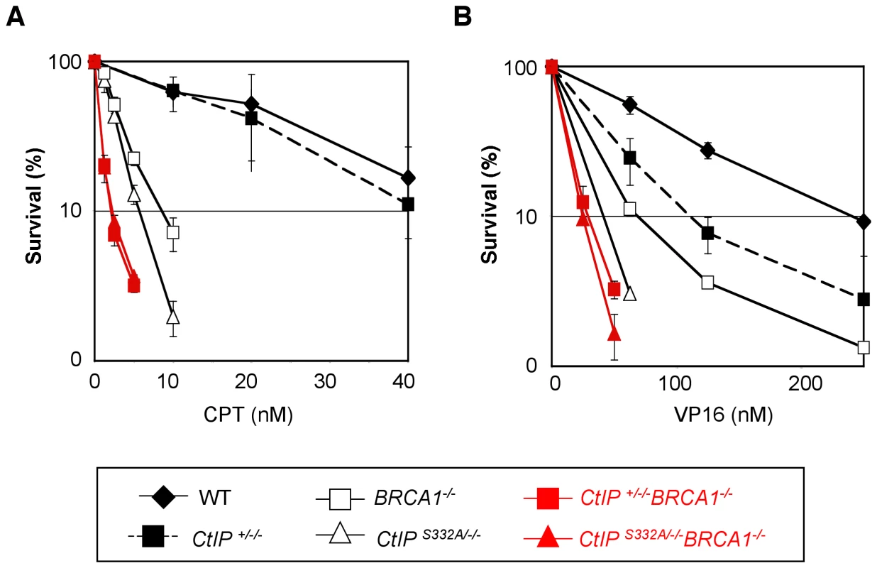
Cellular survival of each genotype was assessed by measuring the amount of ATP in cell lysates after 48-h exposure to CPT (A) or VP16 (B). The survival curves for the CtIPS332A/−/−BRCA1−/− cells and their control CtIP+/−/−BRCA1−/− cells are depicted in red. At least five independent experiments were performed with triplicate samples, and the representative results are shown. The error bars indicate S.D. Discussion
We here show that conditional depletion of CtIP protein led to cellular lethality with increased frequency of chromosomal aberrations in DT40 cells. CtIP depletion abolished the accumulation of RPA and Rad51 at DNA damaged sites, suggesting that it is required for the resection of DSBs during HR, and that this function is essential for the proliferation of cells. These results are in agreement with previous reports [3]. In contrast, the DT40 cells harboring S332A mutation in CtIP showed the accumulation of RPA and Rad51 upon DNA damage, and were able to proliferate with normal kinetics. Remarkably, compared to the CtIP+/−/− cells, the CtIPS332A/−/− clones exhibited significantly increased sensitivity to CPT and VP16, both of which stabilize the Topo-DNA cleavage complex. These observations support the proposition that, in additon to the resection of DSBs, CtIP has the second function, most likely the removal of covalently-bound polypeptides from DSBs. Hence, CtIPS332A/−/− clones are the novel separation-of-function mutants where CtIP-dependent resection is proficient, whereas the second function required for the tolerance to topoisomerase inhibitors is deficient.
In this study, we demonstrated that the inactivation of CtIP in DT40 cells results in cellular death. We speculate that the defective DSB repair during S phase is the primary cause of cellular death rather than the misregulation of RB/E2F pathway [33],[34]. It has been reported that CtIP promotes G1/S progression by releasing RB-imposed repression and by upregulating the genes required for S phase entry such as cyclin D1. MEF from CtIP-deficient mice and NIH3T3 cells transfected with CtIP siRNA arrest at G1 phase of cell cycle. In contrast, DT40 cells that are depleted of CtIP showed a marked reduction in S phase and an increase in sub-G1 population with the spontaneous chromosomal aberrations. We speculate that DT40 cells have a lower threshold to enter the S phase in the presence of DNA damage compared to the other types of cells owing to their character that they lack p53 expression [35] and overexpress c-myc [36].
The phenotype of our CtIP-depleted DT40 cells was remarkably different from that of the CtIP-deficient DT40 cells generated by Hiom's group [37]. Surprisingly, their CtIP-depleted DT40 cells were capable of proliferating. However, we believe that CtIP is essential for cellular proliferation because it has been shown that CtIP works together with Mre11/Rad50/Nbs1 complex in budding and fission yeasts as well as in mammalian cells [3],[6],[38], and the increased spontaneous chromosomal aberrations and cellular death observed in our CtIP-depleted cells are consistent with our previous reports that deficiency of either one of Mre11, Rad50, or Nbs1 was all lethal to DT40 clones [23],[24]. The viability of the CtIP-deficient DT40 cells generated by Hiom's group might be due to the occurrence of suppressor mutations during the disruption of the three allelic CtIP genes. Another possibility is that the disruption of exons 1 and 2 in Hiom's group might still allow the residual expression of an N-terminal-truncated CtIP protein, as is observed for the expression of an N-terminal-truncated Nbs1 protein in patients with Nijmegen syndrome [39].
Another critically different point between our study and Hiom's group is that they conclude that the phosphorylation of CtIP-S332 promotes the resection of DSBs, whereas our data do not support this conclusion. The discrepancy between the two studies may be attributable to the different ways of introducing the S332A mutation into the DT40 cells. They randomly integrated wild-type and CtIPS332A transgenes at different loci in their “CtIP-null” cells, while we inserted the S332A mutant into one of the CtIP allelic genes. This knock-in approach is essential for the accurate quantitative evaluation of HR and NHEJ, because the endogenous promoter expresses CtIP transcripts differently in each phase of the cell cycle, and this differential expression accounts for the reduced usage of HR in the G1 phase in fission yeast [38]. Alternatively, the difference between our results could be because Hiom's group re-introduced human CtIP cDNA (wild type or mutants) instead of that derived from chicken into DT40 cells to create individual clones. The human protein may act differently or incompletely in chicken DT40 cells.
The exact function of BRCA1 in HR is controversial. The discovery of the BRCA1-CtIP interaction has led to a proposal that BRCA1 might facilitate the resection step of HR [11],[37],[40]. However, RPA foci are not completely abolished in BRCA1 mutant cells in these reports, suggesting that ssDNA does form in the absence of functional BRCA1. We found that RPA accumulated at the sites of laser microirradiation in BRCA1−/− and CtIPS332A/−/− cells, while Rad51 focus formation is impaired in BRCA1−/− cells. These results indicate that the BRCA1-CtIP interaction is not involved in the promotion of HR including the resection step, and are in agreement with the idea that BRCA1 facilitates the loading of Rad51 on resected ssDNA as does BRCA2 [1],[29],[41]. Recently, it was found that BRCA1 forms a complex with BRCA2 [42], further supporting the collaborative and overlapping function of BRCA1 and BRCA2. Although we cannot formally exclude the possibility that the RPA accumulation is delayed in BRCA1−/− cells (the extent of RPA accumulation induced by laser irradiation cannot be quantified, and we failed to induce RPA foci by other genotoxic stimuli in DT40 cells), our data, together with the fact that BRCA1 deficiency does not lead to cellular lethality in DT40 cells, indicate that BRCA1 has only a minor role, if any, in the resection step. The discrepancies among researchers may arise from different experimental settings including how BRCA1 is inactivated (by gene targeting, siRNA knockdown, or C-terminal truncation), the cell cycle distribution of each cell type, and the extent of DSB end modifications induced by laser or γ-ray irradiation. Further studies will clarify the differences among each group.
Accumulating evidence indicates that there are two parallel pathways to eliminate chemical modifications from single-strand breaks and DSBs (Figure 7). Firstly, tyrosyl-DNA phosphodiesterase1 (Tdp1) removes polypeptides covalently bound at the 3′ end of DSBs [43]. Polynucleotide kinase 3′-phosphatase (PNKP) and AP endonuclease I (APE1) are also involved in this process. Likewise, PNKP, DNA polymerase β, and aprataxin remove aberrant chemical modifications from the 5′ ends of DSBs [44]. These enzymes may be capable of accurately repairing damaged bases at DSBs. On the other hand, the second pathway involves endonucleases and removes damaged bases along with proximal intact oligonucleotides from the 3′ or 5′ ends of DSBs. Our study showed that this pathway could contribute to cellular tolerance to alkylating agents such as MMS as well as to topoisomerase inhibitors.
Fig. 7. Model for processing the modified DNA ends. 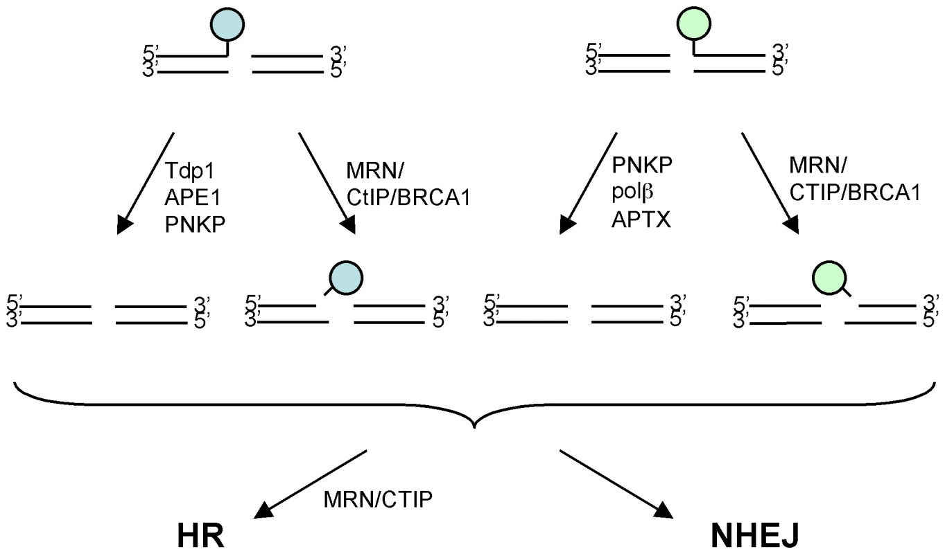
Topoisomerase inhibitors induce chemical modification (indicated by a circle) at 3′ and 5′ ends of DSBs, and thereby inhibit subsequent DSB repair. There are two distinct pathways to eliminate such chemical modifications from DSBs. In the first pathway, tyrosyl-DNA phosphodiesterase1 (Tdp1), polynucleotide kinase 3′-phosphatase (PNKP), and AP endonuclease I (APE1) remove various chemical modifications from the 3′ ends of DSBs, while PNKP, DNA polymerase β (Polβ), and aprataxin (APTX) remove those from the 5′ ends of DSBs (the arrows on the left). In the second pathway, MRN-CtIP-BRCA1 may act as an endonuclease and eliminate oligonucleotides covalently bound to polypeptide (the arrows on the right). The resulting processed DSBs are subject to homologous recombination (HR) and nonhomologous-end-joining (NHEJ)-dependent DSB repair. A well-known precedent involving the second pathway is the Mre11/Rad50/Nbs1-complex-dependent elimination of oligonucleotides as well as the covalently associated topoisomerase-like protein (Spo11) from DSBs during meiotic HR in S. cerevisiae [2]. A more recent study of the S. pombe CtIP mutant (ctp1Δ) showed that the level of Top2 protein covalently bound to DNA in the ctp1Δ mutant increased during treatment with TOP-53, one of the VP16 derivatives, suggesting that Ctp1 plays a role in the endonuclease-dependent removal of covalently-bound polypeptides from the 5′ end of DSBs [20]. Our study indicates that this conclusion is also relevant to vertebrate cells although there are significant differences between vertebrate and yeast systems. First, yeast Ctp1 or Sae2 seem to be important only for the removal of the peptide covalently bound to 5′ of DSB ends as demonstrated for DNA damage induced by TOP-53 or Spo11 [2],[20]. Second, yeast does not have BRCA1 counterpart. BRCA1 is involved in degradation of trapped Topo1 cleavage complexes along with proteasome [45]. We hypothesize that BRCA1 may facilitate the removal of Topo1 by degrading them to small polypeptides, which in turn are removed with oligonucleotides by the nuclease activity of CtIP. In summary, we here show compelling evidence that the collaborative action of BRCA1 and CtIP plays a critical role in the endonuclease-dependent removal of damaged nucleotides from DSBs, and acts on the processed DSBs for subsequent HR and NHEJ.
Materials and Methods
Cell culture
DT40 cells were cultured in RPMI-1640 medium supplemented with 10−5 M β-mercaptoethanol, penicillin, streptomycin, 10% fetal calf serum (FCS), and 1% chicken serum (Sigma, St Louis, MO, USA) at 39.5°C.
Generation of CtIP conditional mutant DT40 cells
To generate CtIP gene disruption constructs, genomic DNA sequences of DT40 cells were amplified using primers 5′-GGATGCGGAGAGGCTTGAAGAGTTTTACAC-3′ and 5′-TTACAGCACAACGATCACATAATCCCGCTC-3′ for the 5′ arm, and 5′-GGAGCTTCTAGCAATACGCGGAACAACTCA-3′ and 5′-GCTTCCCCTCCAATTCTTGACTGAGAATCA-3′ for the 3′ arm. The amplified PCR products were cloned into the pCR2.1-TOPO vector (Invitrogen, CA, USA). The BamHI site in the plasmid that contains the 5′ arm was disrupted by blunt-self ligation. The 1.6-kb HindIII fragment was ligated into the partially-digested HindIII site of the 3.0-kb 3′ arm containing the plasmid. A drug-resistance gene (hisD or bsr) was inserted into the BamHI site of the pCR2.1 vector containing both the 5′ and 3′ arms. To generate CtIP+/−/− cells, linearized CtIP gene-disruption constructs were transfected sequentially by electroporation (BioRad). The genomic DNA of the transfectants was digested with SacI and the targeted clones were confirmed by Southern blot analysis. The 0.5-kb fragment was amplified using primers 5′-GATTGTATGCTTCAGAGGCTCCTGC-3′ and 5′-GAAATTCCCAACTTTAGCTCCCCTTGAC-3′ and used as a probe. To construct the CtIP expression plasmid, chicken the CtIP open reading frame was amplified by PCR, using the primers 5′-GGGGACAAGTTTGTACAAAAAAGCAGGCTTCGAACCATGAATGCGTCTGGGGGAACTTGTG-3′ and 5′-GGGGACCACTTTGTACAAGAAAGCTGGGTCTTATGTCTTCTGCTCTTTGCCTTTTGG-3′, and cloned into a Gateway donor vector, pDONR207 (Invitrogen, CA, USA), by BP reaction. The CtIP gene in the donor vector was transferred to an expression vector (pA-puro) containing the Gateway conversion cassette under the β-actin promoter by LR reaction. To construct a CtIP expression vector under the control of a tetracycline-repressible promoter, CtIP cDNA was amplified by PCR, using the primers 5′-CTCGAGATGAATGCGTCTGGGGGAACTTGTG-3′ and 5′-GTCGACTTATGTCTTCTGCTCTTTGCCTTTTGG-3′, and cloned into pCR2.1-TOPO vector. The XhoI-SalI fragment containing the CtIP cDNA was blunted and cloned into the EcoRV site of a modified pTRE2 vector (Clontech, CA, USA) containing a loxP-flanked puromycin-resistant cassette.
CtIP+/−/− cells were introduced with the tetracycline-controlled trans-activator (tTA) gene through retrovirus infection. Infected cells were sub-cloned, and tTA expression was confirmed by western blot analysis. The resulting tTA-expressing CtIP+/−/− cells were transfected with the pTRE2 puroR/CtIP, and puromycin-resistant clones were selected to isolate the CtIP+/−/−tetCtIP cells. The puromycin-resistance gene was then deleted by transiently expressing the Cre recombinase (Amaxa solution T, program B-23). Puromycin-sensitive CtIP+/−/−tetCtIP cells were transfected with the CtIP gene-disruption construct carrying the puromycin-resistant cassette to generate CtIP−/−/−tetCtIP cells.
Generation of CtIPS332A/−/− mutant cells
The targeting vectors for the CtIP S332A mutants were generated by site-directed mutagenesis. To generate the S332A knock-in vector, genomic DNA was amplified by PCR, using primers 5′-ATTATGCCCCTGAAAGAAGGGAAAC-3′ and 5′-TTTCCTGGGTTTGCTCTTGATTTT-3′, and cloned into the pCR2.1-TOPO vector (Invitrogen, CA, USA). Site-directed mutagenesis was performed using primers 5′-GATTCTCAGGTAGTTGCTCCTGTTTTCGGA-3′ and 5′-TCCGAAAACAGGAGCAACTACCTGAGAATC-3′. The puromycin-resistance gene was inserted into the HpaI site of the resulting plasmid. After transfection of the S332A knock-in vector into the CtIP+/+/− cells, the targeted clones were selected against puromycin and then identified by Southern blot analysis of genomic DNA digested with HindIII. To make probe DNAs, the 0.6-kb fragments were amplified using primers 5′-GACTAACAAAGATCAACCTGTC-3′ and 5′-GTGCATGAGATTTTGGTCGTTG-3′. After the deletion of the puromycin-resistance gene by transiently expressing Cre recombinase by nucleofection (Amaxa, Germany), the third allele of the CtIP gene was disrupted by transfecting the CtIP gene-disruption construct carrying the puromycin-resistance gene. The insertion of the S332A mutation into the endogenous CtIP gene was confirmed by RT-PCR followed by sequencing amplified DNA.
Generation of CtIP+/−/−BRCA1−/− and CtIPS332A/−/−BRCA1−/− clones
The puromycin-resistant cassette in the targeting vector for the BRCA1 gene [31] was replaced with the neomycin-resistant cassette. CtIP+/−/− and CtIPS332A/−/− cells were sequentially transfected with targeting vectors containing the puromycin - and neomycin-resistant gene, and selected against G418 and puromycin, respectively. The clones with the disrupted BRCA1 gene were identified by Southern blot analysis as described previously [31].
Real-time PCR quantification of gene expression
Quantitative real-time PCR was performed in an ABI Prism 7000 sequence detector (Applied Biosystems) using SYBR Green PCR Master Mix reagent (Applied Biosystems) according to the manufacturer's instruction. CtIP cDNA was amplified using primers 5′-GGAATTGGAGGAGCAAAAGCAAC-3′ and 5′-GAAACTCACTGTTGCTCTTTG-3′. The expression level of CtIP was normalized against β-actin using the comparative CT method.
Western blotting analysis
For Western blot analysis, the antibodies specific for CtIP (BL1914, Bethyl, TX, USA), β-actin (Sigma, MO, USA), Rad51 (Ab-1, Calbiochem, CA, USA) were used for detection of each protein. Secondary antibodies were horseradish peroxidase (HRP)-conjugated antibodies to mouse Ig (GE Healthcare, MA, USA) and HRP-conjugated antibody to rabbit Ig (Santa Cruz, CA, USA).
Chromosome aberration analysis
Karyotype analysis was performed as described previously [25]. To measure the number of γ-ray-induced chromosome breaks in mitotic cells, we exposed cells to 2 Gy γ-rays and immediately added colcemid. At 3 hours after irradiation, mitotic cells were harvested and subjected to chromosome analysis.
Measurement of cellular sensitivity to DNA–damaging agents
Methylcellulose colony formation assays were performed as described previously [30],[46]. Since in this assay the plating efficiency of BRCA1-deficient cells was less than 50%, we used a different assay to measure cellular sensitivity to DNA-damaging agents. Cells (1×10³) were seeded into 24-well plates containing 1 ml culture medium per well and the DNA-damaging agents, and then incubated at 39.5°C for 48 hours. To assess the number of live cells, we measured the amount of ATP in the cellular lysates. We confirmed that the number of live cells was closely correlated with the amount of ATP. This ATP assay was carried out with 96-well plates using a CellTiter-Glo Luminescent Cell Viability Assay Kit (Promega Corporation, WI, USA). Briefly, we transferred 100 µl of cell suspension to the individual wells of the plates, placed the plates at room temperature for approximately 30 minutes, added 100 µl of CellTiter-Glo Reagent, and mixed the contents for 2 minutes on an orbital shaker to induce cell lysis. The plate was then incubated at room temperature for 10 minutes to stabilize the luminescent signal. Luminescence was measured by Fluoroskan Ascent FL (Thermo Fisher Scientific Inc., MA, USA).
I-Sce-I–induced gene conversion and targeted integration frequencies
The measurement of homologous recombination frequencies using a SCneo cassette [28] and CENP-H-EGFP was performed as described previously [47]. After the I-Sce-I vector was transfected into the cells, the frequency of neomycin-resistant colony formation was measured.
Synchronization of cells
To enrich DT40 cells in the G1 phase, cells were synchronized by centrifugal counterflow elutriation (Hitachi Industrial, Japan). The cell suspension (∼5×10⁷) was loaded at a flow rate of 11 ml/min into an elutriation chamber running at 2,000 rpm. The first 50 ml was discarded, and the following 100 ml was used as a G1-phase cell fraction.
Microscopy imaging and generation of DNA damage
Fluorescence microscopy was carried out and images were obtained and processed using the IX81 (Olympus, Japan). Cells were cultured in medium containing BrdU (10 µM) for 24–48 h to sensitize them to DSB generation by means of a 405 nm laser from a confocal microscope (FV-1000, Olympus, Japan). During laser treatment, cells were incubated in phenol red-free Opti medium (GIBCO, NY, USA) to prevent the absorption of the laser's wavelength. γ-irradiation was performed using 137C (Gammacell 40, Nordion, Kanata, Ontario, Canada). Antibodies against Rad51 (Ab-1, Calbiochem, CA, USA), FK2 (Nippon Biotest Laboratories, Japan), RPA p32 (GeneTex, TX, USA), rabbit Ig (Alexa 488-conjugated antibody, Molecular Probe, OR, USA), and mouse Ig (Alexa 594-conjugated antibody, Molecular probe, OR, USA) were used for visualization.
Supporting Information
Zdroje
1. BarberLJ
BoultonSJ
2006 BRCA1 ubiquitylation of CtIP: Just the tIP of the iceberg? DNA Repair (Amst) 5 1499 1504
2. NealeMJ
PanJ
KeeneyS
2005 Endonucleolytic processing of covalent protein-linked DNA double-strand breaks. Nature 436 1053 1057
3. SartoriAA
LukasC
CoatesJ
MistrikM
FuS
2007 Human CtIP promotes DNA end resection. Nature 450 509 514
4. PrinzS
AmonA
KleinF
1997 Isolation of COM1, a new gene required to complete meiotic double-strand break-induced recombination in Saccharomyces cerevisiae. Genetics 146 781 795
5. McKeeAH
KlecknerN
1997 A general method for identifying recessive diploid-specific mutations in Saccharomyces cerevisiae, its application to the isolation of mutants blocked at intermediate stages of meiotic prophase and characterization of a new gene SAE2. Genetics 146 797 816
6. LengsfeldBM
RattrayAJ
BhaskaraV
GhirlandoR
PaullTT
2007 Sae2 is an endonuclease that processes hairpin DNA cooperatively with the Mre11/Rad50/Xrs2 complex. Mol Cell 28 638 651
7. HuertasP
JacksonSP
2009 Human CtIP mediates cell cycle control of DNA end resection and double strand break repair. J Biol Chem 284 9558 9565
8. HuertasP
Cortes-LedesmaF
SartoriAA
AguileraA
JacksonSP
2008 CDK targets Sae2 to control DNA-end resection and homologous recombination. Nature 455 689 692
9. MikiY
SwensenJ
Shattuck-EidensD
FutrealPA
HarshmanK
1994 A strong candidate for the breast and ovarian cancer susceptibility gene BRCA1. Science 266 66 71
10. WangB
MatsuokaS
BallifBA
ZhangD
SmogorzewskaA
2007 Abraxas and RAP80 form a BRCA1 protein complex required for the DNA damage response. Science 316 1194 1198
11. ChenL
NieveraCJ
LeeAY
WuX
2008 Cell cycle-dependent complex formation of BRCA1.CtIP.MRN is important for DNA double-strand break repair. J Biol Chem 283 7713 7720
12. YuX
FuS
LaiM
BaerR
ChenJ
2006 BRCA1 ubiquitinates its phosphorylation-dependent binding partner CtIP. Genes Dev 20 1721 1726
13. BermejoR
DoksaniY
CapraT
KatouYM
TanakaH
2007 Top1 - and Top2-mediated topological transitions at replication forks ensure fork progression and stability and prevent DNA damage checkpoint activation. Genes Dev 21 1921 1936
14. WangJC
2002 Cellular roles of DNA topoisomerases: a molecular perspective. Nat Rev Mol Cell Biol 3 430 440
15. PommierY
2006 Topoisomerase I inhibitors: camptothecins and beyond. Nat Rev Cancer 6 789 802
16. AndohT
IshidaR
1998 Catalytic inhibitors of DNA topoisomerase II. Biochim Biophys Acta 1400 155 171
17. NitissJL
2009 Targeting DNA topoisomerase II in cancer chemotherapy. Nat Rev Cancer 9 338 350
18. AdachiN
SuzukiH
IiizumiS
KoyamaH
2003 Hypersensitivity of nonhomologous DNA end-joining mutants to VP-16 and ICRF-193: implications for the repair of topoisomerase II-mediated DNA damage. J Biol Chem 278 35897 35902
19. AdachiN
SoS
KoyamaH
2004 Loss of nonhomologous end joining confers camptothecin resistance in DT40 cells. Implications for the repair of topoisomerase I-mediated DNA damage. J Biol Chem 279 37343 37348
20. HartsuikerE
NealeMJ
CarrAM
2009 Distinct requirements for the Rad32(Mre11) nuclease and Ctp1(CtIP) in the removal of covalently bound topoisomerase I and II from DNA. Mol Cell 33 117 123
21. YamazoeM
SonodaE
HocheggerH
TakedaS
2004 Reverse genetic studies of the DNA damage response in the chicken B lymphocyte line DT40. DNA Repair (Amst) 3 1175 1185
22. SonodaE
HocheggerH
SaberiA
TaniguchiY
TakedaS
2006 Differential usage of non-homologous end-joining and homologous recombination in double strand break repair. DNA Repair (Amst) 5 1021 1029
23. Yamaguchi-IwaiY
SonodaE
SasakiMS
MorrisonC
HaraguchiT
1999 Mre11 is essential for the maintenance of chromosomal DNA in vertebrate cells. Embo J 18 6619 6629
24. NakaharaM
SonodaE
NojimaK
SaleJE
TakenakaK
2009 Genetic evidence for single-strand lesions initiating Nbs1-dependent homologous recombination in diversification of Ig v in chicken B lymphocytes. PLoS Genet 5 e1000356 doi:10.1371/journal.pgen.1000356
25. SonodaE
SasakiMS
BuersteddeJM
BezzubovaO
ShinoharaA
1998 Rad51-deficient vertebrate cells accumulate chromosomal breaks prior to cell death. Embo J 17 598 608
26. PolanowskaJ
MartinJS
Garcia-MuseT
PetalcorinMI
BoultonSJ
2006 A conserved pathway to activate BRCA1-dependent ubiquitylation at DNA damage sites. Embo J 25 2178 2188
27. ZhaoGY
SonodaE
BarberLJ
OkaH
MurakawaY
2007 A critical role for the ubiquitin-conjugating enzyme Ubc13 in initiating homologous recombination. Mol Cell 25 663 675
28. FukushimaT
TakataM
MorrisonC
ArakiR
FujimoriA
2001 Genetic analysis of the DNA-dependent protein kinase reveals an inhibitory role of Ku in late S-G2 phase DNA double-strand break repair. J Biol Chem 276 44413 44418
29. TauchiH
KobayashiJ
MorishimaK
van GentDC
ShiraishiT
2002 Nbs1 is essential for DNA repair by homologous recombination in higher vertebrate cells. Nature 420 93 98
30. TakataM
SasakiMS
SonodaE
MorrisonC
HashimotoM
1998 Homologous recombination and non-homologous end-joining pathways of DNA double-strand break repair have overlapping roles in the maintenance of chromosomal integrity in vertebrate cells. Embo J 17 5497 5508
31. MartinRW
OrelliBJ
YamazoeM
MinnAJ
TakedaS
2007 RAD51 up-regulation bypasses BRCA1 function and is a common feature of BRCA1-deficient breast tumors. Cancer Res 67 9658 9665
32. JiK
KogameT
ChoiK
WangX
LeeJ
2009 A novel approach to screening and characterizing the genotoxicity of environmental contaminants using DNA-repair-deficient chicken DT40 cell lines. Environ Health Perspect 117 1737 1744
33. ChenPL
LiuF
CaiS
LinX
LiA
2005 Inactivation of CtIP leads to early embryonic lethality mediated by G1 restraint and to tumorigenesis by haploid insufficiency. Mol Cell Biol 25 3535 3542
34. LiuF
LeeWH
2006 CtIP activates its own and cyclin D1 promoters via the E2F/RB pathway during G1/S progression. Mol Cell Biol 26 3124 3134
35. TakaoN
KatoH
MoriR
MorrisonC
SonadaE
1999 Disruption of ATM in p53-null cells causes multiple functional abnormalities in cellular response to ionizing radiation. Oncogene 18 7002 7009
36. SwanbergSE
PayneWS
HuntHD
DodgsonJB
DelanyME
2004 Telomerase activity and differential expression of telomerase genes and c-myc in chicken cells in vitro. Dev Dyn 231 14 21
37. YunMH
HiomK
2009 CtIP-BRCA1 modulates the choice of DNA double-strand-break repair pathway throughout the cell cycle. Nature 459 460 463
38. LimboO
ChahwanC
YamadaY
de BruinRA
WittenbergC
2007 Ctp1 is a cell-cycle-regulated protein that functions with Mre11 complex to control double-strand break repair by homologous recombination. Mol Cell 28 134 146
39. MaserRS
ZinkelR
PetriniJH
2001 An alternative mode of translation permits production of a variant NBS1 protein from the common Nijmegen breakage syndrome allele. Nat Genet 27 417 421
40. SchlegelBP
JodelkaFM
NunezR
2006 BRCA1 promotes induction of ssDNA by ionizing radiation. Cancer Res 66 5181 5189
41. BhattacharyyaA
EarUS
KollerBH
WeichselbaumRR
BishopDK
2000 The breast cancer susceptibility gene BRCA1 is required for subnuclear assembly of Rad51 and survival following treatment with the DNA cross-linking agent cisplatin. J Biol Chem 275 23899 23903
42. SySM
HuenMS
ChenJ
2009 PALB2 is an integral component of the BRCA complex required for homologous recombination repair. Proc Natl Acad Sci U S A 106 7155 7160
43. YangSW
BurginABJr
HuizengaBN
RobertsonCA
YaoKC
1996 A eukaryotic enzyme that can disjoin dead-end covalent complexes between DNA and type I topoisomerases. Proc Natl Acad Sci U S A 93 11534 11539
44. CaldecottKW
2008 Single-strand break repair and genetic disease. Nat Rev Genet 9 619 631
45. SordetO
LarochelleS
NicolasE
StevensEV
ZhangC
2008 Hyperphosphorylation of RNA polymerase II in response to topoisomerase I cleavage complexes and its association with transcription - and BRCA1-dependent degradation of topoisomerase I. J Mol Biol 381 540 549
46. OkadaT
SonodaE
YamashitaYM
KoyoshiS
TateishiS
2002 Involvement of vertebrate polkappa in Rad18-independent postreplication repair of UV damage. J Biol Chem 277 48690 48695
47. KikuchiK
TaniguchiY
HatanakaA
SonodaE
HocheggerH
2005 Fen-1 facilitates homologous recombination by removing divergent sequences at DNA break ends. Mol Cell Biol 25 6948 6955
Štítky
Genetika Reprodukční medicína
Článek Activation of Mutant Enzyme Function by Proteasome Inhibitors and Treatments that Induce Hsp70Článek Maternal Ethanol Consumption Alters the Epigenotype and the Phenotype of Offspring in a Mouse ModelČlánek Genetic Dissection of Differential Signaling Threshold Requirements for the Wnt/β-Catenin PathwayČlánek Distinct Type of Transmission Barrier Revealed by Study of Multiple Prion Determinants of Rnq1
Článek vyšel v časopisePLOS Genetics
Nejčtenější tento týden
2010 Číslo 1- Akutní intermitentní porfyrie
- Růst a vývoj dětí narozených pomocí IVF
- Vliv melatoninu a cirkadiálního rytmu na ženskou reprodukci
- Délka menstruačního cyklu jako marker ženské plodnosti
- Intrauterinní inseminace a její úspěšnost
-
Všechny články tohoto čísla
- Irradiation-Induced Genome Fragmentation Triggers Transposition of a Single Resident Insertion Sequence
- A Major Role of the RecFOR Pathway in DNA Double-Strand-Break Repair through ESDSA in
- Kidney Development in the Absence of and Requires
- Modeling of Environmental Effects in Genome-Wide Association Studies Identifies and as Novel Loci Influencing Serum Cholesterol Levels
- Inverse Correlation between Promoter Strength and Excision Activity in Class 1 Integrons
- Activation of Mutant Enzyme Function by Proteasome Inhibitors and Treatments that Induce Hsp70
- Postnatal Survival of Mice with Maternal Duplication of Distal Chromosome 7 Induced by a / Imprinting Control Region Lacking Insulator Function
- The Werner Syndrome Protein Functions Upstream of ATR and ATM in Response to DNA Replication Inhibition and Double-Strand DNA Breaks
- Maternal Ethanol Consumption Alters the Epigenotype and the Phenotype of Offspring in a Mouse Model
- Understanding Gene Sequence Variation in the Context of Transcription Regulation in Yeast
- miR-30 Regulates Mitochondrial Fission through Targeting p53 and the Dynamin-Related Protein-1 Pathway
- Elevated Levels of the Polo Kinase Cdc5 Override the Mec1/ATR Checkpoint in Budding Yeast by Acting at Different Steps of the Signaling Pathway
- Alternative Epigenetic Chromatin States of Polycomb Target Genes
- Co-Orientation of Replication and Transcription Preserves Genome Integrity
- A Comprehensive Map of Insulator Elements for the Genome
- Environmental and Genetic Determinants of Colony Morphology in Yeast
- U87MG Decoded: The Genomic Sequence of a Cytogenetically Aberrant Human Cancer Cell Line
- The MCM-Binding Protein ETG1 Aids Sister Chromatid Cohesion Required for Postreplicative Homologous Recombination Repair
- Genetic Dissection of Differential Signaling Threshold Requirements for the Wnt/β-Catenin Pathway
- Differential Localization and Independent Acquisition of the H3K9me2 and H3K9me3 Chromatin Modifications in the Adult Germ Line
- Genetic Crossovers Are Predicted Accurately by the Computed Human Recombination Map
- Collaborative Action of Brca1 and CtIP in Elimination of Covalent Modifications from Double-Strand Breaks to Facilitate Subsequent Break Repair
- Distinct Type of Transmission Barrier Revealed by Study of Multiple Prion Determinants of Rnq1
- Genome-Wide Association Study Identifies as a Novel Susceptibility Gene for Osteoporosis
- and Regulate Reproductive Habit in Rice
- Nonsense-Mediated Decay Enables Intron Gain in
- Altered Gene Expression and DNA Damage in Peripheral Blood Cells from Friedreich's Ataxia Patients: Cellular Model of Pathology
- The Systemic Imprint of Growth and Its Uses in Ecological (Meta)Genomics
- The Gift of Observation: An Interview with Mary Lyon
- Genotype and Gene Expression Associations with Immune Function in
- The Elongator Complex Regulates Neuronal α-tubulin Acetylation
- Rising from the Ashes: DNA Repair in
- Mis-Spliced Transcripts of Nicotinic Acetylcholine Receptor α6 Are Associated with Field Evolved Spinosad Resistance in (L.)
- BRIT1/MCPH1 Is Essential for Mitotic and Meiotic Recombination DNA Repair and Maintaining Genomic Stability in Mice
- Non-Coding Changes Cause Sex-Specific Wing Size Differences between Closely Related Species of
- Evidence for Pervasive Adaptive Protein Evolution in Wild Mice
- Evolutionary Mirages: Selection on Binding Site Composition Creates the Illusion of Conserved Grammars in Enhancers
- VEZF1 Elements Mediate Protection from DNA Methylation
- PLOS Genetics
- Archiv čísel
- Aktuální číslo
- Informace o časopisu
Nejčtenější v tomto čísle- A Major Role of the RecFOR Pathway in DNA Double-Strand-Break Repair through ESDSA in
- Kidney Development in the Absence of and Requires
- The Werner Syndrome Protein Functions Upstream of ATR and ATM in Response to DNA Replication Inhibition and Double-Strand DNA Breaks
- Alternative Epigenetic Chromatin States of Polycomb Target Genes
Kurzy
Zvyšte si kvalifikaci online z pohodlí domova
Autoři: prof. MUDr. Vladimír Palička, CSc., Dr.h.c., doc. MUDr. Václav Vyskočil, Ph.D., MUDr. Petr Kasalický, CSc., MUDr. Jan Rosa, Ing. Pavel Havlík, Ing. Jan Adam, Hana Hejnová, DiS., Jana Křenková
Autoři: MUDr. Irena Krčmová, CSc.
Autoři: MDDr. Eleonóra Ivančová, PhD., MHA
Autoři: prof. MUDr. Eva Kubala Havrdová, DrSc.
Všechny kurzyPřihlášení#ADS_BOTTOM_SCRIPTS#Zapomenuté hesloZadejte e-mailovou adresu, se kterou jste vytvářel(a) účet, budou Vám na ni zaslány informace k nastavení nového hesla.
- Vzdělávání


