-
Články
- Vzdělávání
- Časopisy
Top články
Nové číslo
- Témata
- Kongresy
- Videa
- Podcasty
Nové podcasty
Reklama- Kariéra
Doporučené pozice
Reklama- Praxe
HP1 Recruitment in the Absence of Argonaute Proteins in
Highly repetitive and transposable element rich regions of the genome must be stabilized by the presence of heterochromatin. A direct role for RNA interference in the establishment of heterochromatin has been demonstrated in fission yeast. In metazoans, which possess multiple RNA–silencing pathways that are both functionally distinct and spatially restricted, whether RNA silencing contributes directly to heterochromatin formation is not clear. Previous studies in Drosophila melanogaster have suggested the involvement of both the AGO2-dependent endogenous small interfering RNA (endo-siRNA) as well as Piwi-interacting RNA (piRNA) silencing pathways. In order to determine if these Argonaute genes are required for heterochromatin formation, we utilized transcriptional reporters and chromatin immunoprecipitation of the critical factor Heterochromatin Protein 1 (HP1) to monitor the heterochromatic state of piRNA clusters, which generate both endo-siRNAs and the bulk of piRNAs. Surprisingly, we find that mutation of AGO2 or piwi increases silencing at piRNA clusters corresponding to an increase of HP1 association. Furthermore, loss of piRNA production from a single piRNA cluster results in genome-wide redistribution of HP1 and reduction of silencing at a distant heterochromatic site, suggesting indirect effects on HP1 recruitment. Taken together, these results indicate that heterochromatin forms independently of endo-siRNA and piRNA pathways.
Published in the journal: . PLoS Genet 6(3): e32767. doi:10.1371/journal.pgen.1000880
Category: Research Article
doi: https://doi.org/10.1371/journal.pgen.1000880Summary
Highly repetitive and transposable element rich regions of the genome must be stabilized by the presence of heterochromatin. A direct role for RNA interference in the establishment of heterochromatin has been demonstrated in fission yeast. In metazoans, which possess multiple RNA–silencing pathways that are both functionally distinct and spatially restricted, whether RNA silencing contributes directly to heterochromatin formation is not clear. Previous studies in Drosophila melanogaster have suggested the involvement of both the AGO2-dependent endogenous small interfering RNA (endo-siRNA) as well as Piwi-interacting RNA (piRNA) silencing pathways. In order to determine if these Argonaute genes are required for heterochromatin formation, we utilized transcriptional reporters and chromatin immunoprecipitation of the critical factor Heterochromatin Protein 1 (HP1) to monitor the heterochromatic state of piRNA clusters, which generate both endo-siRNAs and the bulk of piRNAs. Surprisingly, we find that mutation of AGO2 or piwi increases silencing at piRNA clusters corresponding to an increase of HP1 association. Furthermore, loss of piRNA production from a single piRNA cluster results in genome-wide redistribution of HP1 and reduction of silencing at a distant heterochromatic site, suggesting indirect effects on HP1 recruitment. Taken together, these results indicate that heterochromatin forms independently of endo-siRNA and piRNA pathways.
Introduction
In D. melanogaster, an estimated one-third of the genome is composed of repetitive and noncoding sequences associated with a condensed form of chromatin known as heterochromatin. Heterochromatin is characterized by repeat-rich sequences, hypoacetylation of histone tails, and dimethylation of histone H3 on lysine 9 (H3K9me2) [1]. A conserved nonhistone Heterochromatin Protein 1 (HP1) is a critical component of heterochromatin, localizing predominantly at and near centromeres but also residing at telomeres, and the Y and fourth chromosomes. These regions tend to be rich in transposable elements (TEs), which must be suppressed in order to maintain genomic stability but can serve a cellular function, particularly in the case of Het-A and TART at the telomeres (reviewed in [2]).
The phenomenon of position-effect variegation (PEV) provided the first glimpse into the role of heterochromatin in gene silencing in Drosophila. When a normally euchromatic gene is relocated near heterochromatin, variegated expression results from variable levels of heterochromatin spreading over the gene in each cell. Screens for dominant mutations that either suppress {Suppressor of variegation [Su(var)]} or enhance {Enhancer of variegation [E(var)]} PEV were performed to identify key components of heterochromatin. For example, mutation of Su(var)3-9, which encodes an H3K9 methyltransferase, was identified in a large screen for modifiers of PEV [3]. Accordingly, loss of HP1, encoded by Su(var)2-5, causes increased expression of a gene subject to PEV while an extra copy has the reverse effect [4].
Pioneering genetic and biochemical studies in Schizosaccharomyces pombe have shed considerable light on mechanisms of heterochromatin assembly. The RNA interference (RNAi) machinery was found to play a key role in heterochromatin formation by detecting the transcription of specific DNA repeats located at the mating type locus and the centromere and subsequently nucleating heterochromatin. For example, double-stranded RNAs (dsRNA) produced by bidirectional transcription of pericentromeric repeats are processed by the RNase III endonuclease Dicer1 into short interfering RNAs (siRNAs) [5]. The Argonaute1 PAZ and PIWI domain protein binds these siRNAs as part of the RNA-induced transcriptional silencing complex (RITS) [6]. Loading of RITS with siRNA and recruitment of the complex to the site of dsRNA transcription requires the Clr4 histone methyltransferase, which methylates H3K9 [7]. This methylation mark serves as a binding site for Swi6, a fission yeast homolog of HP1, leading to heterochromatin establishment and spreading. Importantly, heterochromatin can also be nucleated independently of RNAi by other mechanisms. For example, in the absence of RNAi the ATF/CREB stress-activated proteins promote heterochromatin formation at the mating type locus [8], and the Taz1 protein can establish HP1 recruitment to telomeres [9]. These studies exemplify the redundancy of RNAi and additional mechanisms with respect to the formation of heterochromatin.
All RNA silencing pathways are characterized by the activity of an Argonaute effector protein that binds directly to small RNA. The five Argonautes in Drosophila can be divided into two families based on homology. The AGO subfamily includes AGO1 and AGO2, and the Piwi subfamily consists of Piwi, Aubergine (Aub), and AGO3 (reviewed in [10]). AGO1 and AGO2 are expressed throughout the fly while piwi, aub, and AGO3 are expressed mainly, although not exclusively, in the gonad [11]–[13]. AGO1 is required for the microRNA pathway, which regulates mRNA expression and functions chiefly through translational repression. Protecting against exogenous double stranded RNA, AGO2 associates with 21–22 nt siRNA produced by Dicer-2 (Dcr-2), and this pathway is required for viral immunity and a robust RNAi response [14],[15]. In addition, AGO2 also binds endogenous siRNAs (endo-siRNAs), the majority of which silence the expression of TEs outside of the gonad [16]–[19].
Suppression of TEs is especially imperative in the gonad in order to limit the propagation of unwanted mutations and is achieved principally by the activity of the Piwi subfamily proteins. Piwi, Aub, and AGO3 bind to 23–30 nt RNAs termed Piwi-interacting RNAs (piRNAs) that are predominantly derived from genomic locations termed piRNA clusters [20],[21]. These piRNA producing loci are mainly pericentromeric and enriched in transposon sequences. From these and previous studies, it became clear that the piRNA pathway exists to eliminate TE transcripts in the gonad [22]–[24]. Based on comparative sequence analysis of piRNAs immunopurified from the ovary, the “ping-pong” or “amplification loop” model for germline piRNA biogenesis was proposed [20]–[23]. Precursor transcripts from piRNA clusters, derived from either one or both strands [25], give rise to piRNAs bound by Piwi, Aub, or AGO3. Those piRNAs antisense to a homologous TE transcript can result in its cleavage, and this event defines the 5′ end of a secondary piRNA that can then bind and cleave an antisense piRNA cluster transcript, and the cycle can continue. Piwi appears to play a minor role in ping-pong piRNA amplification [25],[26], which is thought to occur primarily in the cytoplasmic nuage where Aub and AGO3 localize [20],[23],[27]. In contrast, Piwi resides in the nucleus [28]. Production of precursor transcripts at certain piRNA clusters that give rise to piRNAs from both sense and antisense strands (dual-strand clusters) is dependent on the germline specific HP1 homolog Rhino [29]. Rhino functions specifically in the ping-pong pathway, acting upstream of Aub and AGO3 but not Piwi.
Piwi independently serves an additional role in the silencing of certain TEs expressed in somatic follicle cells surrounding the ovary. This somatic piRNA pathway depends on Piwi alone and therefore does not undergo ping-pong amplification [25],[26],[30]. The flamenco (flam) piRNA cluster, which controls the gypsy, ZAM, and Idefix retrotransposons [31],[32], is one of the major sites of primary piRNA production [25],[26],[30],[33]. Piwi associates with piRNAs generated by flam and other piRNA clusters and has been proposed to cleave homologous TE transcripts using its Slicer activity [22].
Previous studies suggest that one or more RNA silencing pathways may participate in transcriptional TE silencing by inducing heterochromatin formation. First, mutation of AGO2 results in pleiotropic cellular defects in early embryos including mislocalization of HP1 and the histone H3 variant CID, which binds specifically the centromere [34]. Later in development, AGO2 mutants display mislocalization of HP1 on polytene chromosomes of the larval salivary gland [35]. Additionally, silencing of a pericentromeric transcriptional reporter is relieved when the maternally derived pool of AGO2 is reduced. Despite these defects, AGO2 mutant flies develop normally and are fertile, suggesting that these defects are mild and can be compensated by other mechanisms.
Several pieces of evidence implicate piRNA pathways in establishment or maintenance of heterochromatin in the soma. First, mutation of piwi, aub, or spn-E, encoding an RNA helicase required for the germline piRNA pathway [24],[26], results in defects in heterochromatic silencing and visible changes in heterochromatin localization. These mutants reduce silencing of pericentromeric transcriptional reporters and exhibit mislocalization of HP1 and H3K9me2 in salivary gland polytene chromosomes [36]. Moreover, a recent study identified HP1 as an interactor of Piwi in yeast two-hybrid screens [11]. The two proteins coimmunoprecipitate from embryonic nuclear lysate and display partially overlapping localization patterns in polytene chromosomes. Furthermore, both proteins associate specifically with the chromatin of two transposable elements, 1360 and the F element. Based on their findings, the authors propose that Piwi could serve as a recruitment platform for HP1 binding. This model appears not to be applicable to the 3R-TAS subtelomeric region, a site of Piwi chromatin association and piRNA production [21]. Mutation of piwi results in an increase of HP1 association and an increase of transcriptional silencing at 3R-TAS. It remains an open question whether other sites in the genome could serve as Piwi-dependent HP1 recruitment sites.
In other metazoans, it is similarly unclear whether RNA silencing can establish heterochromatin directly. A recent study in chicken indicates that a 16 kb constitutive heterochromatin domain that separates the folate receptor gene and the β-globin locus is maintained by a Dicer and Argonaute 2 (cAgo2) dependent mechanism [37]. Intriguingly, cAgo2 was shown to associate with the heterochromatic domain by chromatin immunoprecipitation (ChIP) suggesting a direct effect. However, it is not known whether this represents a general mechanism to maintain heterochromatin.
In this study, we investigated whether HP1 association with heterochromatin in Drosophila is mediated by either the AGO2 dependent endo-siRNA pathway or by piwi dependent piRNA pathways. Using transcriptional reporters and ChIP, we show that piRNA clusters are subject to heterochromatic silencing and bound by HP1. Interestingly, mutation of AGO2, piwi or aub results in increased silencing at piRNA clusters and an increase in HP1 association with these loci. Furthermore, loss of piRNA production at a single piRNA locus results in global redistribution of HP1 and a reduction of silencing at a distant heterochromatic site. Therefore, our results indicate that HP1 can associate with chromatin independently of both endo-siRNA and piRNA pathways.
Results
Heterochromatin-dependent transcriptional silencing at piRNA clusters
We sought to determine if HP1 is recruited to heterochromatin by AGO2 or Piwi. The majority of genomic regions that produce the bulk of piRNA, termed piRNA clusters, are pericentromeric and rich in transposable elements [20],[21]. These regions also produce endo-siRNA [16]–[19], and due to their proximity to the centromere, may be heterochromatic and serve as platforms for Argonaute mediated HP1 recruitment.
In order to test genetically whether pericentromeric piRNA clusters are heterochromatic, we examined a collection of fly lines bearing P element transgene insertions inside or in close proximity to four piRNA producing loci, flam, 80EF, 42AB, and 38C. The P elements contain a mini-white transcriptional reporter that was assayed for expression in the adult eye. Genomic locations of these transgene insertions are indicated in relation to previously identified small RNAs immunoprecipitated with Piwi, Aub/AGO3, and AGO2 respectively from various cell types (Figure 1, Figure S1) [16],[17],[20],[21]. Lines harboring P elements inside or in the vicinity of a piRNA cluster exhibit variegating coloration of distinct eye facets similar to PEV, suggesting the presence of variably spreading heterochromatin at their sites of insertion (Figure 2, Table 1). Interestingly, insertions within a piRNA cluster that display high mini-white expression without variegation harbor SUPor-P constructs, which contain Suppressor of Hairy wing [Su(Hw)] insulator sequences that flank and likely protect the mini-white reporter from the effects of surrounding heterochromatin [38].
Fig. 1. Schematic representation of three top piRNA clusters. 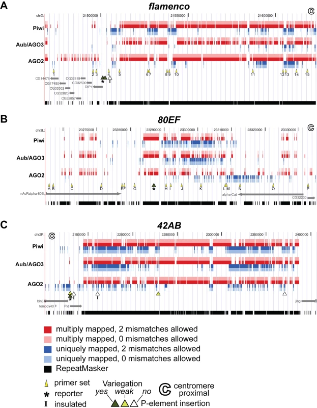
Genomic locations of small RNAs, primer sets used for ChIP, and P element insertions at (A) flam piRNA cluster on chromosome X, (B) 80EF piRNA cluster on chromosome 3L, and (C) 42AB piRNA cluster on chromosome 2R. Sequence datasets derived from previous studies were mapped to the genome using Bowtie software allowing two or zero mismatches [57]. Piwi-immunoprecipitated [20],[21], Aub or AGO3-immunoprecipitated [20] and AGO2-immunoprecipitated [16],[17] reads mapping to multiple locations in the genome are indicated in red (with two mismatches allowed) and pink (with zero mismatches allowed) while uniquely mapping reads are in dark blue (with two mismatches allowed) and light blue (with zero mismatches allowed). Primer sets used for ChIP analysis are indicated by yellow arrowheads. Strongly variegating (dark green triangle), weakly variegating (light green triangle), and non-variegating P elements with high expression levels (white triangle) are indicated. (A) P{EPgy2}DIP1EY02625, (B) PBac{PB}c06482, and (C) P{EPgy2}EY08366 P element insertions are marked by an asterisk. SUPor-P P elements containing insulator sequences are marked by an “I”. Centromere proximal end is marked by a hollow C. RepeatMasker detected sequences are represented in black. Fig. 2. piRNA and endo-siRNA pathway mutants display increased silencing of transcriptional reporters at or near piRNA clusters. 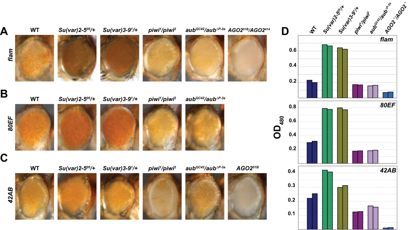
Adult eyes of wild type, Su(var)2-505/+, Su(var)3-91/+, piwi1/piwi2, aubQC42/aubΔP-3a, and/or AGO2 -/AGO2 - mutants carrying a mini-white transgene inserted inside or in close proximity to the (A) flam, (B) 80EF, and (C) 42AB piRNA clusters. AGO251B/AGO2414 mutants are examined in (A) while AGO251B mutants are examined in (C). (D) Levels of eye pigment measured at 480 nm extracted from male heads of the indicated genotypes (A–C). Tab. 1. Expression of mini-white in fly lines harboring P element insertions in four top piRNA clusters. 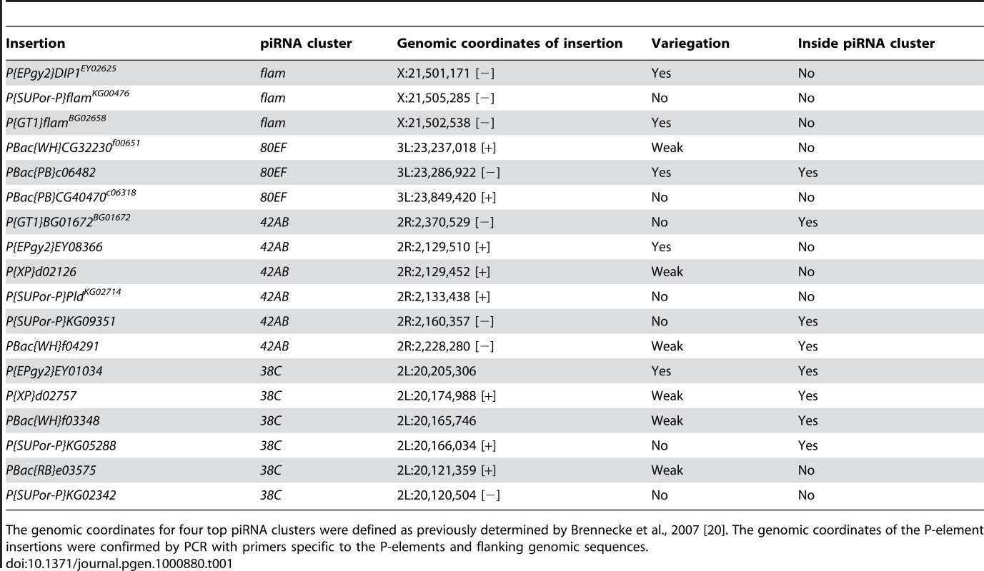
The genomic coordinates for four top piRNA clusters were defined as previously determined by Brennecke et al., 2007 [20]. The genomic coordinates of the P-element insertions were confirmed by PCR with primers specific to the P-elements and flanking genomic sequences. Expression analysis of these transcriptional reporter insertions indicates that piRNA clusters and their immediate vicinity are subject to HP1 dependent silencing. Reporter expression levels of three lines harboring an insertion at flam, 80EF, or 42AB with the most apparent variegation were tested for dependence on heterochromatin. P{EPgy2}DIP1EY02625 is inserted in a gene located on the centromere distal side of the flam piRNA producing locus on the X chromosome (Figure 1A), PBac{PB}c06482 resides within the 80EF cluster on chromosome 3L (Figure 1B), and P{EPgy2}EY08366 borders the centromere proximal edge of the 42AB piRNA locus on chromosome 2R (Figure 1C). In order to test whether these reporters are sensitive to perturbation of heterochromatin, the expression of mini-white was examined in Su(var)2-505/+ and Su(var)3-91/+ dominant loss-of-function mutants, which are compromised for HP1 and H3K9 methyltransferase activity respectively. As expected, decreased silencing of mini-white expression resulting in increased pigmentation was observed for all three insertions in the heterochromatin mutants compared to wild type (Figure 2), suggesting that the vicinity of P element insertion are indeed heterochromatic.
piRNA and endo-siRNA pathway mutants decrease transcription at piRNA clusters
We next tested whether the transcriptional reporters at piRNA clusters are sensitive to perturbations in the piRNA and endo-siRNA silencing pathways. If Piwi were responsible for direct recruitment of HP1 to piRNA clusters, mutation of piwi should increase mini-white expression similarly to disruption of heterochromatin. Surprisingly, piwi1/piwi2 loss-of-function mutants exhibit a substantial loss of reporter expression indicating increased silencing when compared to wild type (Figure 2). Furthermore, aubQC42/aubΔP-3a loss-of-function piRNA pathway mutants result in a similar reduction of mini-white expression. Strikingly, the flam transcriptional reporter expression level was decreased dramatically in the transheterozygous endo-siRNA pathway mutant, AGO251B/AGO2414 compared to wild type (Figure 2A). Similarly, in the AGO251B null mutant, the 42AB transcriptional reporter displays almost complete silencing (Figure 2C). Spectroscopic analysis of extracted eye pigment verifies the overall changes in mini-white expression levels for each genotype compared to wild type (Figure 2D). Additionally, examination of Dcr-2L811fsX mutants shows a similar mild increase in silencing for the transcriptional reporter inserted near flam (Figure S2A). The opposite effects of piRNA and endo-siRNA pathway mutations compared to heterochromatin mutations suggest that these RNA silencing pathways may actually oppose heterochromatin formation at piRNA clusters.
HP1 chromatin association is increased at piRNA clusters in somatic tissues of RNA silencing mutants
In order to further examine the heterochromatic nature of piRNA clusters at higher resolution, ChIP assays were performed in adult heads to assess HP1 association with two piRNA clusters, flam and 80EF, in the soma. Genomic locations of primer sets that uniquely amplify regions spanning these piRNA clusters are indicated in Figure 1A and 1B. As positive controls, primers for two transposable elements known to recruit HP1, TART, a telomere-specific non-LTR retrotransposon, and 1360, a DNA transposon were also tested [39]–[40]. Euchromatic genes hsp26 and yellow were also included in the analysis as negative controls for HP1 association.
In wild type fly heads, HP1 is observed at or near locations that give rise to piRNAs and endo-siRNAs at both flam and 80EF loci. ChIP was performed using α-HP1 antibodies in chromatin prepared from wild type heads, and the amount of DNA associated was determined by quantitative PCR using specific primer sets. As expected, low levels of hsp26 and yellow are immunoprecipitated with HP1, while TART and 1360 levels are enriched above the euchromatic genes by over six-fold (Figure 3). At flam, HP1 associates with the majority of regions that produce high levels of piRNAs or endo-siRNAs approximately two to three-fold over the euchromatic sites (Figure 3A, primer sets 1–15). Similarly, at 80EF, HP1 immunoprecipitates piRNA and endo-siRNA producing regions two to three-fold higher than the negative controls indicating the presence of heterochromatic marks at these loci (Figure 3B, primer sets G-M). Regions flanking these areas display approximately one to two-fold enrichment over euchromatic sites, which may be due to tapering of HP1 spreading (Figure 3B, primer sets A-F and N-P). ChIP using antibodies directed against the chromatin insulator protein Su(Hw) verified its presence at known insulator sequences gypsy and 1A-2 [41] but only background levels at TART, 1360, and piRNA clusters, indicating the specificity of HP1 association at these sites (Figure S3). Rabbit IgG negative control immunoprecipitations yielded negligible amounts of DNA for all sites tested (<0.3% input).
Fig. 3. HP1 associates with chromatin at piRNA clusters, and its levels increase in RNA–silencing mutants. 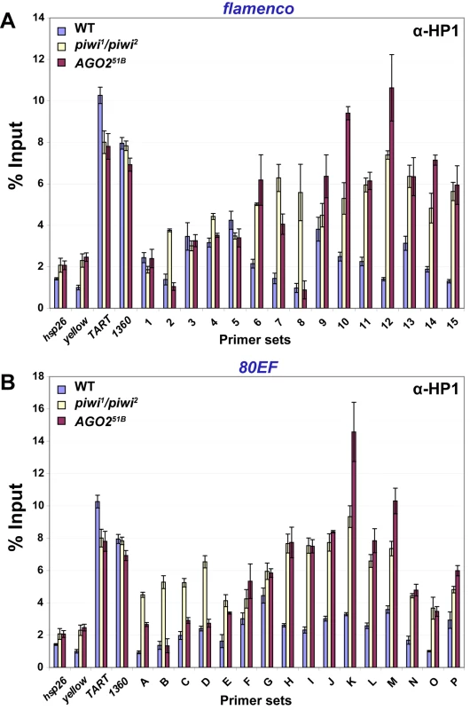
ChIP at (A) flam and (B) 80EF piRNA clusters in wild type (blue), piwi1/piwi2 (yellow), and AGO251B (red) mutants from adult heads with antibodies specific to HP1. Percent input immunoprecipitated is shown for each primer set, and error bars indicate standard deviation of quadruplicate PCR measurements. Consistent with the transcriptional reporter assay, RNA silencing mutants display elevated levels of HP1 at piRNA clusters. ChIP of HP1 was performed in piwi1/piwi2 mutant heads, and similar levels at positive and negative controls were obtained compared to wild type (Figure 3). In contrast, at the flam locus, a two to five-fold increase in HP1 levels is observed at the centromere proximal side of the locus compared to wild type (Figure 3A, primer sets 6–15). Little change in HP1 recruitment is observed at the centromere distal end of flam in piwi1/piwi2 mutants (Figure 3A, primer sets 1–5). At 80EF, HP1 levels increase two to three-fold in piwi1/piwi2 mutants compared to wild type across all primer sets examined (Figure 3B, primer sets A-P).
In order to address differences in strain background and potential accumulation of TEs in piwi mutant strains, we performed ChIP assays comparing piwi1/piwi2 mutants to a piwi1/+ heterozygous strain and obtained similar results (Figure S4).
ChIP experiments performed in AGO251B mutant heads show a similar overall increase of HP1 at piRNA clusters compared to piwi1/piwi2 mutants. Levels of HP1 at hsp26, yellow, TART, and 1360 are similar in AGO251B mutants and wild type while differences are apparent at piRNA clusters (Figure 3). At flam, AGO251B mutants display a two to seven-fold increase of HP1 association with the centromere proximal side compared to wild type (Figure 3A, primer sets 6–15). At the centromere distal end, no significant changes in HP1 levels are detected (Figure 3A, primer sets 1–5). For 80EF, AGO251B mutants show similar levels of HP1 to wild type at the centromere distal end (Figure 3B, primer sets A-D) while an approximately two to five-fold increase of HP1 is detected in the remainder of the regions tested (Figure 3B, primer sets E-P). Moreover, ChIP assays in AGO251B homozygous mutants compared to an AGO251B/+ heterozygous strain produced similar results (Figure S5). Similar to AGO251B mutants, Dcr-2L811fsX mutants show an increase of HP1 at regions that produce small RNAs compared to wild type (Figure S2B and S2C). HP1 protein levels in wild type, piwi1/piwi2, and AGO251B fly heads are similar indicating that the increased chromatin association observed is not due to an increased amount of HP1 (Figure S6). The increased HP1 chromatin association with piRNA clusters in RNA silencing mutants compared to wild type is consistent with increased silencing of P element insertions, and these results suggest that at least some of the observed effects on reporter gene expression in RNA silencing mutants are due to chromatin related events. Taken together, these data suggest an antagonistic effect of Piwi, Aub, and AGO2 on HP1 recruitment to chromatin in somatic tissue.
HP1 also associates with piRNA clusters in ovaries
Given the evidence that transposable elements are mainly silenced in the gonad via piRNA pathways and in the soma via the endo-siRNA pathway, we wanted to determine whether HP1 also associates with piRNA clusters in gonadal tissues. Therefore, we investigated HP1 recruitment to piRNA clusters in wild type ovaries by ChIP. As in heads, low levels of hsp26 and yellow are immunoprecipitated with HP1, whereas TART and 1360 levels are enriched above the euchromatic genes by over ten-fold (Figure 4). At the flam locus, a four to fifteen-fold increase over the euchromatic sites in HP1 levels is observed at most sites at the centromere proximal side of the locus (Figure 4A, primer sets 4–15). Similarly, at 80EF, HP1 immunoprecipitates small RNA producing regions two to twenty-fold higher than euchromatic sites indicating the presence of heterochromatic marks at these loci (Figure 4B, primer sets A-P). Rabbit IgG negative control immunoprecipitations yielded negligible amounts of DNA for all sites tested. We were unable to immunoprecipitate DNA at levels above background from either heads or whole ovaries using multiple antibodies to Piwi, Aub, AGO3, and AGO2 that have been used in previous studies for immunoprecipitation or immunofluorescence (data not shown) [11],[22],[23],[42].
Fig. 4. HP1 associates with chromatin at piRNA clusters in ovaries. 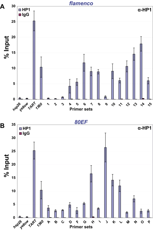
ChIP at (A) flam and (B) 80EF piRNA clusters in wild type ovaries with antibodies specific to HP1 (blue) and normal rabbit IgG (red). HP1 chromatin association is not affected greatly by depletion of Piwi in somatic ovarian follicle cells
We wished to address whether HP1 association with piRNA clusters is dependent on Piwi in the gonad, which express high levels of both proteins. Due to a complete loss of germ cells and the severe underdevelopment of ovary tissue in piwi mutants, it was not possible to obtain enough mutant material to perform ChIP. Therefore, we examined the recruitment of HP1 to chromatin in an ovarian somatic follicle cell line (OSC) that expresses Piwi but not Aub or AGO3 and produces only primary piRNAs, a large proportion of which derive from the flam locus [30]. The majority of Piwi was depleted from OSC cells by siRNA-mediated knockdown, and depletion of Piwi does not affect HP1 or Lamin protein levels compared to mock transfected cells (Figure 5A).
Fig. 5. Depletion of Piwi from ovarian somatic follicle cells does not affect HP1 recruitment to piRNA clusters. 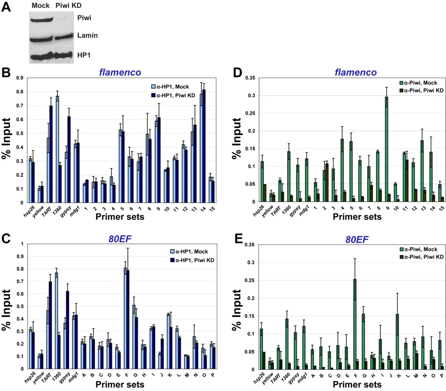
(A) Western blotting of Piwi, HP1 and Lamin in OSC cells that were either mock treated (left lane) or treated with siRNA directed against piwi (right lane, Piwi KD). ChIP at flam (B,D) and 80EF (C,E) piRNA clusters in mock treated and Piwi KD OSC cells with antibodies specific to HP1 (B,C) or Piwi (D,E). Subsequently, we investigated HP1 recruitment to piRNA clusters by ChIP in OSC cells. In mock treated cells, low levels of hsp26 and yellow are immunoprecipitated with HP1, while TART and 1360 levels are enriched above the euchromatic genes by 1.5 - to over two-fold (Figure 5B and 5C). Two additional TEs tested, gypsy and mdg1, are immunoprecipitated at similar levels to TART with HP1 (Figure 5B–5E). At flam, HP1 associates with the piRNA cluster similar to TE levels (Figure 5B and 5C). Despite much lower piRNA production from the 80EF cluster in OSC compared to flam [30], HP1 associates with piRNA producing regions of 80EF at similar levels to flam and TEs (Figure 5C, primer sets A-P). Overall, the HP1 recruitment profile in OSC is similar to that of heads and whole ovaries albeit at lower relative levels. In Piwi knockdown cells, no significant differences are seen for HP1 recruitment to all sites compared to mock treated cells except a two-fold decrease at the 1360 element. Rabbit IgG negative control immunoprecipitations yielded low amounts of DNA for all sites tested (<0.06% and <0.07% input for mock and Piwi knockdown cells, respectively).
Importantly, Piwi association with chromatin is detectable in OSC cells, but its profile differs from that of HP1. In mock treated cells, antibodies directed against Piwi [22] immunoprecipitate euchromatic sites at levels similar to that of TEs (Figure 5D and 5E). Furthermore, the majority of regions producing piRNA at flam is also immunoprecipitated at comparable levels to both euchromatic sites and TEs (Figure 5D). Moreover, levels of Piwi association with 80EF is akin to that of flam, while several sites in both flam and 80EF clusters show particular enrichment of Piwi up to three-fold compared to the average association with other sites tested (Figure 5D and 5E). In Piwi knockdown cells, Piwi chromatin association drops two to five-fold, down to background levels at all sites except for some residual association with two sites in or near the flam locus. Mouse IgG negative control immunoprecipitations yielded low amounts of DNA in comparison to α-Piwi immunoprecipitations in mock treated cells for all sites tested (<0.04% and <0.02% input for mock and Piwi knockdown cells, respectively). We conclude that in ovarian somatic follicle cells, reduction of the total pool of Piwi as well as the chromatin bound fraction does not affect HP1 association with piRNA clusters and has a minimal effect on HP1 association with TE chromatin association.
Loss of piRNA production from a single cluster results in global HP1 mislocalization
We next sought to determine whether loss of piRNA production at a single piRNA cluster would affect HP1 recruitment to chromatin. Previous studies have shown that mutation of various RNA silencing components results in global mislocalization of HP1 on polytene chromosomes [35]–[36]. Mutation of flam has been previously shown to result in loss of piRNA production [20] and upregulation of the gypsy retroelement [32]. In order to obtain a genome-wide view of HP1 chromatin association in flam mutants, we examined the localization of HP1 to highly replicated salivary gland polytene chromosomes from either wild type or flam1 mutant third instar larvae by indirect immunofluorescence using α-HP1 antibodies. In wild type, HP1 localizes predominantly to a concentration of heterochromatin where the centromeres of each chromosome coalesce, termed the chromocenter (Figure 6A, green). In contrast, flam1 mutants display expansion of HP1 at the chromocenter. Spreading of HP1 is apparent on the second and third chromosomes, but not on the X chromosome, where flam is located. As a reference, we also examined the localization of the chromatin insulator protein Mod(mdg4)2.2, which is unchanged in localization between wild type and flam1 (Figure 6A, red). The extent of HP1 chromocenter expansion is comparable to the level of HP1 expansion that we observe in spn-EhlsE1/spn-EhlsE616 mutants (Figure S7). A lesser degree of HP1 expansion was also observed in flamBG02658/flamKG00476 mutants (data not shown). Finally, total HP1 levels are unchanged in flam1 whole flies compared to wild type (Figure S6). These results indicate a global change in HP1 localization resulting from inactivation of a single piRNA cluster.
Fig. 6. Mutation of the flam piRNA cluster results in global HP1 redistribution. 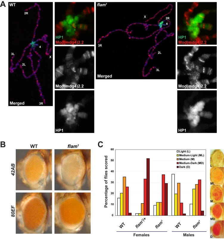
(A) Wild type (left) and flam1 (right) polytene chromosomes stained with antibodies directed against HP1 (green) and a reference protein Mod(mdg4)2.2 (red). DNA is stained with DAPI (blue). Chromosome arms are labeled, and insets of the enlarged chromocenter are shown. (B) Adult eyes of wild type and flam1 mutants carrying a mini-white transgene inserted in 42AB (top row) and 80EF (bottom) piRNA clusters. (C) Degree of eye pigmentation due to expression of the DX1 transgene array at 50C on chromosome 2L, which undergoes repeat-induced heterochromatic silencing, in wild type, flam1/+, and flam1 female flies and wild type and flam1 male flies. Scoring of variegation in the eye is categorized into five groups that range between light (few pigmented facets) to dark (almost all pigmented facets). Percentage of flies falling into each category was graphed. Representative eyes are shown on right. We reasoned that accumulation of HP1 at the chromocenter of flam1 mutants may result in an increase in silencing at pericentromeric sites. Therefore, the expression of transcriptional reporters at 42AB or 80EF piRNA clusters, which are located on different chromosomes from the flam locus, was examined in flam1 mutants. Compared to wild type, flam1 mutants harboring a P element insertion at either 42AB or 80EF piRNA clusters display mildly decreased pigmentation suggesting increased silencing at these distinct pericentromeric loci (Figure 6B).
Mutation of the flam piRNA cluster suppresses heterochromatic silencing at a distant site
Finally, to verify HP1 genome-wide redistribution in flam1mutants, we examined the effect of flam1 on the silencing of a centromere distal heterochromatic site on a different chromosome. The DX1 transgene array consists of seven mini-white P elements with one inverted copy at a normally euchromatic site at 50C on chromosome 2R [43]. Due to this configuration, the array forms ectopic repeat induced heterochromatin and displays a variegated phenotype similar to PEV that is dependent on HP1. Expression of the DX1 array was assessed based on variegation of eye pigmentation in wild type, heterozygous flam1/+, and homozygous or hemizygous flam1 mutants (Figure 6C). Due to a wide range of eye coloration, variegation was scored by categorization into five groups that ranged between Light (few pigmented facets) to Dark (almost all facets pigmented). For females, 3% of wild type was classified as Dark, while 29% of flam1/+ and 52% of flam1 mutants displayed the same high level of pigmentation. In males, 15% of wild type was scored as Medium-Dark or Dark while 40% of flam1 males fell into these categories. These results indicate that mutation of flam can suppress heterochromatic silencing in trans. Taken together with the HP1 centromeric expansion in polytene chromosomes and increased pericentromeric silencing in flam1 mutants, there appears to be a global redistribution of HP1 resulting from the loss of piRNA production from a single locus.
Discussion
In this study, we tested directly whether the Argonautes AGO2 or Piwi recruit HP1 to chromatin. As candidate sites for Argonaute/HP1 interaction, we examined whether piRNA clusters may be heterochromatic using both genetic and molecular approaches. First, P elements inserted at or near pericentromeric piRNA clusters were assayed as transcriptional reporters, and these transgenes were found to display variegated expression that is increased in heterochromatin mutants. Next, ChIP with α-HP1 antibodies showed that HP1 associates with piRNA clusters at levels significantly above euchromatic sites. However, mutation of piwi, aub, or AGO2 leads to a modest increase in silencing of transcriptional reporters as well as an increase of HP1 association at piRNA clusters in heads. In ovarian somatic follicle cells, in which both Piwi and HP1 are highly expressed, depletion of Piwi results in little or no change in HP1 recruitment to piRNA clusters and TEs. Furthermore, loss of piRNA production at a single locus results in expansion of HP1 at the centromere. In these flam1 mutants, silencing of a distant heterochromatic transgene array is reduced, further indicating a global redistribution of HP1 and suggesting indirect effects. Taken together, the results argue against direct recruitment of HP1 or maintenance of its association by AGO2 or Piwi in the soma.
AGO2 and Piwi are not required for HP1 association at piRNA clusters
Several reasons dictated the choice of piRNA clusters as the focus of our analyses. First, both endo-siRNAs and piRNAs are generated from these loci [16]–[21]. Next, we reasoned that at least some piRNA clusters are likely to be heterochromatic because of their strong bias toward TE-rich pericentromeric positions in the genome [20],[21], in close proximity to the vast majority of HP1 localization. In fact, early cloning attempts determined that the flam locus is located in a repetitive, TE rich heterochromatic region [44]. Furthermore, the pericentromeric position of these clusters likely coincides with the transition between euchromatin and heterochromatin, corresponding to the borders of HP1 spreading. This characteristic allows variegation assays, which monitor the variable spreading of HP1 and heterochromatin, to be extremely sensitive. ChIP assays at the borders of HP1 spreading would also likely be optimally sensitive to both local and overall changes in HP1 chromatin association. Finally, piRNA clusters contain enough unique sequence for specific primer design and monitoring by directed ChIP analysis.
Given that AGO2 is the predominant Argonaute expressed outside the gonad that participates in the silencing of TEs in the soma, we tested whether AGO2 could recruit HP1 to chromatin in somatic tissue. Moreover, it has been shown that AGO2 mutants exhibit mislocalization of HP1 [34],[35]. However, our results show that mutation of AGO2 results in a strong increase of silencing of transcriptional reporters at or near piRNA clusters and a mild increase of HP1 chromatin association in heads. Given the extent of increased silencing in the AGO2 mutant compared to piwi or aub mutants, which accumulate HP1 on chromatin to a similar degree, a posttranscriptional step of silencing likely contributes to the negative effects observed on transcriptional reporters. AGO2 mutants show a plethora of cellular defects during early nuclear divisions but develop normally and are fertile suggesting that effects on these various processes as well as HP1 localization are mild or otherwise compensated [34]. Therefore, AGO2 is unlikely to be required for HP1 recruitment in this tissue.
Additionally, we find that HP1 association at piRNA clusters does not depend on the presence of Piwi. Our analysis of piRNA clusters included flam, a primary piRNA cluster, and 80EF, a germline piRNA producing locus. We examined both flam and 80EF clusters in somatic head tissue and ovaries, which are a mixed population of somatic follicle and germline derived cells. In heads, there is no apparent requirement for piwi with respect to HP1 recruitment to the piRNA clusters or to TEs that were examined.
In our study, Piwi chromatin association was detected only in OSC cells, and its presence is dispensable for HP1 chromatin association. The flam piRNA cluster produces high levels of primary piRNA in OSC while 80EF is active for piRNA production in germ cells but not in OSC [25],[26],[30]. Nonetheless, Piwi associates with both the flam and 80EF clusters at comparable levels, suggesting that the amount of piRNA production from a particular locus does not correlate with Piwi chromatin association. Furthermore, the pattern of Piwi chromatin association in OSC differs from that of HP1 in that there is no particular enrichment of Piwi at TEs above euchromatic sites and only a minor accumulation at a few sites in the flam and 80EF piRNA clusters. When Piwi levels were reduced by siRNA knockdown, Piwi chromatin association was essentially abolished but HP1 recruitment was not affected except for a two-fold decrease over the 1360 element. Previous studies suggested that the 1360 element may be responsible for nucleating heterochromatin on the largely heterochromatic fourth chromosome and further showed that mutation of factors representing all RNA silencing pathways, piwi, aub, spn-E, Dcr-1, and Dcr-2, affect 1360 dependent heterochromatic silencing [40],[45]. Unlike the results in adult heads, no accumulation of HP1 over piRNA clusters was detected as a result of Piwi knockdown in OSC cells. This discrepancy may reflect differential effects in distinct cell types or the length of the Piwi knockdown in OSC cells, which was at least adequate to essentially eliminate Piwi chromatin association. In a related but independently derived ovarian somatic follicle cell line (OSS), Piwi and HP1 do not colocalize in the nucleus [33], and this finding supports the conclusion that Piwi does not direct HP1 recruitment in this cell type. Also consistent with our results, HP1 remains localized to the chromocenter in salivary gland polytene chromosomes in piwi null mutants [11],[36]. We conclude that association of HP1 with chromatin can occur independently of AGO2 and piwi in somatic tissue.
A previous study addressed the role of the germline piRNA pathway in HP1 association with transposable elements. The spn-E gene controls predominantly germline piRNA production but does not affect the somatic piRNA pathway [26]. ChIP was used to show that spn-E mutants display significantly decreased levels of H3K9me3 and HP1 at telomeric Het-A but similar to wild type HP1 levels at the I-element and copia TEs, which are distributed throughout the genome [46]. This modest reduction of HP1 at Het-A was apparent in ovaries but not in carcasses, which contain only somatic tissue. One caveat to this study is that ChIP was performed using primers that detect all TEs matching a particular sequence, thus measuring average HP1 and H3K9me levels on TEs across the genome. Nonetheless, this work suggests a limited role for the germline piRNA pathway in HP1 recruitment at the telomere.
Additional candidate platforms for Piwi-dependent HP1 recruitment
Several studies have shown that Piwi associates with at least some heterochromatic sites in the genome, but direct evidence that any of these sites serve as recruitment platforms for HP1 and subsequent spreading is lacking. The best characterized Piwi-associated site is the heterochromatic 3R-TAS subtelomeric region, which generates the abundant Piwi bound 20nt 3R-TAS piRNA. Surprisingly, the role of piwi at this location is transcriptional activation, as piwi mutants display increased transcriptional silencing of a nearby reporter transgene as well as an increase of HP1 association at 3R-TAS [21]. Likewise, we observe a mild corresponding increase in HP1 association and silencing at piRNA clusters in piwi mutants suggesting that piwi function could in fact oppose HP1 recruitment at multiple sites in the genome. Our results are consistent with the possibility that piRNA clusters act as boundaries to the spread of pericentromeric heterochromatin. The mechanism of Piwi dependent transcriptional activation has not been determined, but considering that Piwi interacts with the chromoshadow domain of HP1 [11], Piwi may compete for binding with other HP1 interactors such as Su(var)3–9 that promote heterochromatic silencing.
Functions for piwi outside of the gonad
The majority of Piwi protein is found in both somatic and germline tissues of the gonad, yet piwi clearly exerts an effect on non-gonadal somatic tissues as well. RT-PCR analysis shows that piwi transcript is readily detectable outside the gonad and in somatic cell lines [11],[12], but Piwi protein is difficult to detect [11]. Nevertheless, mutation of piwi suggests important functions for this gene outside of the gonad. For example, piwi is essential for viability, and loss-of-function mutants display a variety of phenotypes manifest in various non-gonadal somatic tissues such as demonstrated in this study and others, which show a requirement for piwi in pairing-dependent silencing, nucleolar integrity, and chromatin insulator function [47]–[50]. For each of these chromatin related studies, it remains a possibility that even a small amount of maternally deposited Piwi could trigger early events in the oocyte or embryo that persist throughout development, manifesting phenotypes visible in adult somatic tissues.
HP1 redistribution in piRNA pathway mutants
Our results along with previous studies have demonstrated that HP1 mislocalizes from the chromocenter in a subset of piRNA pathway mutants. We found that polytene chromosomes of flam1 mutants exhibit expanded HP1 chromocenter distribution. This result is intriguing because the flam1 mutation affects a single piRNA cluster on the X chromosome but HP1 spreading to other chromosomes is apparent. A previous study detected spreading of HP1 to euchromatic arms especially in spn-E mutants [36], and we confirmed this result albeit to a lesser degree, with spreading being comparable to the extent seen in flam1 mutants. Perhaps the increase of TE expression in RNA silencing mutants can stimulate HP1 recruitment and spreading from the centromere, which contains the highest concentration of TEs. In fact, transcription of pericentromeric repeats stimulates RNAi-dependent heterochromatin formation in fission yeast [51]–[53].
Redistribution of HP1 in RNA silencing mutants may indirectly affect silencing at various heterochromatic locations in the genome. Seemingly inconsistent with HP1 spreading, spn-E, aub, and piwi mutants display decreased silencing of P element transgene arrays such as DX1 and single insertions at pericentromeric regions on chromosomes 2 and 4 [36]. In our study, we found that mutation of flam also results in loss of silencing at DX1, which is distant from the flam locus. This reduced silencing in trans could not be due to posttranscriptional events as there are no shared sequences between DX1 and the flam locus. Therefore, we consider the possibility that there exists a finite pool of HP1 that accumulates at the centromere in flam and other RNA silencing mutants at the cost of reduced density and reduced silencing at other heterochromatic regions such as the transgene array, the fourth chromosome, and the telomere. The concept of a limited population of HP1 was suggested previously to explain the finding that the Y chromosome behaves as a suppressor of variegation by acting as a sink for HP1 [43].
Conclusions
Studies in multiple organisms have identified or suggested alternative mechanisms to RNA silencing for the recruitment of HP1 to chromatin. In fission yeast, overlapping and redundant RNAi-dependent and independent mechanisms of heterochromatin formation have been elucidated. In mouse cells, HP1 localization to pericentromeric heterochromatin was found to be RNase A sensitive suggesting that an RNA moiety may be involved in HP1 recruitment [54]. Our data indicate that heterochromatin can form independently of RNA silencing in Drosophila. It will be interesting to determine if any of these alternative mechanisms of heterochromatin formation are conserved throughout evolution.
Materials and Methods
Drosophila stocks
Fly stocks were maintained at 25°C on standard cornmeal medium. Lines containing P{EPgy2}DIP1EY02625 and P{EPgy2}EY08366 were obtained from the Bloomington Drosophila Stock Center, and a line harboring PBac{PB}c06482 was obtained from the Exelixis Collection at Harvard Medical School. Genomic coordinates of these P-element insertions were confirmed by PCR with primers specific to the P-elements and flanking genomic sequences followed by sequencing. For transcriptional reporter assays, transgenes were crossed or recombined into mutant backgrounds and scored against crosses to yw67c23 as a reference. For ChIP and immunofluorescence, Oregon-R was used as a wild type control. The y v f mal flam1/FM3 stock was selected for heterozygous females each generation to prevent mobilization and accumulation of TEs. For the DX1 variegation assay, DX1/CyO was crossed to y w v f mal flam1/FM7c; CyO/Sp flies or yw67c23; CyO/Sp as a reference.
Transcriptional reporter and eye pigmentation assays
Eye pigmentation of 40 to 60 adult males six days of age was examined, and representative eye photos were taken. To quantify overall levels of eye pigmentation, the heads of 25 male flies of each genotype were dissected, and eye pigmentation was measured as previously described [36]. Briefly, heads were homogenized in 0.8 ml of methanol, acidified with 0.1% HCl and centrifuged. The absorbance of the supernatant was measured at 480 nm.
Chromatin immunoprecipitation
Adult fly heads or ovaries were dissected and crosslinked with 1.8% formaldehyde for 20 min at 23°C. Chromatin was fragmented to an average size of 300 bp by sonication and incubated with antibodies overnight at 4°C. Quantitative PCR was conducted on Applied Biosystems Real Time PCR system using SYBR Green incorporation (Affymetrix/USB). Amplicon sizes ranged between 150 and 250 bp. Chromatin was immunoprecipitated with the following antibodies: α-HP1 (Covance), α-Su(Hw), α-Piwi (P3G11, a gift from M. Siomi), and normal rabbit or mouse IgG (Santa Cruz Biotechnology). A recombinant N-terminal His-tagged fusion protein of the N-terminal of Su(Hw) (amino acids 1–218, kind gift of M. Labrador) was purified from E.coli on a nickel-agarose column and used to immunize guinea pigs using standard procedures. Similar results were obtained using the C1A9 α-HP1 antibody (Developmental Studies Hybridoma Bank), but lower quantities of DNA were obtained. Fifty to one hundred fly heads and twenty five to fifty ovaries were used per IP. The quantities of target genomic regions precipitated by different antibodies were calculated as percent input based on four-point standard curves constructed from input DNA for each primer set. Standard deviation of each PCR performed in quadruplicate was calculated to determine the error of measurement. Two independent ChIP samples were analyzed, and similar results were obtained. ChIP primers were designed to be unique, detecting only sequences present in the flam and 80EF piRNA loci and verified by in silico PCR. All primers (Table S1 and Table S2) were checked for both specificity and efficiency by standard agarose gel electrophoresis and real time PCR respectively. Primers to piRNA clusters amplify in the same DNA dilution range as primers specific to hsp26 and yellow single copy genes compared to high copy TE elements (Figure S8). Primers to the flam locus were verified to amplify approximately two-fold more DNA from female compared to male genomic DNA. A detailed version of this protocol is available in Text S1.
Culture of OSC cell line and siRNA knockdowns
The OSC line was maintained and Piwi siRNA knockdown was performed as previously described [30]. Briefly, 3×10⁶ trypsinized cells were resuspended in 0.1 mL of Solution V of the Cell Line Nucleofector Kit V (Amaxa Biosystems) and mixed with 200 pmol of siRNA duplex. Transfection was conducted according to the manufacturer's protocol using the nucleofector program T-029, and the transfected cells were incubated at 25°C for 48 hrs. Protein knockdowns were verified by Western blotting, and ChIP assays were performed on mock and piwi siRNA transfected cells (5×10⁶ cells per IP).
Immunostaining of polytene chromosomes
Preparation and immunostaining of salivary gland polytene chromosomes was performed as described previously [55]. Primary antibodies directed against HP1 (Covance) and Mod(mdg4)2.2 (generated similarly as in [56]) and Alexa Fluor 488 labeled anti-guinea pig or Alexa Fluor 594 labeled anti-rabbit secondary antibodies (Invitrogen-Molecular Probes) were used. The chromosomes were viewed using a Leica epifluorescence microscope and photographed using a Hamamatsu digital camera.
DX1 variegation assay
Eye pigmentation of 100 to 200 flies was scored. The scoring of variegation was categorized into five groups: Light, Medium-Light, Medium, Medium-Dark and Dark corresponding to the percentage of pigmented facets. Percentage of flies falling into each category was graphed. Representative eye photos were taken.
Supporting Information
Zdroje
1. GrewalSI
ElginSC
2007 Transcription and RNA interference in the formation of heterochromatin. Nature 447 399 406
2. MasonJM
FrydrychovaRC
BiessmannH
2008 Drosophila telomeres: an exception providing new insights. Bioessays 30 25 37
3. TschierschB
HofmannA
KraussV
DornR
KorgeG
1994 The protein encoded by the Drosophila position-effect variegation suppressor gene Su(var)3-9 combines domains of antagonistic regulators of homeotic gene complexes. EMBOJ 13 3822 3831
4. EissenbergJC
JamesTC
Foster-HartnettDM
HartnettT
NganV
1990 Mutation in a heterochromatin-specific chromosomal protein is associated with suppression of position-effect variegation in Drosophila melanogaster. Proc Natl Acad SciUSA 87 9923 9927
5. VolpeTA
KidnerC
HallIM
TengG
GrewalSI
2002 Regulation of heterochromatic silencing and histone H3 lysine-9 methylation by RNAi. Science 297 1833 1837
6. VerdelA
JiaS
GerberS
SugiyamaT
GygiS
2004 RNAi-mediated targeting of heterochromatin by the RITS complex. Science 303 672 676
7. NomaK
SugiyamaT
CamH
VerdelA
ZofallM
2004 RITS acts in cis to promote RNA interference-mediated transcriptional and post-transcriptional silencing. Nat Genet 36 1174 1180
8. JiaS
NomaK
GrewalSI
2004 RNAi-independent heterochromatin nucleation by the stress-activated ATF/CREB family proteins. Science 304 1971 1976
9. KanohJ
SadaieM
UranoT
IshikawaF
2005 Telomere binding protein Taz1 establishes Swi6 heterochromatin independently of RNAi at telomeres. Curr Biol 15 1808 1819
10. HutvagnerG
SimardMJ
2008 Argonaute proteins: key players in RNA silencing. Nat Rev Mol Cell Biol 9 22 32
11. Brower-TolandB
FindleySD
JiangL
LiuL
YinH
2007 Drosophila PIWI associates with chromatin and interacts directly with HP1a. Genes Dev 21 2300 2311
12. RehwinkelJ
NatalinP
StarkA
BrenneckeJ
CohenSM
2006 Genome-wide analysis of mRNAs regulated by Drosha and Argonaute proteins in Drosophila melanogaster. Mol Cell Biol 26 2965 2975
13. WilliamsRW
RubinGM
2002 ARGONAUTE1 is required for efficient RNA interference in Drosophila embryos. Proc Natl Acad SciUSA 99 6889 6894
14. WangL
LigoxygakisP
2006 Pathogen recognition and signalling in the Drosophila innate immune response. Immunobiology 211 251 261
15. HammondSM
BoettcherS
CaudyAA
KobayashiR
HannonGJ
2001 Argonaute2, a link between genetic and biochemical analyses of RNAi. Science 293 1146 1150
16. CzechB
MaloneCD
ZhouR
StarkA
SchlingeheydeC
2008 An endogenous small interfering RNA pathway in Drosophila. Nature 453 798 802
17. KawamuraY
SaitoK
KinT
OnoY
AsaiK
2008 Drosophila endogenous small RNAs bind to Argonaute 2 in somatic cells. Nature 453 793 797
18. ChungWJ
OkamuraK
MartinR
LaiEC
2008 Endogenous RNA interference provides a somatic defense against Drosophila transposons. Curr Biol 18 795 802
19. GhildiyalM
SeitzH
HorwichMD
LiC
DuT
2008 Endogenous siRNAs derived from transposons and mRNAs in Drosophila somatic cells. Science 320 1077 1081
20. BrenneckeJ
AravinAA
StarkA
DusM
KellisM
2007 Discrete small RNA-generating loci as master regulators of transposon activity in Drosophila. Cell 128 1089 1103
21. YinH
LinH
2007 An epigenetic activation role of Piwi and a Piwi-associated piRNA in Drosophila melanogaster. Nature 450 304 308
22. SaitoK
NishidaKM
MoriT
KawamuraY
MiyoshiK
2006 Specific association of Piwi with rasiRNAs derived from retrotransposon and heterochromatic regions in the Drosophila genome. Genes Dev 20 2214 2222
23. GunawardaneLS
SaitoK
NishidaKM
MiyoshiK
KawamuraY
2007 A slicer-mediated mechanism for repeat-associated siRNA 5′ end formation in Drosophila. Science 315 1587 1590
24. VaginVV
SigovaA
LiC
SeitzH
GvozdevV
2006 A distinct small RNA pathway silences selfish genetic elements in the germline. Science 313 320 324
25. LiC
VaginVV
LeeS
XuJ
MaS
2009 Collapse of germline piRNAs in the absence of Argonaute3 reveals somatic piRNAs in flies. Cell 137 509 521
26. MaloneCD
BrenneckeJ
DusM
StarkA
McCombieWR
2009 Specialized piRNA pathways act in germline and somatic tissues of the Drosophila ovary. Cell 137 522 535
27. HarrisAN
MacdonaldPM
2001 Aubergine encodes a Drosophila polar granule component required for pole cell formation and related to eIF2C. Development 128 2823 2832
28. CoxDN
ChaoA
LinH
2000 piwi encodes a nucleoplasmic factor whose activity modulates the number and division rate of germline stem cells. Development 127 503 514
29. KlattenhoffC
XiH
LiC
LeeS
XuJ
2009 The Drosophila HP1 homolog Rhino is required for transposon silencing and piRNA production by dual-strand clusters. Cell 138 1137 1149
30. SaitoK
InagakiS
MituyamaT
KawamuraY
OnoY
2009 A regulatory circuit for piwi by the large Maf gene traffic jam in Drosophila. Nature 461 1296 1299
31. DessetS
MeigninC
DastugueB
VauryC
2003 COM, a heterochromatic locus governing the control of independent endogenous retroviruses from Drosophila melanogaster. Genetics 164 501 509
32. Prud'hommeN
GansM
MassonM
TerzianC
BuchetonA
1995 Flamenco, a gene controlling the gypsy retrovirus of Drosophila melanogaster. Genetics 139 697 711
33. LauNC
RobineN
MartinR
ChungWJ
NikiY
2009 Abundant primary piRNAs, endo-siRNAs, and microRNAs in a Drosophila ovary cell line. Genome Res 19 1776 1785
34. DeshpandeG
CalhounG
SchedlP
2005 Drosophila argonaute-2 is required early in embryogenesis for the assembly of centric/centromeric heterochromatin, nuclear division, nuclear migration, and germ-cell formation. Genes Dev 19 1680 1685
35. FagegaltierD
BougeAL
BerryB
PoisotE
SismeiroO
2009 The endogenous siRNA pathway is involved in heterochromatin formation in Drosophila. Proc Natl Acad SciUSA 106 21258 21263
36. Pal-BhadraM
LeibovitchBA
GandhiSG
RaoM
BhadraU
2004 Heterochromatic silencing and HP1 localization in Drosophila are dependent on the RNAi machinery. Science 303 669 672
37. Giles KE, Ghirlando R, Felsenfeld G Maintenance of a constitutive heterochromatin domain in vertebrates by a Dicer-dependent mechanism. Nat Cell Biol 12 94 99; sup pp 91-96
38. RosemanRR
PirrottaV
GeyerPK
1993 The su(Hw) protein insulates expression of the Drosophila melanogaster white gene from chromosomal position-effects. EMBOJ 12 435 442
39. FantiL
DorerDR
BerlocoM
HenikoffS
PimpinelliS
1998 Heterochromatin protein 1 binds transgene arrays. Chromosoma 107 286 292
40. SunFL
HaynesK
SimpsonCL
LeeSD
CollinsL
2004 cis-Acting determinants of heterochromatin formation on Drosophila melanogaster chromosome four. Mol Cell Biol 24 8210 8220
41. ParnellTJ
VieringMM
SkjesolA
HelouC
KuhnEJ
2003 An endogenous suppressor of hairy-wing insulator separates regulatory domains in Drosophila. Proc Natl Acad SciUSA 100 13436 13441
42. MiyoshiK
TsukumoH
NagamiT
SiomiH
SiomiMC
2005 Slicer function of Drosophila Argonautes and its involvement in RISC formation. Genes Dev 19 2837 2848
43. DorerDR
HenikoffS
1994 Expansions of transgene repeats cause heterochromatin formation and gene silencing in Drosophila. Cell 77 993 1002
44. RobertV
Prud'hommeN
KimA
BuchetonA
PelissonA
2001 Characterization of the flamenco region of the Drosophila melanogaster genome. Genetics 158 701 713
45. HaynesKA
CaudyAA
CollinsL
ElginSC
2006 Element 1360 and RNAi components contribute to HP1-dependent silencing of a pericentric reporter. Curr Biol 16 2222 2227
46. KlenovMS
LavrovSA
StolyarenkoAD
RyazanskySS
AravinAA
2007 Repeat-associated siRNAs cause chromatin silencing of retrotransposons in the Drosophila melanogaster germline. Nucleic Acids Res 35 5430 5438
47. PengJC
KarpenGH
2007 H3K9 methylation and RNA interference regulate nucleolar organization and repeated DNA stability. Nat Cell Biol 9 25 35
48. LeiEP
CorcesVG
2006 RNA interference machinery influences the nuclear organization of a chromatin insulator. Nat Genet 38 936 941
49. Pal-BhadraM
BhadraU
BirchlerJA
2002 RNAi related mechanisms affect both transcriptional and posttranscriptional transgene silencing in Drosophila. Mol Cell 9 315 327
50. GrimaudC
BantigniesF
Pal-BhadraM
GhanaP
BhadraU
2006 RNAi components are required for nuclear clustering of Polycomb group response elements. Cell 124 957 971
51. ZofallM
GrewalSI
2006 Swi6/HP1 recruits a JmjC domain protein to facilitate transcription of heterochromatic repeats. Mol Cell 22 681 692
52. ChenES
ZhangK
NicolasE
CamHP
ZofallM
2008 Cell cycle control of centromeric repeat transcription and heterochromatin assembly. Nature 451 734 737
53. KlocA
MartienssenR
2008 RNAi, heterochromatin and the cell cycle. Trends Genet 24 511 517
54. MaisonC
BaillyD
PetersAH
QuivyJP
RocheD
2002 Higher-order structure in pericentric heterochromatin involves a distinct pattern of histone modification and an RNA component. Nat Genet 30 329 334
55. GerasimovaTI
ByrdK
CorcesVG
2000 A chromatin insulator determines the nuclear localization of DNA. Mol Cell 6 1025 1035
56. MongelardF
LabradorM
BaxterEM
GerasimovaTI
CorcesVG
2002 Trans-splicing as a novel mechanism to explain interallelic complementation in Drosophila. Genetics 160 1481 1487
57. LangmeadB
TrapnellC
PopM
SalzbergSL
2009 Ultrafast and memory-efficient alignment of short DNA sequences to the human genome. Genome Biol 10 R25
Štítky
Genetika Reprodukční medicína
Článek vyšel v časopisePLOS Genetics
Nejčtenější tento týden
2010 Číslo 3- Akutní intermitentní porfyrie
- Růst a vývoj dětí narozených pomocí IVF
- Vliv melatoninu a cirkadiálního rytmu na ženskou reprodukci
- Délka menstruačního cyklu jako marker ženské plodnosti
- Intrauterinní inseminace a její úspěšnost
-
Všechny články tohoto čísla
- Parental Genome Dosage Imbalance Deregulates Imprinting in
- Identification and Functional Analysis of the Vision-Specific BBS3 (ARL6) Long Isoform
- HAP2(GCS1)-Dependent Gamete Fusion Requires a Positively Charged Carboxy-Terminal Domain
- Initial Genomics of the Human Nucleolus
- Role of RecA and the SOS Response in Thymineless Death in
- PPS, a Large Multidomain Protein, Functions with Sex-Lethal to Regulate Alternative Splicing in
- Mislocalization of XPF-ERCC1 Nuclease Contributes to Reduced DNA Repair in XP-F Patients
- Transgenic Rat Model of Neurodegeneration Caused by Mutation in the Gene
- Human Population Differentiation Is Strongly Correlated with Local Recombination Rate
- Local-Scale Patterns of Genetic Variability, Outcrossing, and Spatial Structure in Natural Stands of
- Arginylation-Dependent Neural Crest Cell Migration Is Essential for Mouse Development
- HP1 Recruitment in the Absence of Argonaute Proteins in
- MiR-218 Inhibits Invasion and Metastasis of Gastric Cancer by Targeting the Robo1 Receptor
- Bias and Evolution of the Mutationally Accessible Phenotypic Space in a Developmental System
- Papillorenal Syndrome-Causing Missense Mutations in / Result in Hypomorphic Alleles in Mouse and Human
- Rapid Assessment of Genetic Ancestry in Populations of Unknown Origin by Genome-Wide Genotyping of Pooled Samples
- Regulation of Lifespan, Metabolism, and Stress Responses by the SH2B Protein, Lnk
- KRAB–Zinc Finger Proteins and KAP1 Can Mediate Long-Range Transcriptional Repression through Heterochromatin Spreading
- Identification of the Regulatory Logic Controlling Pathoadaptation by the SsrA-SsrB Two-Component System
- Drosophila Xpd Regulates Cdk7 Localization, Mitotic Kinase Activity, Spindle Dynamics, and Chromosome Segregation
- Multiple Signals Converge on a Differentiation MAPK Pathway
- In the Tradition of Science: An Interview with Victor Ambros
- Association of the Polymorphism His615Arg with Melanin Content in East Asian Populations: Further Evidence of Convergent Evolution of Skin Pigmentation
- Fatal Cardiac Arrhythmia and Long-QT Syndrome in a New Form of Congenital Generalized Lipodystrophy with Muscle Rippling (CGL4) Due to Mutations
- Deciphering Normal Blood Gene Expression Variation—The NOWAC Postgenome Study
- Derepression of the Plant Chromovirus Induces Germline Transposition in Regenerated Plants
- PLOS Genetics
- Archiv čísel
- Aktuální číslo
- Informace o časopisu
Nejčtenější v tomto čísle- Deciphering Normal Blood Gene Expression Variation—The NOWAC Postgenome Study
- Transgenic Rat Model of Neurodegeneration Caused by Mutation in the Gene
- Papillorenal Syndrome-Causing Missense Mutations in / Result in Hypomorphic Alleles in Mouse and Human
- Fatal Cardiac Arrhythmia and Long-QT Syndrome in a New Form of Congenital Generalized Lipodystrophy with Muscle Rippling (CGL4) Due to Mutations
Kurzy
Zvyšte si kvalifikaci online z pohodlí domova
Autoři: prof. MUDr. Vladimír Palička, CSc., Dr.h.c., doc. MUDr. Václav Vyskočil, Ph.D., MUDr. Petr Kasalický, CSc., MUDr. Jan Rosa, Ing. Pavel Havlík, Ing. Jan Adam, Hana Hejnová, DiS., Jana Křenková
Autoři: MUDr. Irena Krčmová, CSc.
Autoři: MDDr. Eleonóra Ivančová, PhD., MHA
Autoři: prof. MUDr. Eva Kubala Havrdová, DrSc.
Všechny kurzyPřihlášení#ADS_BOTTOM_SCRIPTS#Zapomenuté hesloZadejte e-mailovou adresu, se kterou jste vytvářel(a) účet, budou Vám na ni zaslány informace k nastavení nového hesla.
- Vzdělávání


