-
Články
Top novinky
Reklama- Vzdělávání
- Časopisy
Top články
Nové číslo
- Témata
Top novinky
Reklama- Kongresy
- Videa
- Podcasty
Nové podcasty
Reklama- Kariéra
Doporučené pozice
Reklama- Praxe
Top novinky
ReklamaThe Protease Cruzain Mediates Immune Evasion
Trypanosoma cruzi is the causative agent of Chagas' disease. Novel chemotherapy with the drug K11777 targets the major cysteine protease cruzain and disrupts amastigote intracellular development. Nevertheless, the biological role of the protease in infection and pathogenesis remains unclear as cruzain gene knockout failed due to genetic redundancy. A role for the T. cruzi cysteine protease cruzain in immune evasion was elucidated in a comparative study of parental wild type - and cruzain-deficient parasites. Wild type T. cruzi did not activate host macrophages during early infection (<60 min) and no increase in ∼P iκB was detected. The signaling factor NF-κB P65 colocalized with cruzain on the cell surface of intracellular wild type parasites, and was proteolytically cleaved. No significant IL-12 expression occurred in macrophages infected with wild type T. cruzi and treated with LPS and BFA, confirming impairment of macrophage activation pathways. In contrast, cruzain-deficient parasites induced macrophage activation, detectable iκB phosphorylation, and nuclear NF-κB P65 localization. These parasites were unable to develop intracellularly and survive within macrophages. IL 12 expression levels in macrophages infected with cruzain-deficient T. cruzi were comparable to LPS activated controls. Thus cruzain hinders macrophage activation during the early (<60 min) stages of infection, by interruption of the NF-κB P65 mediated signaling pathway. These early events allow T. cruzi survival and replication, and may lead to the spread of infection in acute Chagas' disease.
Published in the journal: . PLoS Pathog 7(9): e32767. doi:10.1371/journal.ppat.1002139
Category: Research Article
doi: https://doi.org/10.1371/journal.ppat.1002139Summary
Trypanosoma cruzi is the causative agent of Chagas' disease. Novel chemotherapy with the drug K11777 targets the major cysteine protease cruzain and disrupts amastigote intracellular development. Nevertheless, the biological role of the protease in infection and pathogenesis remains unclear as cruzain gene knockout failed due to genetic redundancy. A role for the T. cruzi cysteine protease cruzain in immune evasion was elucidated in a comparative study of parental wild type - and cruzain-deficient parasites. Wild type T. cruzi did not activate host macrophages during early infection (<60 min) and no increase in ∼P iκB was detected. The signaling factor NF-κB P65 colocalized with cruzain on the cell surface of intracellular wild type parasites, and was proteolytically cleaved. No significant IL-12 expression occurred in macrophages infected with wild type T. cruzi and treated with LPS and BFA, confirming impairment of macrophage activation pathways. In contrast, cruzain-deficient parasites induced macrophage activation, detectable iκB phosphorylation, and nuclear NF-κB P65 localization. These parasites were unable to develop intracellularly and survive within macrophages. IL 12 expression levels in macrophages infected with cruzain-deficient T. cruzi were comparable to LPS activated controls. Thus cruzain hinders macrophage activation during the early (<60 min) stages of infection, by interruption of the NF-κB P65 mediated signaling pathway. These early events allow T. cruzi survival and replication, and may lead to the spread of infection in acute Chagas' disease.
Introduction
Trypanosoma cruzi is the parasitic agent of Chagas' disease that affects approximately 12 million people throughout Latin America (WHO). Current chemotherapy with nifurtimox and benznidazole is unsatisfactory due to severe side effects that require medical supervision [1]–[3]. T. cruzi infection is classically transmitted by an insect vector, the reduviid bug [4]. Parasites disseminate from the insect bite site and, in the most common clinical course of Chagas' disease, infect cardiac myocytes leading to acute myocarditis or chronic infection with relentless cardiac failure. Fulminant disease is commonly seen with HIV infection or immunosuppression. In patients with immunodeficieny, parasites may be found in many organs, and a highly fatal meningoencephalitis often ensues. These observations led us to hypothesize that T. cruzi successfully evades the host immune response, and may in fact utilize unresponsive macrophages as a means of egress from the insect bite site prior to dissemination to other cell types [5]. While the specific mechanisms of immune evasion by T. cruzi remain largely unknown, several reports have suggested that the major protease of T. cruzi, cruzain, (a.k.a. cruzipain, GP57/51) is a key factor [6], [7].
Cruzain plays a biological role in cell remodeling during transformation of the insect epimastigote stage of T. cruzi to infectious metacyclic [8]–[11]. Scharfstein and collaborators found cruzain involvement during trypomastigote infection by the proteolytic release of kinin from host cells surfaces and activation of bradykinin receptors [12]–[14]. More recent reports suggested that cruzain mediates anti-apoptotic mechanisms in T. cruzi-infected myocardiocytes in vitro [15]. The biological role of cruzain in the intracellular amastigote stage of T. cruzi and in Chagas' disease pathogenesis remains nevertheless largely unknown. Assessments of the role of cruzain in T. cruzi pathogenicity have been hampered by genetic redundancy and the failure of gene deletion attempts [9], [12], [16], [17]. Successful knockout of the cruzain gene cluster has not been achieved to date leading to speculation that deletion of the cruzain coding genes might be lethal. Chemical knockout with cysteine protease inhibitors was also lethal for T. cruzi [18]. Although the role of auto-proteolysis in cruzain activation was confirmed, these studies did not allow elucidation of the biological role of this protease in human disease. Duschak et al. [19], [20] observed lower cruzain activity and protease sequence alterations in attenuated strains as compared to their virulent parental T. cruzi, while other authors found infectivity is not dependent on cruzain expression or activity [21]. An alternative approach to study the biological role of this vital gene product is to examine the phenotype of protease-deficient organisms. We generated a protease deficient T. cruzi that retained less than 1% of cruzain activity of the wild type parental clone [22]. We now report that cruzain deficient T. cruzi rapidly activate host macrophages via NF-κB P65 and are unable to survive intracellularly within macrophages. In contrast, infection with wild type parasites appears to induce cruzain-mediated proteolysis of NF-κB P65 leading to unresponsiveness of the host macrophage during early (<60 minutes) infection. This immune evasion mechanism may be critical for T. cruzi survival during early natural infection with a low number of trypmastigotes.
Materials and Methods
Parasites and cells
Wild type CA-I/72 T. cruzi was isolated from an Argentinean chronic chagasic patient and cloned [23]. Cruzain-deficient, and cysteine protease inhibitor (K11777)-resistant (CA-I/KR; KR) T. cruzi were derived from parental CA-I/72 parasites [22]. The cysteine protease inhibitor K11777 (K777, N-Methyl-Pip-F-hF-VSΦ) was kindly provided by J. Palmer, Celera, CA [18], [24]. Epimastigotes were maintained at 26°C in Brain-heart tryptose (BHT) medium [25] with 10% heat inactivated fetal calf serum (FCS), and with the addition of 20-fold the lethal dose (200 µM) of K11777 for phenotypic cruzain-deficient CA-I/KR.
Bovine embryo skeletal muscle cells (BESM) cells were a kind gift of J. A. Dvorak, NIH. Mammalian stages of wild type (WT) CA-I/72 were maintained in BESM cells with RPMI-1640 medium and 5% heat inactivated horse serum (HS) at 37°C [26]. Intracellular cruzain-deficient CA-I/KR T. cruzi were maintained serially in BESM as above but with the addition of 5% heat inactivated FCS, 5% heat inactivated horse serum, and 10 µM K11777. Long-term culture at higher concentrations of the inhibitor was toxic for BESM cells. For some experiments, the mammalian stages of cruzain-deficient CAI/KR were also cultured for 4 passages (2 months) in the absence of inhibitor. The approximate duration of the intracellular cycle of wild type CA-I/72 (4.5 days) [23] and CA-I/KR (18 days) was estimated in infected BESM cells by contrast phase microscopy and by fluorescence microscopy of propidium iodine stained slides. Cruzain-deficient parasites were unable to survive and develop within macrophages.
J774 mouse macrophages were from the UCSF cell culture facility. J774 cells were cultured as above and in some experiments irradiated (1000 Rad) 24 h prior to use to arrest cell division. Normal peritoneal macrophages were collected from C3H mice (The Jackson Lab).
Ethics statement
This study was carried out in strict accordance with the Guide for the Care and Use of Laboratory Animals of the National Institutes of Health. The protocol was approved by the Institutional Animal Care and Use Committee of the University of California, San Francisco (AN079928-02A).
Protease labeling
WT and cruzain-deficient epimastigotes were washed three times in PBS, counted in a Coulter Counter Multisizer 3 (Beckman) and taken to equal numbers of parasites/ml, lysed by 5 cycles of freeze-thawing, centrifuged at 14,000 xg for 1 h at 4°C [22], and frozen at −70°C until used. Fractions (105 epimastigotes/lane) were reacted with the iodinated inhibitor DCG04 [27], [28] and electrophoresed. Resolved molecular species were then isolated and analyzed by nano LC/MS, CID spectra, and/or fractionation by MALDI-TOF at the Protein Facility, UCSF. Protein fragments (9-17 aa long) were analyzed by NCBI blast.
Immunoelectron microscopy (IEM)
BESM cells infected with intracellular amastigotes of WT or cruzain-deficient T. cruzi were processed for IEM with a specific anti-cruzain polyclonal antibody as previously described [24]. Briefly, T. cruzi-infected BESM cells were collected by centrifugation, washed twice with PBS and fixed for 2 hours at 4°C with 2% paraformaldehyde-0.05% glutaraldehyde in 0.1 M phosphate buffer, pH 7.4. Cells were then cryoprotected, frozen, sectioned, and immunolabeled sequentially with rabbit polyclonal anti-cruzain Ab and goat anti-rabbit IgG-10 nm gold-labeled Ab (dilutions 1∶250 and 1∶500, respectively). Thin sections were observed in a Tecnai 10 (FEI Co.) electron microscope. Gold-labeled cruzain localizing to the amastigote cell surface was quantified in micrographs of intracellular parasites in three independent experiments (n = 55 wild type amastigotes; n = 55 cruzain-deficient amastigotes). The cell perimeter of sectioned intracellular amastigotes was measured with Openlab software (Improvision), and cruzain density was expressed as the number of gold labeled cruzain particles/µm of parasitic cell membrane. Results were statistically analyzed (t test).
Western blots (WB)
J774 macrophages were seeded onto 12 well tissue culture plates for 24 h prior to infection with WT or cruzain-deficient T. cruzi as appropriate. Infection was performed at a low ratio of 0.5 parasites/macrophage. Some infected cultures were treated with cysteine protease inhibitor. Controls consisted of J774 macrophage cultures that were uninfected, treated with purified LPS (150 ng/ml LPS, Sigma) [29], or treated with K11777. Samples were collected at 1 min, 30 min, 60 min, and 150 min, and at 48 h post-infection. Monolayers were washed twice with cold PBS, solubilized in 1 ml cold PBS with1% Triton X-100, scraped, aliquoted, immediately snap-frozen, and stored at −70°C until used. Samples were centrifuged at 14,000 xg for 30 min at 4°C and supernatants taken to identical protein concentration. Samples (1 mg protein/lane) were heated at 70°C for 5 min and resolved by Nupage electrophoresis (10% BisTris gels at 200 V, Mops Buffer) (Novex, Invitrogen) prior to transferring to PVDF membranes (35 V for 2 h). Blots were blocked overnight with 1% BSA (Sigma). Samples were blotted with rabbit anti–NF-κB P65 Ab, rabbit anti - iκB Ab, mouse anti-phosphorylated (∼P) iκB Ab (Santa Cruz Biotech, CA) and anti actin Ab (Cal Biochem). All methods were according to manufacturer's instructions (Santa Cruz Biotech, CA). Additional controls were similarly infected with Leishmania mexicana, a kind gift of J. Mottram (University of Glasgow, UK), for 48 hours. Results were confirmed in duplicate experiments according to methods described by Ma and colleagues [30]–[32]. Briefly, 5×10⁶ cells per point (1, 10, 30, and 60 min) were infected with T. cruzi trypomastigotes as above (ratio 0.5 parasites/cell). Non-infected macrophages either untreated (MΦ) or treated with LPS, and/or K11777 as indicated (10, 20, 40 and 60 min) were also used as controls. Macrophages were washed twice with PBS, scraped, transferred to eppendorf tubes, and centrifuged prior to lysis with NP40 lysis buffer (0.1% NP40, 1 mM Na vanadate and 5 mM Na fluoride−50 mM HEPES). Nuclei were then pelleted by centrifugation (20 min at 14,000 xg, 4°C) and discarded, and the supernatants were boiled in Laemli buffer for 10 min immediately prior to SDS-PAGE/WB (Cell Signalling Technology) [30], [31]. Membranes were developed sequentially with anti ∼PiκB Ab (Cell Signalling), anti-iκB Ab (Cell Signalling), and anti-actin Ab (Southern Biotech) [30].
Confocal microscopy
Duplicates of samples described above for WB and appropriate controls were simultaneously seeded onto 12 well tissue culture plates containing sterile round cover glasses. At 1 min, 30 min, 60 min, and 150 min post-infection, cells were washed, fixed with fresh 4% paraformaldehyde in PBS, rinsed in PBS, treated with 0.1% Triton X-100 in PBS for 5 min, rinsed, and processed for confocal microscopy with anti -mouse NF-κB P65Ab (Dil 1/100; Santa Cruz Biotech, CA) followed by Alexa 488 labeled secondary Ab and propidium iodine (Molecular Probes). Results are from 3–7 independent experiments (n = 2–3 slides per treatment). Confocal images were acquired with a Leica Laser Confocal microscope TCS-NP, using Leica software and identical parameters for all samples, namely, a 100X objective with a numerical aperture of 1.4 in a 1024×1024 format, a pinhole of 0.7 airy, and a Z section of 0.2 µm.
For colocalization studies of cruzain and NF-κB P65, macrophages infected for 30 min with WT T. cruzi and treated or not with K11777, or similarly infected with cruzain-deficient T. cruzi and treated or not with K11777, and non-infected controls were fixed and stained sequentially with rabbit anti-NF-κB P65 Ab (Dil 1/100; Santa Cruz Biotech) and Alexa 488-labeled secondary Ab, followed by rabbit polyclonal anti-cruzain antibody (Dil 1/100) [24] previously labeled with Alexa 594 according to manufacturer's instructions (Molecular Probes).
For confocal studies of IL12 expression, macrophages treated or not with K11777 were infected with WT or cruzain-deficient T. cruzi for 1 h (ratio 0.5 trypomastigotes/macrophage) followed by overnight incubation with fresh RPMI medium at 37°C. Cultures were then exposed to 150 ng/ml purified LPS for 2 h (Sigma), followed by 5 µg/ml BFA (Molecular Probes) for 1 h at 37°C. Appropriate controls were included in three independent experiments (n = 2 per sample). Slides were then fixed as above, and simultaneously stained with anti-IL12 antibody (1/100) (R&D Systems) [33] for 1 h followed by secondary Cy2-labeled secondary Ab (dil 1/1000) (Biomed) and propidium iodine (PI) (Dil 1/5000) (Molecular Probes). Coverslips were mounted with Vecta-Shield (Vector Lab). All slides were observed and documented on the same day in a Leica microscope, model TCS-NP, using Leica software and identical confocal parameters as described above. The microscopist analyzed all slides blindly. Fluorescence was then analyzed quantitatively with Openlab software (Improvision). Fluorescence intensity was recorded at random (n = 12 per sample) and statistically analyzed (t test).
For other additional studies, macrophages infected with WT and cruzain-deficient CA-I/KR, and uninfected controls were stained with a macrosialin (mouse CD68 homolog) specific Ab (Serotec) (Dil 1/200) followed by a CY2 affinity-pure donkey anti-rat IgG (Jackson Immuno-Research lab) secondary Ab and PI (Molecular Probes).
Proteolysis of NF-κB P65
Human recombinant NF-κB P65 (rNF-κB P65) (Active Motif, Japan) expressed in E. coli from a full-length cDNA clone has a 14 aa deletion at the C-term. One µl of rNF-κB P65 was diluted to 150 nM in 10 µl of 1x buffer (with 5 µl 1M DTT/ml or 5 mM DTT) and reacted or not with recombinant cruzain at 150 nM (1/1) and serial dilutions (1/10 to 1/1000) in 100 mM Na Acetate Buffer pH 5.5 for 2 h at 37°C. Human rNF-κB P65 was also reacted with sonicates from 2.5×10⁵ wild type CA-I/72 or cruzain deficient CA-I/KR parasites as above. Samples were then resolved by WB as above with anti - NF-κBP65 Ab.
T. cruzi in vitro assay
To better understand the effect of macrophage activation on parasite intracellular development, macrophages were infected with WT T. cruzi (ratio 0.5 trypomastigotes/cell) for 1 h at 37°C. Cultures included untreated controls, and macrophages treated with 150 nM purified LPS [29] as follows: LPS was added 1 h prior to T. cruzi infection, concomitantly with infection, or 1 h after infection. Cultures were treated with LPS for up to 48 h. Duplicate cultures were fixed with 4% paraformaldehyde at 1 h (to), 24 h, and 48 h post-infection. Cells were stained with PI and the mean number of intracellular parasites/cell was estimated in 200 cells per slide (n = 3 slides/treatment) in two independent experiments [26]. Results were analyzed statistically (t test).
Cruzain and L-arginase assays
Cruzain activity was determined in extracts of WT and cruzain-deficient CA-I/KR epimastigotes as previously described [22]. For L-arginase determinations [7], [34], J774 uninfected controls and macrophages infected with WT or cruzain-deficient trypomastigotes were used. Cells were collected 48 h post-infection and samples fixed in 4% paraformaldehyde for counting in a Coulter Counter Multisizer 3 (Beckman). Aliquots corresponding to 105cells/50 µl were prepared by duplicate for L-arginase assays as described by Stempin et al. [7] Briefly, cells were lysed in 0.1% Triton X-100 buffer with protease inhibitors, centrifuged at 14,000 xg for 30 min at 4°C, and supernatants stored at −70°C. L-arginase activity in supernatants was determined as described [7], [34]. Urea (µg/ml) was measured at 540 nm. Results from three independent experiments (n = 3 per sample) were analyzed statistically with Prism 4 software.
Results
Cruzain-deficient T. cruzi
The cysteine protease inhibitor K11777 is cidal for WT T. cruzi. By exposing CA-I/72 T. cruzi to gradual step-wise micromolar increases in K11777 concentration over a period of 2 years, we generated a K11777-resistant and cruzain-deficient T. cruzi that retains negligible (<1%) protease activity as detected by a fluorescent protease substrate [22]. Both parental WT and cruzain-deficient parasites are clonal populations [22]. Cruzain-deficient T. cruzi have remained resistant to K11777 for 14 years even after passage through animals. The mechanism of drug resistance has been described in epimastigotes and is due to secretion of inactive, unprocessed cruzain [22]. The IC50 for CA-I/72 epimastigotes is 3–5 µM K11777 while cruzain-deficient epimastigotes are routinely maintained in 20-fold the lethal dose of K11777 (200 µM). IC50 values for intracellular wild type and cruzain-deficient amastigotes are 0.8–1 µM and 12 µM K11777, respectively.
Differing from parental CA-I/72 T. cruzi that are lethal at doses of ≥103 trypomastigotes, cruzain-deficient parasites are unable to establish infection in normal mice even at doses of 106 trypomastigotes and are only lethal in a severely immunodeficient Rag 1 −/ − mouse model of Chagas' disease (Doyle, unpublished data). These results will be independently submitted for publication.
An active site affinity tag identifies processed active proteases of wild type and cruzain deficient T. cruzi
To identify and compare proteases in WT and cruzain-deficient T. cruzi, we used a functional proteomic method developed to profile protease targets in crude cellular extracts [27], [28]. The iodinated probe DCG04, allowed the specific identification of mature, active cruzain only in parental WT epimastigotes following SDS-PAGE (Figure 1). Cruzain-deficient parasites expressed unprocessed inactive cruzain with the prodomain attached as previously shown by WB [22] but no active mature protease. The identity of cruzain was confirmed by MS/MALDI and NCBI blast.
Fig. 1. An active site affinity tag identifies processed active proteases of wild type and cruzain deficient T. cruzi. 
Iodinated DCGO4 (0.1, 1, 10, and-50 µM) was reacted with epimastigote extracts (105 epimastigotes/lane). Processed, active cruzain (47.5 kD) was identified as the major protease in wild type T. cruzi. Higher molecular species identified as unprocessed cruzain by MALDI but no active protease were observed in cruzain-deficient parasites at 50 µM DCG04. To confirm decreased cruzain expression also in the intracellular pathogenic amastigote stage we performed comparative, quantitative IEM. A statistically significant (p<0.1) three-fold decrease in expression of membrane bound cruzain was confirmed for intracellular cruzain-deficient amastigotes as compared to WT controls (Figure 2).
Fig. 2. IEM confirms lack of surface-associated cruzain. 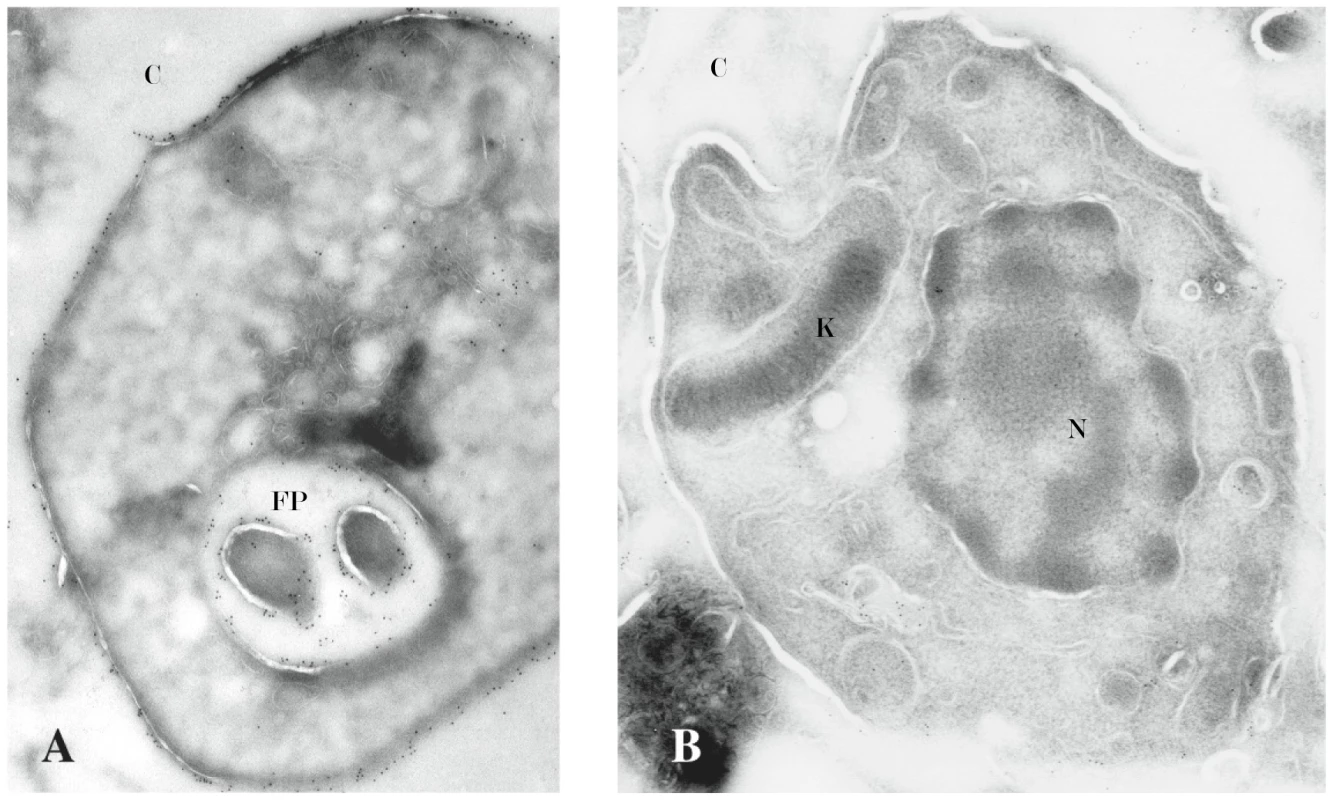
A cruzain-specific gold-labeled secondary antibody identified cruzain on the cell surface of intracellular wild type and cruzain-deficient T. cruzi amastigotes. A 3-fold (p<0.1) reduction in gold-labeled Ab detection of cruzain was present on the cell surface and flagellar pocket of cruzain-deficient CA-I/KR (B) versus wild type amastigotes (A) (45,000X). Results are representative of three independent experiments. N, nucleus; K, kinetoplast; FP, flagellar pocket; C, cytoplasm of BESM cell. Transcription factor NF-κB P65 is only activated in macrophages infected with cruzain-deficient parasites
It has been proposed that proteases of L. mexicana cleave NF-κB to facilitate immune evasion [35]. We therefore assayed NF-κB activation in cells recently (≤60 min) infected by cruzain deficient parasites. We compared macrophage activation signaling pathways [36]–[39] as a consequence of early infection with WT versus cruzain-deficient T. cruzi. The infection of a cell population by T. cruzi follows a binomial distribution, with few cells very heavily infected while most cells remain non-infected. Moreover, infection with a high ratio of parasites per cell results in premature rupture of heavily infected macrophages. To prevent the premature release of intracellular parasites that may secondarily activate macrophages in the population, we used a very low infection ratio of 0.5 parasites per cell.
To detect macrophage activation, we performed WB targeting an NF-κB family member and its specific inhibitor [40]–[45]. Similar results were obtained with two methods and Abs from the two different vendors. WB analyses with anti-NF-κB P65 Ab (Figure 3A) showed expression in macrophages infected with WT T. cruzi at 1 min and 30 min post-infection (lanes 1, 4) was similar to uninfected controls (lane 3), and lower (lane 8) at 60 min post-infection. Phosphorylation of iκB is indicative of cell activation [40], [45]. Samples for WB were either heated to 70°C for 5 min (Santa Cruz Biotech. methods) or boiled for 10 minutes immediately prior to SDS-PAGE to dissociate complexes [30]–[32]. Samples blotted with specific anti ∼PiκB Abs from different commercial sources and using different protocols confirmed negligible ∼P iκB indicative of unresponsiveness in macrophages infected with WT T. cruzi for up to 60 min and uninfected controls (Figure 3B). In contrast, ∼P iκB indicative of activation was confirmed in macrophages infected with cruzain-deficient T. cruzi as early as 1 min post-infection. Additional macrophage controls infected with cruzain-deficient T. cruzi cultured for 2 months without K11777 showed intermediate results as these parasites only regained partial cruzain activity, and were not investigated further [22]. Additional controls were uninfected macrophages treated or not with LPS and/or K11777 (K77) (Figure 3D). In L. mexicana infected controls [32], NF-κB P65 degradation was still observed 48 h post-infection (data not shown).
Fig. 3. Cruzain-deficient (KR) but not WT parasites activate NF-κB. 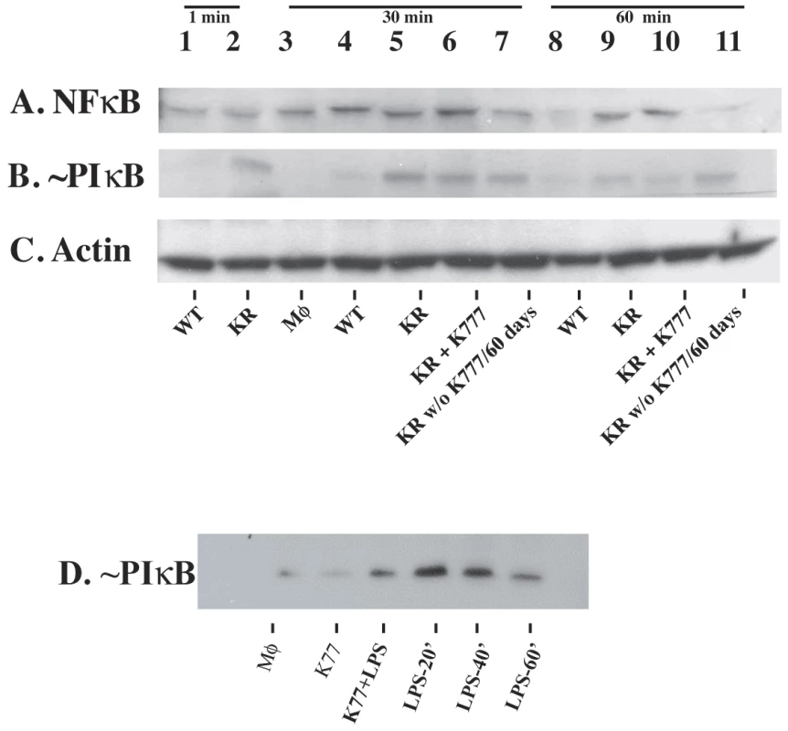
Uninfected controls and mouse macrophages (MΦ) infected with WT or KR T. cruzi were infected for ≤ 1 min (lanes 1, 2), 30 minutes (lanes 3–7) and 60 minutes (lanes 8–11). Cell extracts were western blotted with: A, anti-NF-κB P65 Ab; B, anti ∼P-iκB; C, anti-actin Ab. MΦ were infected as follows: lane #1, WT; lane #2, KR; lane #3, uninfected MΦ; lane #4, WT; lane #5, KR; lane #6, KR with 10 µM K11777; lane #7, KR cultured without K777 for 2 months; lane #8, WT; lane #9, KR; lane #10, KR with 10 µM K11777; lane #11, KR cultured without K11777 for 2 months. Results are representative of six independent experiments using two different methods. Controls (D) were MΦ treated or not with K11777 and/or LPS. NF-κB is translocated to the nucleus only in macrophages infected with cruzain-deficient parasites
To further investigate macrophage responses to WT versus cruzain deficient T. cruzi-infection, we performed confocal studies. NF-κB P65 localization was cytoplasmic in macrophage controls (Figure 4E). NF-κB P65 localization was also cytoplasmic in macrophages infected with WT T. cruzi for 1, 30, and 60 min post-infection confirming unresponsiveness to T. cruzi infection. Interestingly, NF-κB P65 covered the surface of WT amastigotes as early as 30 min post-infection (Figure 4A–B). Similar results were observed in BESM cells and thioglycolate elicited peritoneal mouse macrophages (data not shown). In contrast, intense NF-κB P65 label indicative of cell activation localized to the nucleus of macrophages infected with cruzain-deficient T. cruzi (Figure 4C–D) (yellow fluorescence), and no NF-κB P65 was seen bound to parasites for up to 60 min. Activation also occurred in macrophage controls infected with WT T. cruzi in the presence of 10 µM K11777 (data not shown).
Fig. 4. NF-κB P65 localizes to the cell surface of wild type parasites. 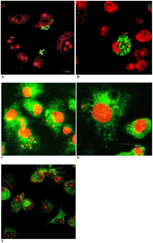
Localization of NF-κB P65 (green fluorescence) was investigated in mouse macrophages. Parasite and host cell DNA was labeled with PI (red). Non-infected macrophages showed an evenly scattered distribution of cytoplasmic NF-κB P65 (E). Intracellular WT- T. cruzi appeared coated with macrophage NF-κB P65 (A, B). In contrast, abundant cytoplasmic (green fluorescence) and nuclear (yellow fluorescence) NF-κB P65 occurred in macrophages infected with cruzain deficient T. cruzi (C–D). Macrophages were infected for 30 min (A, C), and 60 min (B, D). Results are representative of four independent experiments. Colocalization of NF-κB and cruzain
Colocalization of NF-κB P65 (green fluorescence) and cruzain (red fluorescence) on the cell surface of intracellular parasites occurred in macrophages infected with WT T. cruzi even during trypomastigote to amastigote transformation (Figure 5A–C). Intracellular WT T. cruzi treated with the trypanocidal inhibitor K11777 for 30 min showed marked cruzain accumulation presumably in the parasitic Golgi compartment [24] and no recruitment of NF-κB P65 to the parasite surface (Figure 5D, arrow).
Fig. 5. Colocalization of cruzain and NF-κB P65. 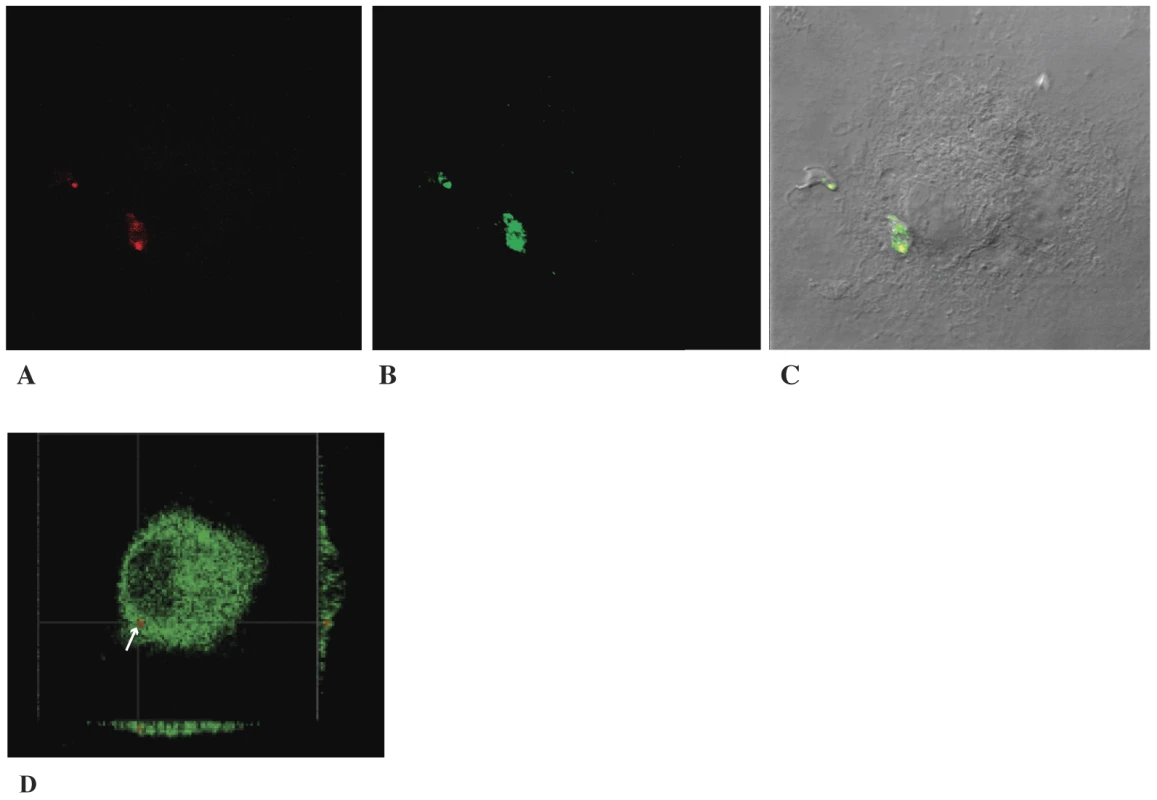
A–C. Representative examples of colocalization of labeled cruzain (A) (red fluorescence) and NF-κB P65 (B) (green fluorescence) in the flagellar pocket region and/or cell surface of interiorized transforming parasites (C, merge). D. Lack of colocalization of accumulated cruzain (red fluorescence) and NF-κB P65 (green) in a macrophage infected as above with WT T. cruzi and treated with 10 µM K11777 that induces accumulation of unprocessed zymogen in the Golgi compartment (arrow) (22). Proteolysis of human rNF-κB by cruzain
Human rNF-κB P65 was proteolytically cleaved into two major fragments by native cruzain expressed by WT (Figure 6) but not by proteases expressed by cruzain-deficient T. cruzi. Similarly, recombinant cruzain degraded human r-P65 at equimolar concentrations under the experimental conditions used but not in the presence of 10 µM K11777.
Fig. 6. Proteolytic cleavage of r-human NF-κB P65 by wild type- and r-cruzain but not by proteases expressed by cruzain-deficient T. cruzi. 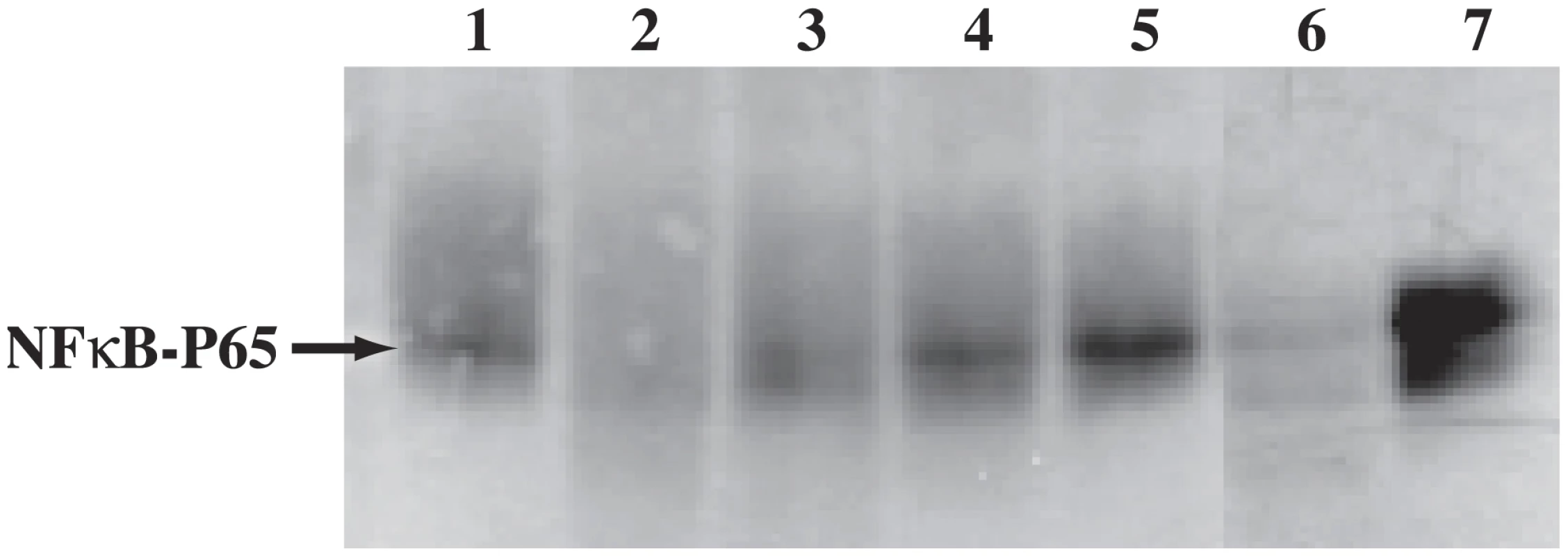
Recombinant human NF-κB P65 was treated or not with cruzain or parasite extracts. Lane 1, untreated control; lane 2, r-cruzain (1∶1 molar ratio); lanes 3–4: dilutions 1∶10 and 1∶100 of r-cruzain, respectively; lane 5, r-cruzain (1∶1 molar ratio) and 10 µM K11777; lane 6, WT cruzain; lane 7, proteases in cruzain-deficient T. cruzi. Results are representative of two independent experiments. T. cruzi development in macrophages
To understand the effect of macrophage activation [37], [42] on T. cruzi intracellular survival and development, we performed quantitative in vitro assays [26]. T. cruzi divided normally in control macrophages and the mean number of parasites per cell (P/cell) increased from ∼0.5 at to to 2.8 at 48 h (Figure 7) [23]. Activation of macrophages with purified LPS 1h prior to or concomitantly with infection resulted in parasite death. T. cruzi developed well in macrophages treated with LPS 1h post-infection; the lower mean P/cell probably results from death of parasites still trapped within the parasitophorus vacuole when activation occurred. Thus macrophage unresponsiveness in early infection (<60 min) is crucial for parasite survival and intracellular development.
Fig. 7. Macrophage activation prevents T. cruzi intracellular development. 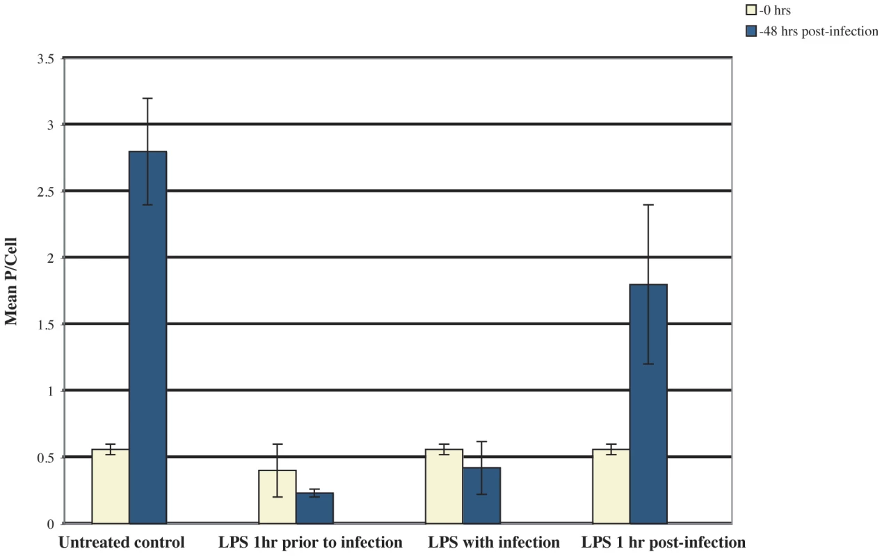
Mouse macrophages were infected with wild type T. cruzi and LPS was added as indicated. Mean parasites/cell were determined at 1 h and 48 h post-infection. Parasite death occurred when LPS was added prior to and concomitantly with T. cruzi trypomastigotes. Results are representative of two independent experiments (n = 3 per treatment). IL12 expression
To confirm macrophage unresponsiveness to infection with WT T. cruzi infection, we next investigated IL12 expression by confocal microscopy followed by fluorescence quantification with Improvision software. Macrophages infected with WT T. cruzi, and subsequently activated with LPS followed by BFA treatment, showed negligible cytoplasmic IL12 (Figure 8A). A significant increase (P<0.1) in IL12 accumulation occurred in macrophages infected with wild type T. cruzi and treated with the inhibitor K11777 that prevents cruzain activity (Figure 8B). Similarly, activation and significant IL12 accumulation occurred in macrophages infected with cruzain-deficient T. cruzi (Figure 8C).
Fig. 8. IL-12 expression. 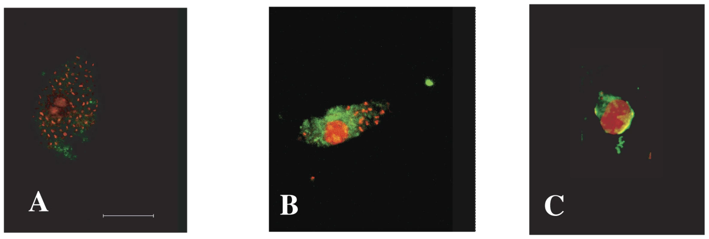
No significant IL12 expression was detected in macrophages infected with wild type T. cruzi while cells treated with 10 µM K11777 had significantly higher cytoplasmic IL12 levels. Increased IL12 expression also occurred in macrophages infected with cruzain-deficient T. cruzi. Results are representative of three independent experiments. A. Wild type-infected control. B. Macrophage infected wild type T. cruzi and treated with 10 µM K11777. C. Macrophage infected with cruzain-deficient T. cruzi. L - arginase assays
Other investigators have reported increased L-arginase activity induced by cruzain treatment of cells [7]. Only WT parasites induced 2-5 fold higher L-arginase activity in host macrophages while cruzain-deficient parasites failed to increase enzymatic activity (P<0.01). Representative values from one of 3 independent experiments are 3.13±0.02 µg urea/ml for macrophages infected with WT parasites, 1.56±0.02 µg urea/ml for macrophages infected with cruzain-deficient parasites, and 1.5±0.01 µg urea/ml for uninfected macrophages.
Discussion
A complex and dynamic scenario of host cell-parasite interactions is becoming apparent as pathogens use different mechanisms to subvert host-cell signaling pathways or modulate their kinetics during intracellular development. Several reports show that T. cruzi modulates signaling pathways in mammalian cells [15], [43], [44]. Some authors propose that activation of NF-κB proteins may modulate tissue specificity, as muscle cells that preferentially harbor T. cruzi do not become activated [37] while others detect muscle and endothelial cell activation post-infection [45], [46]. Our interest was to investigate the role of the protease cruzain during early infection (<60 min) of macrophages by T. cruzi, and in particular on the nuclear factor NF-κB P65, using the inhibitor K11777 and phenotypic cruzain knockouts [22].
A major role of the NF-κB family is the regulation of aspects of the innate and adaptive immune responses. NF-κB members control the transcription of genes encoding cytokines and antimicrobial molecules, as well as genes regulating cell differentiation, survival, proliferation and apoptosis [38]. The complex response depends on the cell type and on the nature, duration and intensity of the activating signal. NF-κB complexes are normally inactive in the cytoplasm and upon activation they enter the nucleus to modulate gene expression. Membrane receptors (e.g. TRL) and other signaling regulators are also involved [38]–[42], [43]–[52].
To compare infection with WT versus cruzain-deficient T. cruzi, we performed WB of NF-κB P65 and ∼P iκB (Figure 3) [39], [47] that showed unresponsiveness of macrophages infected with WT parasites. Confocal studies showed that WT parasites rapidly sequestered NF-κB P65 onto their cell surface (Figure 4, A–B). Similar events occurred during early infection of peritoneal mouse macrophages and BESM cells (data not shown). Cruzain localized to the cell surface of WT intracellular amastigotes (Figure 2) [53] and colocalized with NF-κB P65 (Figure 5). Treatment with K11777 caused accumulation of unprocessed and inactive protease in the Golgi [24] and abrogated NF-κB P65 sequestration by the parasite (Figure 5D). Degradation of NF-κB P65 was noted 60 min after infection with wild-type T. cruzi (Figure 3) and native cruzain degraded recombinant nuclear factor (Figure 6). Moreover, inhibition of IL12 confirmed macrophage unresponsiveness in early WT T. cruzi infection (Figure 8A).
In contrast, macrophages infected with cruzain-deficient T. cruzi became rapidly activated via NF-κB P65 (Figure 3, KR) and intense nuclear localization of P65 occurred shortly after infection (Figure 4C–D) highlighting a role for cruzain in the modulation of host cell signaling pathways. As anticipated, infection with cruzain-deficient parasites induced IL12 accumulation (Figure 8C).
Preventing macrophage activation is crucial for a successful infection. Indeed, wild type T. cruzi were killed when macrophages were activated with LPS prior to or during infection (Figure 7), presumably while parasites are still contained within the parasitophorus vacuole [54]–[57] and are susceptible to cidal macrophage products. Other macrophage metabolic pathways were also modulated by T. cruzi. Infection with WT but not cruzain-deficient T. cruzi significantly increased L-arginase activity [6], [7]. A similar phenomenon occurs during Leishmania infection [58]. We also observed down-modulation of macrosialin, the murine analog of human CD68, in cells infected with WT T. cruzi (not shown). Further studies may identify other host cell molecules modulated by T. cruzi [59]–[63].
Experimental and clinical evidence shows a correlation between the severity of Chagas' disease and the persistence of T. cruzi within tissues [64]–[65]. NF-κB complexes are important for the development and function of both innate and adaptive immune responses triggered by pathogens [38], [51]. Metabolic pathways of the innate immune system induced in early infection have important consequences in the evolution of the disease as they play a role in the control of T. cruzi replication, tissue distribution and degree of parasitism. As infection progresses, an adaptive immune response develops with production of high levels of inflammatory cytokines, IFNγ and NO radicals [38], [59], [66]–[71]. In the case of T. cruzi, TLR2 and TLR9 receptors mediate cellular activation [43], [59], [66], [72]–[73].
Our results show that macrophages infected with WT T. cruzi remained unresponsive in early infection, and support the hypothesis that intracellular pathogens are uniquely protected from macrophage activation [38]. Confirming our results, expression profiling of bone marrow macrophages infected with T. cruzi revealed very few transcriptional changes during the first 12 hours. At 24 hours post infection, both macrophages and fibroblasts express some interferon-regulated genes [62], [74], [75]. The L. mexicana cruzain homologue, “cpb”, likewise degrades NF-κB P65, IκBα, and IκBβpreventing macrophage activation [35]. Similar strategies to suppress immune responses have been found in the coccidian parasite Theileria [76], and in poliovirus and other picornavirus as viral proteases cleave NF-κB P65 generating inactive products [77]. We then hypothesize that cruzain plays a key role by degrading NF-κB P65 and hindering activation of innate phagocytes recruited to the bite site [5]. Macrophage unresponsiveness would favor parasite survival in early infection and delay the onset of the immune response. A successful macrophage infection results in the production of hundreds of infectious T. cruzi. For example a single CA-I/72 parasite originates ∼130 infectious trypomastigotes in just 4.5 days [23]. Once the host immune response is triggered on [65]–[67], parasite development would be restricted to permissive tissues such as muscle cells [37], [71]. Thus cruzain may function during the early events of macrophage infection favoring immune evasion by T. cruzi.
Zdroje
1. UrbinaJA 2010 Specific chemotherapy of Chagas disease: relevance, current limitations and new approaches. Acta Trop 115 55 68
2. BucknerFSNavabiN 2010 Advances in Chagas disease drug development: 2009-2010. Curr Opinion Infect Dis 23 609 616
3. McKerrowJHDoylePSEngelJCPodustLMRobertsonSA 2009 Two approaches to discovering and developing new drugs for Chagas disease. Mem Inst Oswaldo Cruz 104 263 269
4. ChagasC 1982 Coletanea de Trabalhos Cientificos 1909-1913. Editora Universidade de Brasilia 237 335
5. SchusterJPSchaubGA 2000 Trypanosoma cruzi: skin-penetration kinetics of vector derived metacyclic trypomastigotes. Int J Parasitol 30 1475 1479
6. GiordanengoLGuinazuNStempinCFretesRCerbanFM 2002 Cruzipain, a major Trypanosoma cruzi antigen, conditions the host immune response in favor of parasite. Eur J Immunol 32 1003 1011
7. StempinCSTanosTBCosoOACerbánFM 2003 Arginase induction promotes Trypanosoma cruzi intracellular replication in Cruzipain-treated J774 cells through the activation of multiple signaling pathways. Eur J Immunol 34 200 209
8. TomasAMKellyJM 1994 Transformation as an approach to functional analysis of the major cysteine protease of Trypanosoma cruzi. Biochem Soc Trans 22 90S
9. TomasAMKellyJM 1996 Stage-regulated expression of cruzipain, the major cysteine protease of Trypanosoma cruzi is independent of the level of RNA. Mol Biochem Parasitol 76 91 103
10. TomasAMMilesMAKellyJM 1997 Overexpression of cruzipain, the major cysteine proteinase of Trypanosoma cruzi, is associated with enhanced metacyclogenesis. Eur J Biochem 244 596 603
11. YongVSchmitzVVannier-SantosMAde LimaAPLalmanachG 2000 Altered expression of cruzipain and a cathepsin B-like target in a Trypanosoma cruzi cell line displaying resistance to synthetic inhibitors of cysteine-proteinases. Mol Biochem Parasitol 109 47 59
12. Lima A PAdos ReisCGFServeauCLalmanachGJulianoL 2001 Cysteine protease isoforms from Trypanosoma cruzi, cruzipain 2 and cruzain, present different substrate preference and susceptibility to inhibitors. Mol Biochem Parasitol 114 41 52
13. LimaAPAlmeidaPCTersariolLLSchmitzVSchmaierAH 2002 Heparan sulfate modulates kinin release by Trypanosoma cruzi through the activity of cruzipain. J Biol Chem 277 5875 5881
14. ScharfsteinJSchmitzVMorandiVCapellaMMLimaAP 2000 Host cell invasion by Trypanosoma cruzi is potentiated by activation of bradykinin B(2) receptors. J Exp Med 192 1289 1300
15. AokiMPCanoRCPellegriniAVTanosTGuinazuNL 2006 Different signaling pathways are involved in cardiomyocyte survival induced by a Trypanosoma cruzi glycoprotein. Microbes Infect 8 1723 1731
16. EakinAEMillsAAHarthGMcKerrowJHCraikCS 1992 The sequence, organization, and expression of the major cysteine protease (cruzain) from Trypanosoma cruzi. J Biol Chem 267 7411 7420
17. CazzuloJJ 1999 Cruzipain, major cysteine proteinase of Trypanosoma cruzi: sequence and genomic organization of the codifying genes. Medicina (B Aires) 59 7 10
18. EngelJCDoylePSHsiehIMcKerrowJH 1998 Cysteine protease inhibitors cure an experimental Trypanosoma cruzi infection. J Exp Med 188 725 734
19. DuschakVGCiaccioMNassertJRBasombrioMA 2001 Enzymatic activity, protein expression, and gene sequence of cruzipain in virulent and attenuated Trypanosoma cruzi strains. J Parasitol 87 1016 1022
20. DuschakVGRiarteASeguraELLaucellaSA 2001 Humoral immune response to cruzipain and cardiac dysfunction in chronic Chagas' disease. Immunol Letters 78 135 142
21. PaivaCNSouto-PadronTCostaDAGattassCR 1998 High expression of a functional cruzipain by a non-infective and non-pathogenic Trypanosoma cruzi clone. Parasitology 117 483 940
22. EngelJCTorres GarciaCHsiehIDoylePSMcKerrowJH 2000 Upregulation of the secretory pathway in cysteine protease inhibitor-resistant Trypanosoma cruzi. J Cell Science 113 1345 1354
23. EngelJCDoylePSDvorakJA 1985 Trypanosoma cruzi: Biological characterization of clones derived from chronic chagasic patients. II. Quantitative analysis of the intracellular cycle. J Protozool 32 80 83
24. EngelJCDoylePSPalmerJHsiehIBaintonDF 1998 Cysteine protease inhibitors alter Golgi complex ultrastructure and function in Trypanosoma cruzi. J Cell Sci 111 597 606
25. EngelJCde CazzuloBMFStoppaniOAMCanattaJJBCazzuloJJ 1987 Aerobic glucose fermentation by Trypanosoma cruzi axenic culture amastigote-like forms during growth and differentiation to epimastigotes. Mol Bioch Parasitol 26 1 10
26. DoylePSDvorakJAEngelJC 1984 Trypanosoma cruzi: Quantification and analysis of the infectivity of cloned stocks. J Protozool 31 280 283
27. BogyoMVerhelstSBellingard-DubouchaudVTobaSGreenbaumDC 2000 Selective targeting of lysosomal cysteine proteases with radiolabeled electrophilic substrate analogs. Chem Biol 7 27 38
28. GreenbaumDCMedzihradszkyKFBurlingameABogyoM 2000 Epoxide electrophiles as activity-dependent cysteine protease profiling and discovery tools. Chem Biol 7 569 581
29. HirschfeldMMaYWeisJHVogelSNWeisJJ 2000 Cutting edge: repurification of lipopolysaccharide eliminates signaling through both human and murine toll-like receptor 2. J Immunol 165 618 622
30. TurerEETavaresRMMortierEHitotsumatsuOAdvinculaR 2008 Homeostatic MyD88-dependent signals cause lethal inflammation in the absence of A20. J Exp Med 205 451 464
31. HitotsumatsuOAhmadRETavaresRMWangMPhilpottD 2008 The ubiquitin-editing enzyme A20 restricts nucleotide-binding oligomerization domain containing 2-triggered signals. Immunity 28 381 390
32. TavaresRMTurerEELiuCLAdvinculaRScapiniP 2010 The Ubiquitin Modifying Enzyme A20 Restricts B Cell Survival and Prevents Autoimmunity. Immunity 33 181 191
33. QuinonesMAhujaSKMelbyPCPateLReddickRL 2000 Preformed membrane-associated stores of interleukin (IL)-12 are a previously unrecognized source of bioactive IL-12 that is mobilized within minutes of contact with an intracellular parasite. Exp Med 192 507 515
34. CorralizaIMCampoMLSolerGModollelM 1994 Determination of arginase activity in macrophages: a micromethod. J Immunol Methods 174 231 235
35. CameronPMcGachyAAndersonMPaulACoombsGH 2004 Inhibition of lipopolysaccharide-induced macrophage IL-12 production by Leishmania mexicana amastigotes: the role of cysteine peptidases and the NF-kappaB signaling pathway. J Immunol 173 3297 3304
36. GhoshS 1999 Regulation of inducible gene expression by the transcription factor NF-κB. Immunol Res 19 183 189
37. HallBSTamWSenRPereiraME 2000 Cell-specific activator of nuclear factor kappa B by the parasite Trypanosoma cruzi promotes resistance to intracellular infection. Mol Biol Cell 11 153 160
38. HaydenMSWestAPGhoshS 2006 NF-κB and the immune response. Oncogene 25 6758 6780
39. ChenYValleeSWuJVuDSondekJ 2004 Inhibition of NF-κB activity by IκBb in association with κB-ras. Mol Cell Biol 24 3048 3056
40. DiDonatoJAMercurioFKarinM 1995 Phosphorylation of iκBá precedes but is not sufficient for its dissociation from NF-κB. Mol Cell Biol 15 1302 1311
41. MarienfeldRFPalkowitschLGhoshS 2006 Dimerization of the iκB kinase-binding domain of NEMO is required for tumor necrosis factor alpha-induced NF-κB activity. Mol Cell Biol 26 9209 9219
42. De PlaenIHanXBLiuXHsuehWGhoshS 2006 Lipopolysaccharide induces CXCL2/macrophage inflammatory protein-2 gene expression in enterocytes via NF-κB activation: independence from endogenous TNF-α and platelet activation factor. Immunology 118 153 163
43. PetersenCAKrumholzKABurleighBA 2005 Toll-like receptor 2 modulates IL-1B dependent cardiomyocyte hypertrophy triggered by Trypanosoma cruzi. Inf Immun 73 6974 6980
44. PetersenCAKrimholzKACarmenJSinaiAPBurleighBA 2006 Trypanosoma cruzi infection and nuclear factor kappa B activation prevent apoptosis in cardiac cells. Inf Imm 74 1580 1587
45. BaXShivali GuptaSDavidsonMGargNJ 2010 Trypanosoma cruzi Induces the Reactive Oxygen Species-PARP-1-RelA Pathway for Up-regulation of Cytokine Expression in Cardiomyocytes. J Biol Chem 285 11596 11606
46. HuangHCalderonTMBermanJWBraunsteinVLWeissLM 1999 Infection of Endothelial Cells with Trypanosoma cruzi Activates NF-κB and Induces Vascular Adhesion Molecule Expression. Infect Immun. 67 5434 5440
47. WuCGoshS 2003 Differential phosphorylation of the signal-responsive domain of iκBα and iκBβ by IκB kinases. J Biol Chem 34 31980 31987
48. BaeuerlePABaltimoreD 1996 NF-κB ten years after. Cell 87 113 120
49. IsraelAKroemerG 2006 NF-κB in life/death decisions: and introduction. Cell Death Differ 13 685 686
50. PerkinsNDGilmoreTD 2006 Good cop, bad cop: the different faces of NF-κB. Cell Death Differ 13 759 772
51. LiouHCHsiaCY 2003 Distinctions between c-Rel and other NF-κB proteins in immunity and disease. Bioassays 25 767 780
52. NelsonDEIhekwabaAECElliotMJohnsonJRGibneyCA 2004 Oscillations in NF-κB signaling control the dynamics of gene expression. Science 306 704 708
53. NascimentoAEde SouzaW 1996 High resolution localization of cruzipain and Ssp4 in Trypanosoma cruzi by replica staining label fracture. Biol Cell 86 53 58
54. BurleighBAAndrewsNW 1995 The mechanisms of Trypanosoma cruzi invasion of mammalian cells. Ann Rev Microbiol 49 175 200
55. BurleighBA 2005 Host cell signaling and Trypanosoma cruzi invasion: do all roads lead to lysosomes? Sci STKE 293 36 43
56. LeyVAndrewsNWRobbinsESNussenzweigV 1998 Amastigotes of Trypanosoma cruzi sustain an infective cycle in mammalian cells. J Exp Med 168 649 659
57. KimaPEBurleighBAAndrewsNW 2000 Surface-targeted lysosomal membrane glyocoprotein-1 (Lamp-1) enhances lysosome exocytosis and cell invasion by Trypanosoma cruzi. Cell Microbiol 2 477 486
58. GaurURobertsSCDalviRPCorralizaIUllmanB 2007 An Effect of Parasite-Encoded Arginase on the Outcome of Murine Cutaneous Leishmaniasis. J Immunol 179 8446 8453
59. CamposMACloselMValenteEPCardosoJEAkiraS 2004 Impaired production of proinflammatory cytokines and host resistance to acute infection with Trypanosoma cruzi in mice lacking functional myeloid differentiation factor 88. J Immunol 172 1711 1718
60. GordonS 2003 Alternative activation of macrophages. Nature Rev Immunol 3 23 35
61. NoëlWRaesGGhassabehGHDe BaetselierPBeschinA 2004 Alternatively activated macrophages during parasite infections. Tr Parasitol 20 126 133
62. ZhangSKimCCBatraSMcKerrowJHLokeP 2010 Delineation of diverse macrophage activation programs in response to intracellular parasites and cytokines. PLoS Negl Trop Dis 4 e648
63. GradoniLAscenziP 2004 Nitric oxide and anti-protozoon chemotherapy. Parasitol 46 101 103
64. ZhangLTarletonRL 1999 Parasite persistence correlates with disease severity and localization in chronic Chagas' disease. J Infect Dis 180 480 486
65. TarletonR 2001 Parasite persistence in the etiology of Chagas’ disease. Int J Parasitol 31 549 553
66. GazzinelliRTRopertCCamposMA 2004 Role of the Toll/interleukin-1 receptor signaling pathway in host resistance and pathogenesis during infection with protozoan parasites. Imm Rev 201 9 14
67. TarletonRLGrusbyMJZhangL 2000 Increased susceptibility of Stat4-deficient and enhanced resistance in Stat6-deficient mice to infection with Trypanosoma cruzi. J Immunol 165 1520 1525
68. ReedSG 1998 In vivo administration of recombinant IFN-gamma induces macrophage activation, and prevents acute disease, immune suppression, and death in experimental Trypanosoma cruzi infections. J Immunol 140 4342 4347
69. KumarSTarletonRL 2001 Antigen-specific Th1 but not Th2 cells provide protection from lethal Trypanosoma cruzi infection in mice. J Immunol 166 4596 4603
70. BergeronMOlivierM 2006 Trypanosoma cruzi-Mediated IFN-Inducible Nitric Oxide Output in Macrophages Is Regulated by iNOS mRNA Stability. J Immunol 177 6271 80
71. HuangHPetkovaSBCohenAWBouzahzahBChanJ 2003 Activation of transcription factors AP-1 and NF-κB in murine chagasic myocarditis. Inf Immun 71 2859 2867
72. BaficaASantiagoHCGoldszmidRRopertCGazzinelliRT 2006 Cutting edge: TLR9 and TLR2 signaling together account for MyD88-dependent control of parasitemia in Trypanosoma cruzi infection. J Immunol 177 3513 3519
73. RopertCCloselMChavesACLGazzinelliRT 2003 Inhibition of a p38/Stress-Activated Protein Kinase-2-Dependent Phosphatase Restores Function of IL-1 Receptor-Associated Kinase-1 and Reverses Toll-Like Receptor 2 - and 4-Dependent Tolerance of Macrophages. J Immunol 171 1456 1465
74. Vaena de AvalosSBladerIJFisherMBoothroydJCBurleighBA 2002 Immediate early response to Trypanosoma cruzi infection involves minimal modulation of host cell transcription. J Biol Chem 277 639 644
75. BurleighBA 2004 Probing Trypanosoma cruzi biology with DNA microarrays. Parasitol 128 3 10
76. HeusslerVTRottenbergDSSchwabRKüenziPFernandezPC 2002 Hijacking of Host Cell IκK Signalosomes by the Transforming Parasite Theileria. Science 298 1033 1036
77. NesnanovNChumakivKMNeznanovaLAlmasanABanerjeeAK 2005 Proteolytic cleavage of the P65-RelA subunit of NF-κB during poliovirus infection. J Biol Chem 280 24153 24158
Štítky
Hygiena a epidemiologie Infekční lékařství Laboratoř
Článek Hostile Takeover by : Reorganization of Parasite and Host Cell Membranes during Liver Stage EgressČlánek A Trigger Enzyme in : Impact of the Glycerophosphodiesterase GlpQ on Virulence and Gene ExpressionČlánek An EGF-like Protein Forms a Complex with PfRh5 and Is Required for Invasion of Human Erythrocytes byČlánek Th2-polarised PrP-specific Transgenic T-cells Confer Partial Protection against Murine ScrapieČlánek Alterations in the Transcriptome during Infection with West Nile, Dengue and Yellow Fever Viruses
Článek vyšel v časopisePLOS Pathogens
Nejčtenější tento týden
2011 Číslo 9- Stillova choroba: vzácné a závažné systémové onemocnění
- Perorální antivirotika jako vysoce efektivní nástroj prevence hospitalizací kvůli COVID-19 − otázky a odpovědi pro praxi
- Diagnostika virových hepatitid v kostce – zorientujte se (nejen) v sérologii
- Choroby jater v ordinaci praktického lékaře – význam jaterních testů
- Diagnostický algoritmus při podezření na syndrom periodické horečky
-
Všechny články tohoto čísla
- Unconventional Repertoire Profile Is Imprinted during Acute Chikungunya Infection for Natural Killer Cells Polarization toward Cytotoxicity
- Envelope Deglycosylation Enhances Antigenicity of HIV-1 gp41 Epitopes for Both Broad Neutralizing Antibodies and Their Unmutated Ancestor Antibodies
- Co-opts GBF1 and CERT to Acquire Host Sphingomyelin for Distinct Roles during Intracellular Development
- Nrf2, a PPARγ Alternative Pathway to Promote CD36 Expression on Inflammatory Macrophages: Implication for Malaria
- Robust Antigen Specific Th17 T Cell Response to Group A Streptococcus Is Dependent on IL-6 and Intranasal Route of Infection
- Targeting of a Chlamydial Protease Impedes Intracellular Bacterial Growth
- The Protease Cruzain Mediates Immune Evasion
- High-Resolution Phenotypic Profiling Defines Genes Essential for Mycobacterial Growth and Cholesterol Catabolism
- Plague and Climate: Scales Matter
- Exhausted CD8 T Cells Downregulate the IL-18 Receptor and Become Unresponsive to Inflammatory Cytokines and Bacterial Co-infections
- Maturation-Induced Cloaking of Neutralization Epitopes on HIV-1 Particles
- Murine Gamma-herpesvirus Immortalization of Fetal Liver-Derived B Cells Requires both the Viral Cyclin D Homolog and Latency-Associated Nuclear Antigen
- Rapid and Efficient Clearance of Blood-borne Virus by Liver Sinusoidal Endothelium
- Hostile Takeover by : Reorganization of Parasite and Host Cell Membranes during Liver Stage Egress
- A Trigger Enzyme in : Impact of the Glycerophosphodiesterase GlpQ on Virulence and Gene Expression
- Strain Specific Resistance to Murine Scrapie Associated with a Naturally Occurring Human Prion Protein Polymorphism at Residue 171
- Development of a Transformation System for : Restoration of Glycogen Biosynthesis by Acquisition of a Plasmid Shuttle Vector
- Monalysin, a Novel -Pore-Forming Toxin from the Pathogen Contributes to Host Intestinal Damage and Lethality
- Host Phylogeny Determines Viral Persistence and Replication in Novel Hosts
- BC2L-C Is a Super Lectin with Dual Specificity and Proinflammatory Activity
- Expression of the RAE-1 Family of Stimulatory NK-Cell Ligands Requires Activation of the PI3K Pathway during Viral Infection and Transformation
- Structure of the Vesicular Stomatitis Virus N-P Complex
- HSV Infection Induces Production of ROS, which Potentiate Signaling from Pattern Recognition Receptors: Role for S-glutathionylation of TRAF3 and 6
- The Human Papillomavirus E6 Oncogene Represses a Cell Adhesion Pathway and Disrupts Focal Adhesion through Degradation of TAp63β upon Transformation
- Analysis of Behavior and Trafficking of Dendritic Cells within the Brain during Toxoplasmic Encephalitis
- Exposure to the Viral By-Product dsRNA or Coxsackievirus B5 Triggers Pancreatic Beta Cell Apoptosis via a Bim / Mcl-1 Imbalance
- Multidrug Resistant 2009 A/H1N1 Influenza Clinical Isolate with a Neuraminidase I223R Mutation Retains Its Virulence and Transmissibility in Ferrets
- Structure of Herpes Simplex Virus Glycoprotein D Bound to the Human Receptor Nectin-1
- Step-Wise Loss of Bacterial Flagellar Torsion Confers Progressive Phagocytic Evasion
- Complex Recombination Patterns Arising during Geminivirus Coinfections Preserve and Demarcate Biologically Important Intra-Genome Interaction Networks
- An EGF-like Protein Forms a Complex with PfRh5 and Is Required for Invasion of Human Erythrocytes by
- Non-Lytic, Actin-Based Exit of Intracellular Parasites from Intestinal Cells
- The Fecal Viral Flora of Wild Rodents
- The General Transcriptional Repressor Tup1 Is Required for Dimorphism and Virulence in a Fungal Plant Pathogen
- Interferon Regulatory Factor-1 (IRF-1) Shapes Both Innate and CD8 T Cell Immune Responses against West Nile Virus Infection
- A Small Non-Coding RNA Facilitates Bacterial Invasion and Intracellular Replication by Modulating the Expression of Virulence Factors
- Evaluating the Sensitivity of to Biotin Deprivation Using Regulated Gene Expression
- The Motility of a Human Parasite, , Is Regulated by a Novel Lysine Methyltransferase
- Phosphodiesterase-4 Inhibition Alters Gene Expression and Improves Isoniazid – Mediated Clearance of in Rabbit Lungs
- Restoration of IFNγR Subunit Assembly, IFNγ Signaling and Parasite Clearance in Infected Macrophages: Role of Membrane Cholesterol
- Protease ROM1 Is Important for Proper Formation of the Parasitophorous Vacuole
- The Regulated Secretory Pathway in CD4 T cells Contributes to Human Immunodeficiency Virus Type-1 Cell-to-Cell Spread at the Virological Synapse
- Rerouting of Host Lipids by Bacteria: Are You CERTain You Need a Vesicle?
- Transmission Characteristics of the 2009 H1N1 Influenza Pandemic: Comparison of 8 Southern Hemisphere Countries
- Th2-polarised PrP-specific Transgenic T-cells Confer Partial Protection against Murine Scrapie
- Sequential Bottlenecks Drive Viral Evolution in Early Acute Hepatitis C Virus Infection
- Genomic Insights into the Origin of Parasitism in the Emerging Plant Pathogen
- Genomic and Proteomic Analyses of the Fungus Provide Insights into Nematode-Trap Formation
- Influenza Virus Ribonucleoprotein Complexes Gain Preferential Access to Cellular Export Machinery through Chromatin Targeting
- Alterations in the Transcriptome during Infection with West Nile, Dengue and Yellow Fever Viruses
- Protease-Sensitive Conformers in Broad Spectrum of Distinct PrP Structures in Sporadic Creutzfeldt-Jakob Disease Are Indicator of Progression Rate
- Vaccinia Virus Protein C6 Is a Virulence Factor that Binds TBK-1 Adaptor Proteins and Inhibits Activation of IRF3 and IRF7
- c-di-AMP Is a New Second Messenger in with a Role in Controlling Cell Size and Envelope Stress
- Structural and Functional Studies on the Interaction of GspC and GspD in the Type II Secretion System
- APOBEC3A Is a Specific Inhibitor of the Early Phases of HIV-1 Infection in Myeloid Cells
- Impairment of Immunoproteasome Function by β5i/LMP7 Subunit Deficiency Results in Severe Enterovirus Myocarditis
- HTLV-1 Propels Thymic Human T Cell Development in “Human Immune System” Rag2 gamma c Mice
- Tri6 Is a Global Transcription Regulator in the Phytopathogen
- Exploiting and Subverting Tor Signaling in the Pathogenesis of Fungi, Parasites, and Viruses
- The Next Opportunity in Anti-Malaria Drug Discovery: The Liver Stage
- Significant Effects of Antiretroviral Therapy on Global Gene Expression in Brain Tissues of Patients with HIV-1-Associated Neurocognitive Disorders
- Inhibition of Competence Development, Horizontal Gene Transfer and Virulence in by a Modified Competence Stimulating Peptide
- A Novel Metal Transporter Mediating Manganese Export (MntX) Regulates the Mn to Fe Intracellular Ratio and Virulence
- Rhoptry Kinase ROP16 Activates STAT3 and STAT6 Resulting in Cytokine Inhibition and Arginase-1-Dependent Growth Control
- Hsp90 Governs Dispersion and Drug Resistance of Fungal Biofilms
- Secretion of Genome-Free Hepatitis B Virus – Single Strand Blocking Model for Virion Morphogenesis of Para-retrovirus
- A Viral Ubiquitin Ligase Has Substrate Preferential SUMO Targeted Ubiquitin Ligase Activity that Counteracts Intrinsic Antiviral Defence
- Membrane Remodeling by the Double-Barrel Scaffolding Protein of Poxvirus
- A Diverse Population of Molecular Type VGIII in Southern Californian HIV/AIDS Patients
- Disruption of TLR3 Signaling Due to Cleavage of TRIF by the Hepatitis A Virus Protease-Polymerase Processing Intermediate, 3CD
- Quantitative Analyses Reveal Calcium-dependent Phosphorylation Sites and Identifies a Novel Component of the Invasion Motor Complex
- Discovery of the First Insect Nidovirus, a Missing Evolutionary Link in the Emergence of the Largest RNA Virus Genomes
- Old World Arenaviruses Enter the Host Cell via the Multivesicular Body and Depend on the Endosomal Sorting Complex Required for Transport
- Exploits a Unique Repertoire of Type IV Secretion System Components for Pilus Assembly at the Bacteria-Host Cell Interface
- Recurrent Signature Patterns in HIV-1 B Clade Envelope Glycoproteins Associated with either Early or Chronic Infections
- PLOS Pathogens
- Archiv čísel
- Aktuální číslo
- Informace o časopisu
Nejčtenější v tomto čísle- HTLV-1 Propels Thymic Human T Cell Development in “Human Immune System” Rag2 gamma c Mice
- Hostile Takeover by : Reorganization of Parasite and Host Cell Membranes during Liver Stage Egress
- Exploiting and Subverting Tor Signaling in the Pathogenesis of Fungi, Parasites, and Viruses
- A Viral Ubiquitin Ligase Has Substrate Preferential SUMO Targeted Ubiquitin Ligase Activity that Counteracts Intrinsic Antiviral Defence
Kurzy
Zvyšte si kvalifikaci online z pohodlí domova
Autoři: prof. MUDr. Vladimír Palička, CSc., Dr.h.c., doc. MUDr. Václav Vyskočil, Ph.D., MUDr. Petr Kasalický, CSc., MUDr. Jan Rosa, Ing. Pavel Havlík, Ing. Jan Adam, Hana Hejnová, DiS., Jana Křenková
Autoři: MUDr. Irena Krčmová, CSc.
Autoři: MDDr. Eleonóra Ivančová, PhD., MHA
Autoři: prof. MUDr. Eva Kubala Havrdová, DrSc.
Všechny kurzyPřihlášení#ADS_BOTTOM_SCRIPTS#Zapomenuté hesloZadejte e-mailovou adresu, se kterou jste vytvářel(a) účet, budou Vám na ni zaslány informace k nastavení nového hesla.
- Vzdělávání


