-
Články
- Vzdělávání
- Časopisy
Top články
Nové číslo
- Témata
- Kongresy
- Videa
- Podcasty
Nové podcasty
Reklama- Kariéra
Doporučené pozice
Reklama- Praxe
THE USE OF NONINVASIVE DIAGNOSTIC METHODS IN THE ASSESSMENT OF POSTURAL CHANGES IN UNIVERSITY STUDENTS
The purpose of the study was to extend knowledge about the parametrization of traction spiral stabilization using muscle chains in the educational process. The participants were 26 students aged 19 to 25 years who attended the Technical University in Košice. After getting information about the aim and content of the research the students participated in pretest and posttest sessions aimed to assess postural parameters (Shoulder Height Left, Shoulder Height Right, Shoulder 2 Shoulder Caliper, Neck Back Height, Neck Front Height) using the full body 3D scanner TC2 NX 16. The experimental factor was an intervention exercise program based on the SM system (SM – stabilization, mobilization = SPS method – Spiralstabilization of the spine). This program took place over 11 weeks during physical education classes. However, the effect of this variable on postural parameters was difficult to determine. Therefore, the individual analysis of collected data seemed to be necessary. Our practical experience has shown that individual approach plays an important role. The correlations between particular pairs of parameters were determined using the Pearson’s correlation coefficient. The results showed a significant correlation between SHL and S2SC (Shoulder Height Left and Shoulder 2 Shoulder Caliper) during the stabilization phase (posttest). The interpreted data and actually increasing number of movement patterns in university students create the need to study the anatomical structures both theoretically and practically. Therefore, it will be necessary to design and administer a compensatory exercise intervention for the population concerned.
Keywords:
movement pattern, muscle chains, posture, spine stabilization, Spinal Mouse®, full body 3D scanner
Authors: Miroslava Barcalová 1,3; Jozef Živčák 1; Rút Lenková 2; Ľuboš Vojtaško 2,3; Erika Liptáková 4; Viktória Rajťúková 1; Viktória Krajňáková 1
Authors place of work: Technical University in Košice, Faculty of Mechanical Engineering, Department of Biomedical Engineering and Measurement, Košice, Slovakia 1; University of Prešov, Faculty of Sports, Department of Sports Kinanthropology, Prešov, Slovakia 2; Technical University in Košice, Department of Physical Education, Košice, Slovakia 3; Technical University in Košice, Faculty of Economics, Department of Applied Mathematics and Business Informatics, Košice, Slovakia 4
Published in the journal: Lékař a technika - Clinician and Technology No. 1, 2017, 47, 23-29
Category: Původní práce
Summary
The purpose of the study was to extend knowledge about the parametrization of traction spiral stabilization using muscle chains in the educational process. The participants were 26 students aged 19 to 25 years who attended the Technical University in Košice. After getting information about the aim and content of the research the students participated in pretest and posttest sessions aimed to assess postural parameters (Shoulder Height Left, Shoulder Height Right, Shoulder 2 Shoulder Caliper, Neck Back Height, Neck Front Height) using the full body 3D scanner TC2 NX 16. The experimental factor was an intervention exercise program based on the SM system (SM – stabilization, mobilization = SPS method – Spiralstabilization of the spine). This program took place over 11 weeks during physical education classes. However, the effect of this variable on postural parameters was difficult to determine. Therefore, the individual analysis of collected data seemed to be necessary. Our practical experience has shown that individual approach plays an important role. The correlations between particular pairs of parameters were determined using the Pearson’s correlation coefficient. The results showed a significant correlation between SHL and S2SC (Shoulder Height Left and Shoulder 2 Shoulder Caliper) during the stabilization phase (posttest). The interpreted data and actually increasing number of movement patterns in university students create the need to study the anatomical structures both theoretically and practically. Therefore, it will be necessary to design and administer a compensatory exercise intervention for the population concerned.
Keywords:
movement pattern, muscle chains, posture, spine stabilization, Spinal Mouse®, full body 3D scannerIntroduction
The primary source of scientific knowledge in the field of functional motor system disorders is the concept of the functional pathology of the motor system introduced by Lewit and Janda [1], [2]. The knowledge has been significantly extended by Kolář [3], [4], [5], who, in his studies, systematized the hierarchy of the functional muscle disorders from the viewpoint of postural ontogeny. The main preventive element is active physical exercises, which affect the motor system and form the content base for dealing with the existing vertebrogenic difficulties.
Muscle imbalance is a state of deteriorated balance between the shortened and weakened muscles. This does not concern one muscle only because muscles work as a functional unit. Muscle imbalance may be considered one of the primary causes of chronic pain in the motor system and spinal disorders. Also, muscle imbalance has adverse effect on body posture, movement stereotypes, muscle coordination, which leads to increased susceptibility to injury and limited range of movement [6].
Extensive research into health-related and fitness parameters in university students has shown that the overall fitness of university students is decreasing; however, the number of health disorders is increasing. Students, especially females, suffer from frequent back pain, headache, upper respiratory tract disease, cardiovascular disease, and so forth. Students show diminishing levels of physical fitness and motor performance [7]. Specific exertion sustained by university students within the study of theoretical subjects leads to increased occurrence of muscle imbalances, decrease in overall physical fitness levels and increase in obesity rates [8]. Research conducted on the sample of 1,900 university students who attended two universities in Košice in 2012–2013 showed that more than 22% of men and 41% of women suffered from a certain type of back pain [9]. A pilot study dealing in detail with postural parameters in university students was conducted at the Trnava University in 2011. The authors of the study examined the head, neck, and shoulders posture, spine curvature in the sagittal plane, spine curvature in the cervical region, shape of the spine in the frontal plane and shoulder height. The research showed several deviations from the reference values concerning the spine and postural parameters [10]. These findings are becoming the stimulus for detailed analysis of these parameters in the respective population group, more thorough research and designing of preventive measures to eliminate negative factors affecting health.
Movement patterns
The movement patterns are studied by osteopaths, posture analysts as well as chiropractors, with different opinions about the origination of the movement patterns. As reported by Mitchell Jr. and Greenman, there must be a generally applicable pattern. The authors justify their conclusion by the fact that when suffering from motor system dysfunctions different body parts adjust always according to the same movement pattern [11]. In physiology, the whole human organism follows certain patterns. This applies to the process of walking and breathing. The endocrine system is also a good example. The holistic principle of embryologic, physiologic and neurologic principles explains these patterns. In their book, Richter and Hebgen present a model of muscle chains, which follow two movement patterns, that is, flexion and extension [11]. Smíšek assumes that it is necessary to form optimal movement patterns through spiral stabilization of the spine [12]. By analyzing the spiral muscle chains in detail Smíšek seeks ways of developing the muscle corset, which is stabilized during everyday activities and sports activities as well. Sedentary lifestyle, sports and exercise programs with flexion movement pattern (forward flexion) and oblique and horizontal axes lead to the overload and degeneration of the spine [11]. Among other authors who dealt with the muscle chains, for instance, are Magik Ledvina, Thomas Myers and Kurt Tittel [13].
Traction spiral stabilization of the spine
Functional stabilization and mobilization of the spine together with the activation of spiral stabilization through movement patterns in particular spirals /spiral stabilization of the spine/ is arranged in the SM system of exercise. This system leads to active compensation in an effort to achieve desired muscle strengthening effect and to stretch the muscles of the spine, trunk, limbs and girdles. B applying the reciprocal inhibition (active relaxation) when stretching the muscles and by activating the agonists on one side leads to the relaxation of antagonists on the other side. Stretching of reciprocally inhibited muscles is much more effective than stretching. Muscle chaining (cooperation of muscles during movement), with the muscle chain being activated, adds up due to muscle contraction within the chain and this leads to a much more effective use of the muscle system than through the increase of muscle volume. The proprioceptive stimulation of the foot is the postural response of feet to loading in the standing position. This leads to the increased activity of abdominal muscles, which subsequently stabilize the spine [12].
Spiral muscle chaining causes the contraction of the abdominal region and produces a lift-traction force, which pulls the spine upward and allows the intervertebral discs to regenerate.
The traction manual methods of treatment and the adjustments of particular spine segments have been studied by Kaltenborn during his long-time practice.
Fig. 1: Descriptive and functional anatomy of muscle chains [17]. ![Fig. 1: Descriptive and functional anatomy of muscle chains [17].](https://pl-master.mdcdn.cz/media/image/57ad533abd05dea11f02d29d48a9db62.jpg?version=1537794091)
Muscle chains
A muscle chain refers to a group of muscles, the coordinated activity of which during movement maintains body stability, which leads to the correct execution of movement. SM system is based on anatomically defined spiral muscle chains. As much as 50 muscle chains have been described to date. The function of these muscle chains is referred to as spiral stabilization. There are certain principles governing the activity of muscle chains:
PNF effect - the activity of particular muscle is higher during the activity of the whole muscle chain than during isolated work. The knowledge of muscle chains is useful in the treatment of disc herniation, complications after spine surgery, scoliosis and joint dysfunctions. Spiral stabilization, the SM system, is a causal treatment of disc herniation [6].
Dynamic spiral muscle chains
Spiral muscle chain LD –A, B, C, D, E, F, G, H, I, J – latissimus dorsi /latissimus dorsi muscle/ LD attenuates the ES chain in reciprocal inhibition
Spiral muscle chain SA – A, B, C, D, E – serratus anterior /latissimus dorsi muscle/
Spiral muscle chain PM – A, B, C, D – pectoralis major /pectoral muscle/
Spiral muscle chain TR – A, B, C, D, E - trapezius /trapezius muscle/ traction stretching of the trunk upward together with LD –B [15].
Vertical muscle chains
ES – erector spinae – spinal erector
QL–A quadratus lumborum muscle
IP-B iliopsoas – iliopsoas muscle
RA rectus abdominis muscle - maintain trunk stability at rest
Vertical muscle chaining creates a compressive force acting downward, which leads to vertical stabilization of movement. This is the primary source of back pain. Vertically stabilized movement is the source of pain, when the increased tension compresses the vertebrae against each other and decreases the inhibitory function of intervertebral discs [17].
Descriptive and functional anatomy of muscle chains
Particular dynamic muscle chains work together synergistically in the so-called auxiliary forces, that is, traction, stabilization and rotation. It is necessary to understand these spirals so that people could deal with movement and body use to avoid postural deformations [17]. It is necessary to deal with movement and body use in order not to perform movement in the sagittal plane only, or not to perform two-dimensional movement, but to have individuals create an image of three-dimensional and spiral nature of movement [20].
Functional relationships between muscle chains, the so-called RECIPROCAL INHIBITION, develop during active recruitment of spiral dynamic chains, when the vertical dynamic chains relax reciprocally.
The activity of muscle chains and the functionality of particular muscle spirals may be determined by placing electrodes on respective muscles of muscle chains with arm extended and the weight passing over the opposite leg. Musculus trapezius muscle and musculus erector spinae are relaxed in reciprocal inhibition. The electromyogram is shown in Fig. 2.
Fig. 2: Polyelectromyographic examination of muscle chains. Reciprocal inhibition of the vertical chain of ES-erector spinae by the active spiral chain of TR - trapezius [21]. ![Fig. 2: Polyelectromyographic examination of muscle chains. Reciprocal inhibition of the vertical chain of ES-erector spinae by the active spiral chain of TR - trapezius [21].](https://pl-master.mdcdn.cz/media/image/cd1606f2c45f84439aa5c8a8710a9c23.jpg?version=1537796914)
Noninvasive diagnostic methods
One of the noninvasive methods applied to assess postural parameters is the full body 3D scanner method and Spinal Mouse® method shown in Fig. 3.
Fig. 3: Spinal Mouse<sup>®</sup> [30]. ![Fig. 3: Spinal Mouse<sup>®</sup> [30].](https://pl-master.mdcdn.cz/media/image/b65366a1d07ad1af0770d2b6fad55972.jpg?version=1537796119)
Diagnostic testing using the Spinal Mouse®
Spinal Mouse® allows the assessment of spine disorders, body posture and mobility. This method is a noninvasive measurement technique without any side effects on the patient’s body. Therefore, Spinal Mouse method may be applied across all age categories, including children, pregnant women and elderly people. Combined with the computer program the Spinal Mouse® assesses spine curvatures without applying radiation and checks spine alignment, segmental and global angles in the sagittal and frontal planes and spine mobility. Measurements are easily, quickly and accurately performed [22]. The results are displayed graphically providing simple and easy to understand information about the patient. The report includes a 3D graphic display of the spine and a table with angle values for vertebral pairs at both segmental and global levels. The software visualizes “red flags” for indications such as hypomobile and hypermobile vertebral joints and all deviations from reference values [23]. Mikuľáková et al. [26] applied this method to monitor the occurrence of axial organ defects in athletes. The authors found occurrences of qualitative changes in the postural system. Clinically relevant data collected using the Spinal Mouse® device reveal deviations from reference values. This method is standardly applied and its main advantage is that it is noninvasive [27]. The use of Spinal Mouse® in assessing curvatures, deformation and spine mobility in patients showed high degree of reliability of collected data at retest for both planes – sagittal and frontal [29].
Full body 3D scanners and their use
The development of computer and digital technologies has allowed the processing of a high volume of data collected by using various imaging methods. One of the methods applied to collect data about object surfaces is 3D scanning. In addition to portable scanners, experts in the biomedical practice also use the full body scanners. The advantage of full body scanners is that complete data about the shape and dimensions of the scanned object, especially a person, may be obtained in a short period of time [31]. The orthopedists use full body scanning to scan the trunk in order to assess body posture and defects in spine curvatures, to visualize trunk asymmetry, and to assess scoliosis, thoracic hypo - or hyperkyphosis and lumbar hypo - or hyperlordosis. Simple storage of collected data allows the monitoring of the progress or stagnation in the treatment of respective conditions. Another advantage of the scanning methods is that both the patient and the personnel are not exposed to the X-ray radiation, which is common when using conventional scanning of the firm tissue. Full body scanning is a noninvasive, safe and particularly fast scanning method.
The 3D scanning method is one of the diagnostic methods applied in the domain of sports. With respect to the type of sport, the assessment of scanned data allows the adjustment of the training program. The training effectiveness may be controlled via collecting 3D body images before and after training sessions. Training devices must fit the athlete perfectly. The 3D body scan may help to determine the individual bicycle seat height, which will increase the riding efficiency. The collected data make it possible to manufacture various types of tailor-made protective gear usable, for instance, in contact sports.
TC2 NX 16 scanning method
The principle of full body scanner is based on scanning the body surface, and it is necessary that the scanned person wear as little clothing as possible. In addition to wearing as little clothes as possible, the clothes must be tight and single-colored (light color).
The quality of the scan is primarily determined by person’s correct position during scanning. It is necessary to ask the person being scanned to stand at the marked spots, to grasp the handles on both sides of the scanner and to maintain an upright position during the entire scanning period. In case the person assumes an improper position, the scanner provides incorrect data. After the scanning process is finished, the collected data (reconstructed model) need to be visually controlled, before the person is asked to leave the scanning room. The curtains surrounding the scanning room must be drawn to achieve adequate light conditions during the entire scanning period.
The scanning procedure when using the TC2 full body scanner
- Scanning itself – the projection of light rays onto the scanned person’s body
- Point clouds – “raw” data about the scanned person
- Creating segments – the software modifies the point clouds to assess individual segments
- Assessment – the application of longitudinal and circumferential measures with the modified scanned model (see Fig. 4).
The device offers a variety of formats for the storage of collected data (*.rbd; *.vrml; *.obj; *.orf.).
Fig. 4: The scanning procedure when using the TC2 full body scanner. 
Methods
Measurement methods
To collect data about the effect of the SM traction exercise system, we used the 3D scanning method, namely the TC2 NX16 full body scanner. The scanner was calibrated before every measurement. Before the scanning the students received instructions about bringing underwear meeting the scanning conditions. Participants with longer hair had to tie back their hair in order to prevent the hair from covering any part of the body. During the scanning process itself, the participants received instruction in the proper position within the scanning room of the 3D scanner. The disadvantage is that the scanned subject cannot be visually controlled because the scanner is closed during the entire scanning period.
Fig. 5 shows lengths in the basic position at pretest and posttest. This information, which provides visible progress of the university student measured, was used to determine the effect of the experimental factor, that is, the SM exercise system.
Fig. 5: The use of full body scanner in the progressive position of the participant. 
To determine the effect of the intervention experimental factor, that is, the SM exercise system, pretest data were obtained from 50 participants, of whom 11 were men and 39 were women. The testing sessions based on the use of the 3D whole-body scanner took place at the Department of Biomedical Engineering and measurement, Faculty of Mechanical Engineering, Technical University in Košice. We hypothesized positive correlations of the axial body position and the effect of traction exercise and the basic awareness of the correct axial position assumed by the participants. We also hypothesized positive correlations between the symmetrical shoulder position expressed as absolute value and higher values of horizontal distance between shoulders at posttest. The data at both pretest and posttest were evaluated for 26 university students (17 women and 9 men). Muscle chains, which have been analyzed in the paper introduction, are an integral part of the individual exercises included in the SM exercise system. The thorough application of reciprocal inhibition (active relaxation) when stretching muscles leads to more effective exercise, which follows basic principles of the motor system. Spiral muscle chaining causes the contraction of the abdominal region and produces a lift-traction force, which pulls the spine upward and allows the intervertebral discs to recover [13].
The group of university students participated in 11 exercise sessions during the one semester and performed exercises aimed to improve traction spiral stabilization. The students participated in group exercise sessions, which made the correction of individual participants difficult.
Methods of data collection
We obtained paired data by measuring lengths in the basic position at pretest and posttest. We compared the absolute lengths of shoulder height and the sagittal plane of body axis in the anterior and dorsal anatomical position. The following parameters were measured:
- SHL – Shoulder Height Left,
- SHR - Shoulder Height Right,
- S2SC - Shoulder 2 Shoulder Caliper (straight line),
- HBH – Neck Back Height,
- NFH - Neck Front Height
Evaluation of collected data
Before the analysis was conducted if there were correlations between particular pairs of parameters in the observed differences (pretest/posttest):
The differences observed were as follows:
- SHL (pretest – posttest): positive difference means progress in traction stabilization
- SHR (pretest – posttest): positive difference means progress in traction stabilization
- S2SC (posttest – pretest): positive difference means a positive change in the participant’s axial position
- NBH (pretest – posttest): positive difference means a positive change - progress in traction stabilization
- NFH (posttest – pretest): positive difference means a positive change - progress in traction stabilization
As shown in the matrix of scatter plots in Fig. 6, we may hypothesize the existence of correlation between some of the paired parameters.
Fig. 6: The scatter plot showing correlations between paired variables – differences in observed parameters. The ellipsis represents the area, where, with 95% probability, the values of the whole population (values of changes in the observed parameters for all potential students – participants in the SM exercise system). The narrower ellipse shows stronger correlation between variables. The ellipsis direction demonstrates an either direct or indirect correlation between variables. 
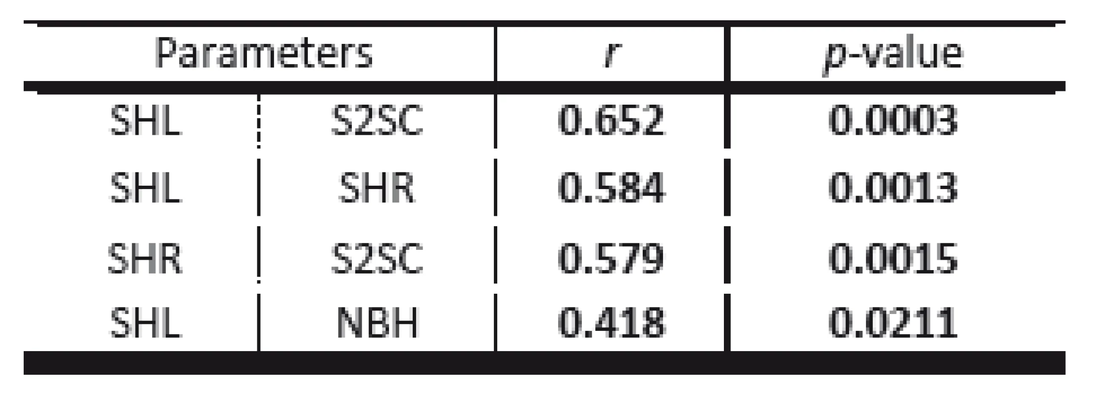
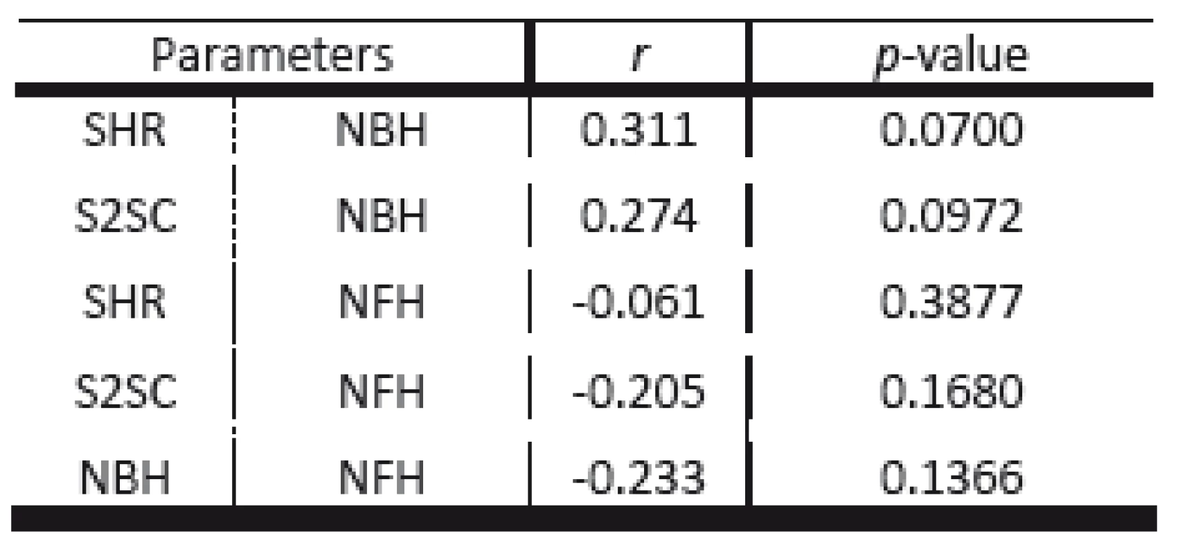
This correlation was quantified using the Pearson’s correlation coefficient (r). The statistical significance of the correlation coefficients was computed at α = 0.05 significance level. The results are shown in Tables 1 and 2 and significant correlations are high-lighted in bold (p < 0.05) (Tab. 1).
Tab. 1. Paired parameters with significant correlations. 
SHL – Shoulder Height Left, SHR - Shoulder Height Right, S2SCShoulder 2 Shoulder Caliper (straight line), HBH – Neck Back Height Tab. 2. Paired parameters with nonsignificant correlations. 
SHL – Shoulder Height Left, SHR - Shoulder Height Right, S2SCShoulder 2 Shoulder Caliper (straight line), HBH – Neck Back Height, NFH - Neck Front Height. There were four significant positive correlations between parameters, with the strongest correlation between the SHL and S2SC (r = 0.652; p = 0.0003).
There were no significant correlations between the other parameters. Table 2 shows the r and p-values for particular correlations.
Conclusion
Spiral stabilization of muscle chains is an exercise process, which consists of the phases of stabilization, traction and rotation. We need to realize that individual phases require specific amounts of exercise and adaptation time. According to our previous experience what needs to be emphasized is the individual approach towards exercisers because new movement patterns will not change and adjust without thoroughly correcting the movement and the work of individual muscle groups. The scientific and technological development has brought about changes in the body posture and reciprocal inhibition of people’s muscle chains. Static vertical chains are constantly overloaded, which considerably affects body posture and causes pain leading to discomfort in life. The course of the exercise plan and the achieved results showed that students who exercised reached the stabilization phase.
The evidence gathered to date has shown that the stabilization phase in the ration of SHL/S2SC is a significant progress. It should be noted that exercise performed during the traction phase needs to be longer and more intense. The practice has shown that students are aware of the need to move. More aware individuals want to attend physical education classes despite the fact that participation in these classes after studying at the Technical University in Košice for two semesters becomes optional. The primary necessity is the precise setting of a correct, physically functional body position when participating in any type of exercise process. We feel that university students should improve their knowledge of anatomy and develop accuracy of movement coordination. This role may be fulfilled through spiral stabilization, the SM exercise system, which adjusts incorrectly acquired movement patterns. Upright body posture is negatively affected by the predominant sedentary lifestyle based on decreasing volume of natural movement and increasing amount of static workload. Therefore, the beginners who decide to take start exercising should be individually coordinated when performing movements.
Acknowledgement
This study has been supported by the grant project KEGA 063TUKE-4/2016 “Metrologic processing of biomedical data using 3D scanning systems for educational purposes”.
The study was conducted within the project KEGA 044PU-4/2016 “Innovation of health-oriented educational tools for future teachers of physical and sports education and experts in sports and health”.
Mgr. Miroslava Barcalová
Department of Biomedical Engineering and Measurement
Faculty of Mechanical Engineering
Technical University in Košice
Letná 9,
042 00 Košice,
Slovakia
E-mail: miroslava.barcalova@tuke.sk
Tel.: +421 905 680 959
Zdroje
[1] LEWIT, K.: Rehabilitace u bolestivých poruch pohybové soustavy. Rehabilitace a fyzikální lékařství, 1, 2001, 4–17.
[2] JANDA, V.: Základy kliniky funkčních (nepatetických) hybných poruch. Brno, Česká republika: Ústav pro další vzdělávání středních zdravotnických pracovníků, 1984.
[3] KOLÁŘ, P.: Systematizace svalových dysbalancí z pohledu vývojové kineziologie. Rehabilitace a fyzikální lékařství, 8(4), 20011, 52–164.
[4] KOLÁŘ, P.: Vadné držení těla z pohledu posturální ontogeneze. Pediatrie pro praxi, 3, 2002, 106–109.
[5] KOLÁŘ, P.: Rehabilitace v klinické praxi. Praha: Galén, 2009.
[6] SMÍŠEK, R., SMÍŠKOVÁ, K., SMÍŠKOVÁ, Z. (2014): Léčba výhťezu medziobratlového disku bez operace, Praha 2014. ISBN 978-80-87568-43-9.
[7] HRČKA, J., 2009: Kapitoly zo športovej zdravovedy vysokoškoláka. Žilinská univerzita, Žilina. ISBN 978-80-554-0096-9.
[8] NOVOTNÁ, V. (2010): Problematika tělesné výchovy na vysokých školách. Dostupné na internete: www.radavs.cz/prilohy/12p7TVnaVS_Novotna.doc.
[9] BARCALOVÁ, M., VOJTAŠKO, Ľ., ŽIVČÁK, J.: Do WHR and BMI have an impact on back pain occurrence in university students’ community. In: Teoretyczne i praktyczne uwarunkowania kultury fizycznej i turystyki. Czestochowa: Wydawnictwo im. Stanislawa Podobińskiego Akadémii im Jana Dlugosza w Częstochovie, 2015 s. 145-155. - ISBN 978-83-7455-442-8.
[10] HRČKA, J., KOVÁŘOVÁ, M., BEŇAČKA, J., 2011.: Pohybová aktivita edukantov fyzioterapie vo voľnom čase a jej reflexia na vybraných zdatnostných a zdravotných charakteristikách. Trnava: Univerzita sv. Cyrila a Metoda, 2011 : 151. ISBN 978-80-8105-323-8.
[11] BARTÍK, P. (2002): Zdravotná telesná výchova I. Banská Bystrica: Pedagogická fakulta Univerzity Mateja Bela, 2002, s 32. 71.
[12] SMÍŠEK, R. (2015): Vzdělávací a rehabilitační centrum Smíšek, Na Úbočí 10, 182 00 Praha 8, 29.6. 2015, Osobná komunikácia.
[13] SMÍŠEK, R., SMÍŠKOVÁ, K., SMÍŠKOVÁ, Z. (2012a): Kondice, regenerace SPORT prevence, kompenzace, [CD nosič] Praha 2012, ISBN 978-80-87568-18-7.
[14] SMÍŠEK, R., SMÍŠKOVÁ, K., SMÍŠKOVÁ, Z. (2012b): Svalové řetězce, [CD nosič] Praha 2012, ISBN 978-80-87568-03-3.
[15] Špirálni stabilizace páteře. [9-11-2016] http://www.spiralstabilization.com/cz
[16] SMÍŠEK, R., SMÍŠKOVÁ, K., SMÍŠKOVÁ, Z. (2011a): Spirální stabilizace páteře: Léčba a prevence bolestí zad. Vydal MUDr. Richard Smíšek v marci 2011, ISBN 978-80-904292-0-8.
[17] SMÍŠEK, R., SMÍŠKOVÁ, K., SMÍŠKOVÁ, Z. (2011b): Učitel metody - SM - System, Vydal MUDr. Richard Smíšek, [CD nosič] Praha 2011, ISBN 978-80-904292-4-6.
[18] KALTENBORN, F.M.: Manual Mobilization of the Joints, Volume III: Traction-Manipulation of the Extremities and Spine, 2008.
[19] SMÍŠEK, R., SMÍŠKOVÁ, K., SMÍŠKOVÁ, Z. (2012): Svalové řetězce, [CD nosič] Praha 2012, ISBN 978-80-87568-15-6.
[20] DIMON, T.: Anatomie těla v pohybu, Vydavateľstvo: Pragma, 2009, ISBN: 9788073491918.
[21] SMÍŠEK, R., SMÍŠKOVÁ, K., SMÍŠKOVÁ, Z. (2016): Stabilizační svalové řetězce. ISBN: 978-80-87568-77-4.
[22] Kociová, K., Mikuľáková, W. 2011: Kineziologická analýza axiálneho systému človeka pomocou zariadenia Spinal Mouse. In Molisa 8. Prešov, Grafotlač, s.r.o, Prešov. 2011. s. 59–66.
[23] Laguna, M. 2001 Spinal Mouse [online]. 2001. [cit.2013.01.11] Dostupné na internete: <http://www.flare.ch/aditus/main.html>.
[24] Glaus, K. SpinalMouse [online]. 2010. [cit.2013.01.05] Dostupné na internete: < http://www.idiag.ch/en/customer-service/support/>.
[25] Dohi, E. SpinalMouse® [online]. 2013. [cit.2013.02.08] Dostupné na internete: <http://spinalmouse.ro/en/contact/>.
[26] MIKUĽÁKOVÁ, W et al. (2016). Monitoring výskytu porúch v oblasti osového orgánu u basketbalistov a volejbalistov. In: Acta Facultatis exercitationis corporis universitatis Presoviensis, Prešov : Vydavateľstvo Prešovskej univerzity, 2016, S. 122-130, ISBN 978-80-555-1701-8.
[27] MIKUĽÁKOVÁ, W., LABUNOVÁ, E., KENDROVÁ, L., KOCIOVÁ, K.: The incidence of changes in the axial skeleton of young athletes - In: Scientific review of physical culture [elektronický zdroj] Vol. 3, no. 4 (2013), online, s. 70-75. ISSN 2083-8581.
[28] MIKUĽÁKOVÁ, W., ŽIVČÁK, J., ELIÁŠOVÁ, A., KOVAĽOVÁ, E., LABUNOVÁ, E., KENDROVÁ, L.: Monitoring výskytu porúch osového orgánu u študentov dentálnej hygieny = Monitoring the occurrence of the axial organ defects in dental hygiene students In: Lékař a technika. Vol. 45, no. 3 (2015), s. 69–74. ISSN 0301-5491.
[29] TAPALIDOU, A., et al. 2014: Evaluation of the reliability of a new non-invasive method for assessing the functionality and mobility of the spine. In: Acta of Bioengineering and Biomechanics, Vol. 16, No. 1, 2014. DOI: 10.5277/abb140114.
[30] SPINAL MOUSE®. 2013. Software user Guide.
[31] RAJŤÚKOVÁ, V. et al.: Metodika merania na celotelovom 3D skenery a možnosti aplikácie, In: Lékař a technika. Vol. 43, no. 4 (2013), p. 5–9 . - ISSN 0301-5491, 2013.
[32] MOLKENSTRUCK, S., WINKELBACH, S., WAHL, M.F.: 3D Body Scanning in a Mirror Cabinet, DAGM 2008, 284-293.
[33] SIMONENKO, J., CUK, I.: Reliability and Validity of NX-16 3D Body Scanner. Int. J. Morphol., 34(4):1506-1514, 2016.
[34] ŽIVČÁK, J. a kol., 2007: Biomechanika človeka I. Prešov: ManaCon. ISBN 978-80-89040-30-8.
[35] ŽIVČÁK, J. a kol., 2007: Biomechanika človeka II. Prešov: ManaCon. ISBN 978-80-89040-31-5.
[36] ŽIVČÁK, J., Knežo, D., 2008: Biomechanika hybnosti. Prešov: Grafotlač. Prvé vydanie. ISBN 978-80-8068-608-6.
Štítky
Biomedicína
Článek vyšel v časopiseLékař a technika

2017 Číslo 1-
Všechny články tohoto čísla
- THE EFFECT OF FLUID ACCUMULATION IN STOMACH ON ELECTRICAL IMPEDANCE TOMOGRAPHY IMAGE OF LUNGS
- EFFECT OF STERILIZATION ON MECHANICAL PROPERTIES OF COLLAGEN-BASED COMPOSITE TUBES
- BIOFEEDBACK AS A NEUROBIOMECHANICAL ASPECT OF POSTURAL FUNCTION
- THE USE OF NONINVASIVE DIAGNOSTIC METHODS IN THE ASSESSMENT OF POSTURAL CHANGES IN UNIVERSITY STUDENTS
- USING PVDF FILMS AS FLEXIBLE PIEZOELECTRIC GENERATORS FOR BIOMECHANICAL ENERGY HARVESTING
- Lékař a technika
- Archiv čísel
- Aktuální číslo
- Informace o časopisu
Nejčtenější v tomto čísle- THE USE OF NONINVASIVE DIAGNOSTIC METHODS IN THE ASSESSMENT OF POSTURAL CHANGES IN UNIVERSITY STUDENTS
- BIOFEEDBACK AS A NEUROBIOMECHANICAL ASPECT OF POSTURAL FUNCTION
- USING PVDF FILMS AS FLEXIBLE PIEZOELECTRIC GENERATORS FOR BIOMECHANICAL ENERGY HARVESTING
- THE EFFECT OF FLUID ACCUMULATION IN STOMACH ON ELECTRICAL IMPEDANCE TOMOGRAPHY IMAGE OF LUNGS
Kurzy
Zvyšte si kvalifikaci online z pohodlí domova
Autoři: prof. MUDr. Vladimír Palička, CSc., Dr.h.c., doc. MUDr. Václav Vyskočil, Ph.D., MUDr. Petr Kasalický, CSc., MUDr. Jan Rosa, Ing. Pavel Havlík, Ing. Jan Adam, Hana Hejnová, DiS., Jana Křenková
Autoři: MUDr. Irena Krčmová, CSc.
Autoři: MDDr. Eleonóra Ivančová, PhD., MHA
Autoři: prof. MUDr. Eva Kubala Havrdová, DrSc.
Všechny kurzyPřihlášení#ADS_BOTTOM_SCRIPTS#Zapomenuté hesloZadejte e-mailovou adresu, se kterou jste vytvářel(a) účet, budou Vám na ni zaslány informace k nastavení nového hesla.
- Vzdělávání


