-
Články
Top novinky
Reklama- Vzdělávání
- Časopisy
Top články
Nové číslo
- Témata
Top novinky
Reklama- Kongresy
- Videa
- Podcasty
Nové podcasty
Reklama- Kariéra
Doporučené pozice
Reklama- Praxe
Top novinky
ReklamaEffect of linear accelerator settings on evaluation of dosimetric verification of VMAT plans
Background and purpose:
Comparison of the measured and the calculated 2D dose distributions in the planning system is one of the standard methods of VMAT (volumetric-modulated arc therapy) plans verification in radiotherapy. The aim of this paper is to assess the ability of the QA (quality assurance) method used at the University Hospital Olomouc to detect significant errors in the linear accelerator settings and to estimate clinical impact of the undetected changes.Materials and methods:
To study this issue we chose a method based on introducing simulated errors in gantry and MLC (multi-leaf collimator) settings into 10 clinical VMAT plans of the prostate and the head and neck patients. The ability of the verification system to recognize changes in prescribed setting is - for the purpose of this paper - characterized by a maximum error, in which the two dose distributions are evaluated as sufficiently identical within the parameters of gamma analysis. VMAT plans with the maximum undetected error were recalculated back into the real patient anatomy in order to assess the potential clinical impact.Results:
The verification method including 3%/3mm gamma analysis criteria together with Octavius II system is able to detect errors larger than 2mm in MLC and 3° in gantry settings in the head and neck VMAT plans and 3mm and 5° for prostate VMATs. In comparison with the original plan, dose recalculations with these errors in the settings back into the real patients showed differences in dose distributions.Conclusion:
Comparison of dose volume histograms for the original plan and the plans recalculated with implemented errors indicate that this system of verification of VMAT plans could cover up clinically relevant errors.Keywords:
Volumetric modulated arc therapy (VMAT), Octavius QA system, gamma analysis
Authors: Václav Novák; Ivo Přidal; David Gremlica
Authors place of work: Czech Republic ; Department of Medical Physics and Radiation Protection, University Hospital Olomouc
Published in the journal: Lékař a technika - Clinician and Technology No. 4, 2015, 45, 110-114
Category: Původní práce
Summary
Background and purpose:
Comparison of the measured and the calculated 2D dose distributions in the planning system is one of the standard methods of VMAT (volumetric-modulated arc therapy) plans verification in radiotherapy. The aim of this paper is to assess the ability of the QA (quality assurance) method used at the University Hospital Olomouc to detect significant errors in the linear accelerator settings and to estimate clinical impact of the undetected changes.Materials and methods:
To study this issue we chose a method based on introducing simulated errors in gantry and MLC (multi-leaf collimator) settings into 10 clinical VMAT plans of the prostate and the head and neck patients. The ability of the verification system to recognize changes in prescribed setting is - for the purpose of this paper - characterized by a maximum error, in which the two dose distributions are evaluated as sufficiently identical within the parameters of gamma analysis. VMAT plans with the maximum undetected error were recalculated back into the real patient anatomy in order to assess the potential clinical impact.Results:
The verification method including 3%/3mm gamma analysis criteria together with Octavius II system is able to detect errors larger than 2mm in MLC and 3° in gantry settings in the head and neck VMAT plans and 3mm and 5° for prostate VMATs. In comparison with the original plan, dose recalculations with these errors in the settings back into the real patients showed differences in dose distributions.Conclusion:
Comparison of dose volume histograms for the original plan and the plans recalculated with implemented errors indicate that this system of verification of VMAT plans could cover up clinically relevant errors.Keywords:
Volumetric modulated arc therapy (VMAT), Octavius QA system, gamma analysisIntroduction
VMAT (volumetric-modulated arc therapy) refers to an arc irradiation technique with an intensity modulated beam. Since it adds extra degrees of freedom to dose delivery, e.g. dose rate or gantry speed variation, it is more complex in comparison with the non-dynamic conformal or Step&Shoot techniques. Even a relatively small uncertainty arising from changes in linear accelerator settings could have a significant impact on the final dose distribution. Hence, dosimetric verification of every patient´s VMAT plan is an integral part of the standard radiotherapy treatment procedure at the University Hospital Olomouc.
During the daily use of a medical linear accelerator, physicists can encounter various deviations from the correct settings. The typical mechanical errors include especially errors in the field size setting and an inaccurate calibration of gantry or a collimator angle. The aim of this paper is therefore to evaluate the ability of the QA (quality assurance) method used at the University Hospital Olomouc to detect significant errors in the field size and the gantry angle settings and to assess the possible clinical impact of undetected changes at VMAT delivery.
Materials and methods
Equipment
All measurements were performed on Elekta Synergy linear accelerator with MLCi2 collimator system (Elekta, Crawley, UK). Flat ionization chamber matrix Seven29 (PTW, Germany) inserted in octagonal phantom Octavius II (PTW, Germany) was used for the purpose of VMAT plan verification as it is used for daily QA routine at the University Hospital Olomouc. Octavius II phantom is made of polystyrene (density 1.04 g/cm3) with air-filled lower part, which serves to compensate for different detector sensitivity when irradiated from the bottom directions (Fig. 1). Seven29 detector is composed of 729 plane-parallel vented ionization chambers on the area of 27×27cm2, each with a sensitive volume of 125cm3. The chambers are spaced 10mm center-to-center. [1]
Fig. 1. Octavius II CT scan with inserted Seven29 detector (labeled with red rectangle), lower part of the phantom with air gap. 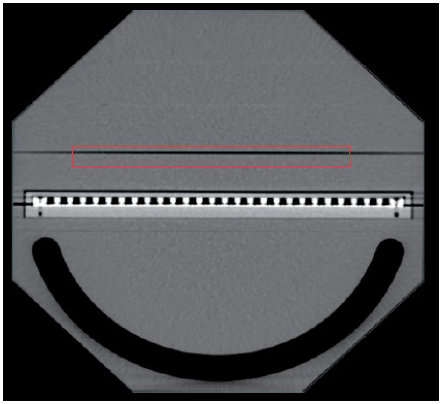
Patients and plans
Five head and neck (energy 6MV) and five prostate (energy 18MV) VMAT plans were selected to test the effect of inaccuracies in the linear accelerator settings. Study of the effect of gantry and MLC (multi-leaf collimator) settings on dose distribution delivery was based on modification in the output parameters of the planning system. These parameters are used to control the radiotherapy treatment procedure and are ordered in groups forming a sequence of control points. Each control point includes information about the linear accelerator position (gantry angle) and the individual leaf position or the number of monitor units (MU). Therefore, off-sets were introduced into all of the control points to simulate uncertainties in linear accelerator settings. This was realized through modification of the original VMAT plan output files, using the homemade script written in Matlab software.
At first, leaf coordinates in all control points were adjusted to extend field size gradually from 1mm to 5mm. These relatively small changes could not be proved in a simpler, conformal treatment technique. On the contrary, dose distribution at VMAT plans is very often delivered through a large number of small segments (up to 1×1cm2). Thus, the error of a few millimeters in the field size can result in considerable changes in the dose distribution within the patient. Hereby, ten altered plans were measured and compared to the original.
The second “variable” under consideration, which could considerably affect the treatment plan, was gantry angle. In a similar way, the gantry angle in each control point was varied from 1° to 10° and the effect of this change on the result of VMAT plan verification was followed.
Analysis
Comparison of the measured dose distributions with those calculated in the planning system was made in Verisoft software version 6.0 (PTW, Germany). The simplest and most direct method is simple comparison of dose difference at each point of measurement. However, this practice can produce large errors in steep dose gradients even with minimal inaccuracy in the detector position. For this reason more robust procedure of dose distribution comparison was used in this paper. Gamma analysis takes into account not only dose differences but also spatial displacement [2, 3]. At the University Hospital Olomouc tolerances of 3% and 3 mm are used, respectively.
Plans with the highest error, which still met the criteria of the gamma analysis, were recalculated back into the patient´s anatomy in order to compare dose volume histograms (DVHs) of target volume and organs at risk (specific for given diagnosis) for the original and modified plan.
Results
Results of the measurements performed with Octavius II verification phantom are listed in Tab. 1 and 2 for the head and neck and the prostate cases respectively. They include the highest errors in the linear accelerator settings tolerated with the gamma analysis of the dose distribution measured in the Seven29 plane. Results show higher tolerance to uncertainties in gantry angle and field size for the irradiation of pelvis than for the head and neck region.
Tab. 1. Measurements for 6MV energy performed with Octavius II phantom – head and neck case. 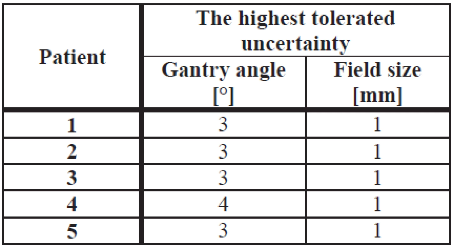
Tab. 2. Measurements for 18MV energy performed with Octavius II phantom – prostate case. 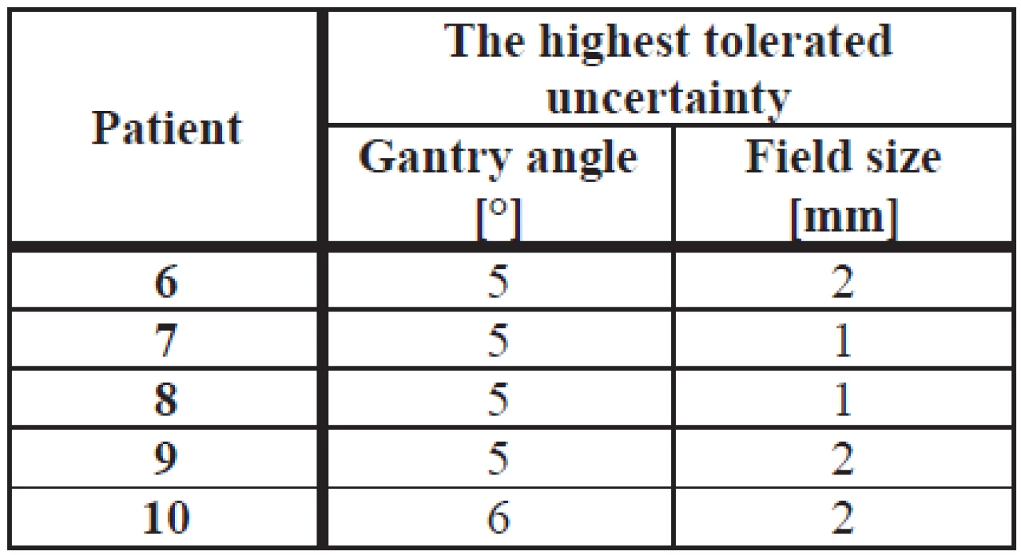
VMAT plans with the highest tolerated error were subsequently recalculated back to the patient anatomy (dose distributions can be seen in Fig. 2 and 4), and DVHs for every patient were compared to estimate potential risk of the treatment. With regard to the results similar for all patients with the given diagnosis, DVHs comparison for only one patient for that case are shown in Fig. 3 and 5.
Fig. 2. Dose distribution comparison of the original plan (a) to the plan with 3° gantry angle change (b) and to 1 mm field size change (c) for the head and neck case (6MV energy). 
Fig. 3. DVHs comparison of the original plan to the plan with 3° gantry angle change and to 1 mm field size change for the head and neck case (6MV energy). 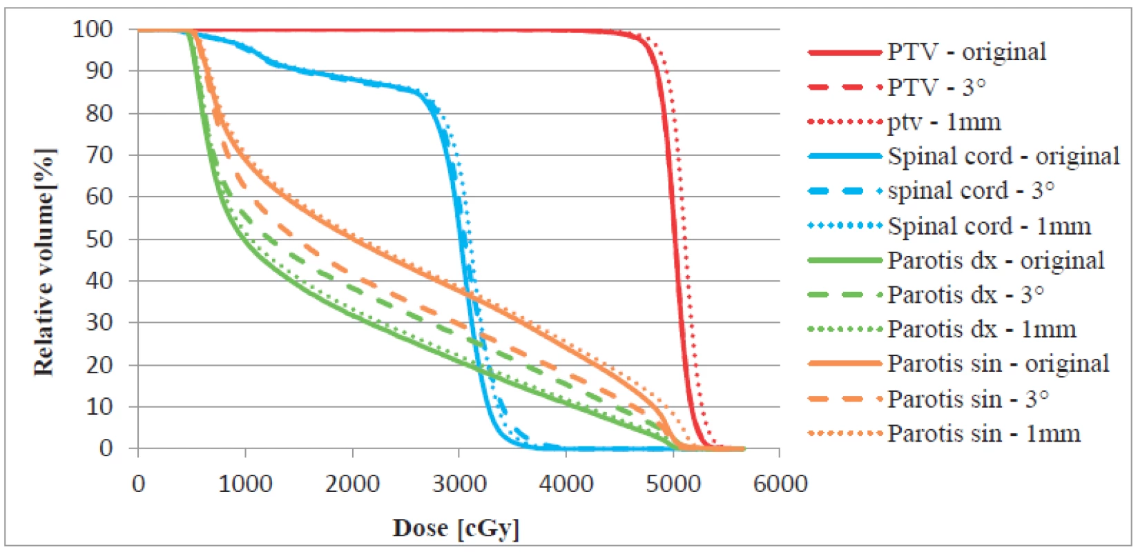
Fig. 4. Dose distribution comparison of the original plan (a) to the plan with 5° gantry angle change (b) and to 2 mm field size change (c) for the prostate case (18MV energy). 
Fig. 5. DVHs comparison of the original plan to the plan with 5° gantry angle change and to 2 mm field size change for the prostate case (18MV energy). 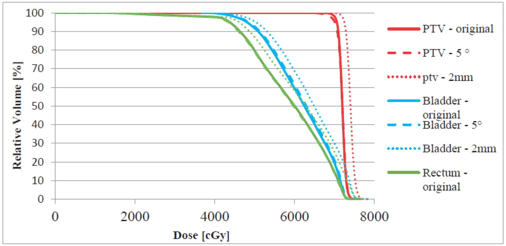
DVHs comparison shows that for patients with prostate cancer diagnosis extension by 1–2 mm in field size could cause an increase in the average and also in maximum dose of 2–3Gy in the planning target volume (PTV), rectum and bladder. On the other hand, influence of gantry angle change of up to 5° appears to be negligible in this anatomic site.
At the University Hospital Olomouc treatment plan quality is evaluated also in terms of organs-at-risk dose limits. One of these limits is set for bladder at V70<20% (V70 is volume that receives dose of 70Gy, in this case less than 20% of the bladder volume can receive dose of 70 Gy) and for rectum at V65<35%. Both the original plan and the gantry angle changed plan met these criteria. In plans with the field size extended to 1–2 mm the limit for bladder was exceeded by 0.5–11%, i.e. V70 = 20.5–31%. Similar results appeared in case of rectum irradiation. Four of five plans exceeded the limit for rectum by 3–8%, i.e. V65 = 38–43%.
Introducing uncertainties of 1mm in field size to treatment plans of the head and neck cases resulted again in a possible increase in the average and maximum dose by more than 1Gy in PTV and in spinal cord. It seems that this anatomical site is more sensitive to gantry angle inaccuracies. DVHs comparison indicates that the change by 3° increases the average dose to one parotid by 1–3.5Gy and decreases it by the same value to the second parotid. Maximum dose to the spinal cord was also raised by 1Gy at this angle change. Although the doses were increased by relatively high values, with no patient with disease in this site, tolerance doses were not exceeded for organs at risk.
Discussion
VMAT plan comparison was realized only on a small group of patients. Nevertheless, anatomy and contouring of critical structures are usually very similar with patients with the same diagnosis. Likewise, the results of the gamma analysis evaluations were also similar, and the same relation can be assumed in case of additional measurements.
Influence of the inaccuracies proved to be dependent on the irradiated location. The plans in the head and neck region seem to be more sensitive to a systematic shift of the gantry angle and to the field size settings than the prostate plans. This is probably due to generally higher complexity of these plans. Head and neck region contains smaller structures which are closer together. Besides, PTV shape is often very irregular, which results in higher number of smaller segments. Increase in the field size by 1mm is then, with regard to the size of individual segments, relatively large. It is supposed that for these cases this all leads to higher sensitivity of VMAT plan´s verification to inaccuracies in the gantry angle and field size.
Recalculations of the changed plans back into the patient´s CT data set show that these uncertainties evaluated by gamma analysis as sufficiently identical with the original plan, might cause delivery of clinically relevant difference from the original dose distribution (see Fig. 3 and 5). Therefore, tolerance of 2mm to field size settings used for conformal radiotherapy techniques and used also e.g. in completion certificate of the Elekta company or in older SÚJB (State Office for Nuclear Safety) recommendations [4] could not be considered sufficient.
Increase in sensitivity to inaccuracies can be achieved with a stricter gamma analysis criterion, e.g. 2%/2mm. However, it is then necessary to count with a higher rate of unsuccessful results of verification.
Conclusion
The efficiency to reveal clinically significant uncertainties in linear accelerator settings during VMAT plan verification was tested. The system of verification used at the University Hospital Olomouc is supposed to be able to detect errors in the gantry angle from 4° for the head and neck and from 6° for the prostate disease.
These errors can be clinically relevant especially in the first region where they increase the maximum dose to the spinal cord and vary the average dose to parotids. Head and neck plans verification appears to be more sensitive also to the field size setting. Plans with errors greater than 2mm did not meet gamma analysis criteria. With respect to organs at risk, increase in field size affects mainly the integral dose.
Systematic introduction of errors into dosimetric verified VMAT plans has disclosed limits of the verification method. The above DVH comparisons show that the gamma analysis criterion of 3%/3mm in connection with the given verification method may not be sufficient to detect important inaccuracies. It is possible to suggest tightening this criterion, and, consequently, pay more attention to the position of hot or cold spots in the evaluated dose distribution and check if they are in clinically relevant area or at the edge of irradiated volume.
Ing. Václav Novák
Department of Medical Physics and Radiation Protection
University Hospital Olomouc
I. P. Pavlova 185/6, 779 00, Olomouc
Czech Republic
Email address: vaclav.novak@fnol.cz
Zdroje
[1] Hussein M., A critical evaluation of the PTW 2D-Array seven29 and Octavius II phantom for IMRT and VMAT verification, Journal of applied clinical medical physics, 2013.
[2] Low D., A technique for the quantitative evaluation of dose distributions, Medical Physics, 1998.
[3] Depuydt T., A quantitative evaluation of IMRT dose distributions: refinement and clinical assessment of the gamma evaluation, Radiotherapy and Oncology, 2002.
[4] State Office for Nuclear Safety: Recommendation: Introduction of quality assurance in using significant sources of ionizing radiation in radiotherapy – linear accelrators for 3D conformal radiotherapy and IMRT, Prague, 2006.
Štítky
Biomedicína
Článek vyšel v časopiseLékař a technika

2015 Číslo 4-
Všechny články tohoto čísla
- Multi-electrode microfluidic platform for protein detection using electrochemical impedance spectroscopy
- Effect of linear accelerator settings on evaluation of dosimetric verification of VMAT plans
- Spectral analysis of atrial components of ablation catheter signals during slow pathway ablation for typical atrioventricular nodal reentrant tachycardia
- MERANIE PARAMETROV ELEKTROMAGNETICKÝCH POLÍ PRI POUŽÍVANÍ PROSTRIEDKOV MOBILNEJ KOMUNIKÁCIE V ŠKOLSKOM PROSTREDÍ
- Lékař a technika
- Archiv čísel
- Aktuální číslo
- Informace o časopisu
Nejčtenější v tomto čísle- MERANIE PARAMETROV ELEKTROMAGNETICKÝCH POLÍ PRI POUŽÍVANÍ PROSTRIEDKOV MOBILNEJ KOMUNIKÁCIE V ŠKOLSKOM PROSTREDÍ
- Spectral analysis of atrial components of ablation catheter signals during slow pathway ablation for typical atrioventricular nodal reentrant tachycardia
- Multi-electrode microfluidic platform for protein detection using electrochemical impedance spectroscopy
- Effect of linear accelerator settings on evaluation of dosimetric verification of VMAT plans
Kurzy
Zvyšte si kvalifikaci online z pohodlí domova
Autoři: prof. MUDr. Vladimír Palička, CSc., Dr.h.c., doc. MUDr. Václav Vyskočil, Ph.D., MUDr. Petr Kasalický, CSc., MUDr. Jan Rosa, Ing. Pavel Havlík, Ing. Jan Adam, Hana Hejnová, DiS., Jana Křenková
Autoři: MUDr. Irena Krčmová, CSc.
Autoři: MDDr. Eleonóra Ivančová, PhD., MHA
Autoři: prof. MUDr. Eva Kubala Havrdová, DrSc.
Všechny kurzyPřihlášení#ADS_BOTTOM_SCRIPTS#Zapomenuté hesloZadejte e-mailovou adresu, se kterou jste vytvářel(a) účet, budou Vám na ni zaslány informace k nastavení nového hesla.
- Vzdělávání


