-
Medical journals
- Career
Congenital isolated juvenile xanthogranuloma of the sole – a unique case report in a newborn
Authors: S. Saoud 1,2; N. Karich 3; G. Belmaati Cherkaoui 1; H. Eladak 1; F. Zouaidia 4; A. A. Oufkir 1
Authors‘ workplace: Department of Burns and Reconstructive Surgery, Mohammed VI Oujda Hospital, Morocco 1; Research Laboratory in Medical Sciences, Faculty of Medicine and Pharmacy of Oujda, Mohammed I University, Morocco 2; Department of Pathology, Mohammed VI Oujda Hospital, Morocco 3; Department of Pathology, University Hospital Ibn Sina, Rabat, Morocco 4
Published in: ACTA CHIRURGIAE PLASTICAE, 66, 1, 2024, pp. 27-30
doi: https://doi.org/10.48095/ccachp202427Introduction
Juvenile xanthogranuloma (JXG) is a rare benign disorder. It was first described by Adamson in 1905 in a report of the case of a 2-year-old boy using the term congenital xanthoma multiplex [1]. Seven years later, McDonagh described five similar cases [2], but it was not until the 1950s that the name JXG was officially used by Helwig and Hackney in a series of 140 cases [3].
Over 24,000 juvenile soft tissue masses were analyzed by the Kiel Pediatric Tumor department, and the incidence of JXG was approximately 0.5%. It is however thought to be much higher, as small isolated lesions may be underdiagnosed. A male predominance has been noted in childhood estimated as 1.5 : 1 [4].
JXG generally appears within the first 2 years of life; most of these lesions are self-limiting and the majority resolve on their own through childhood. Due to its clinical diversity, a histology study is necessary for accurate diagnosis.
Case report
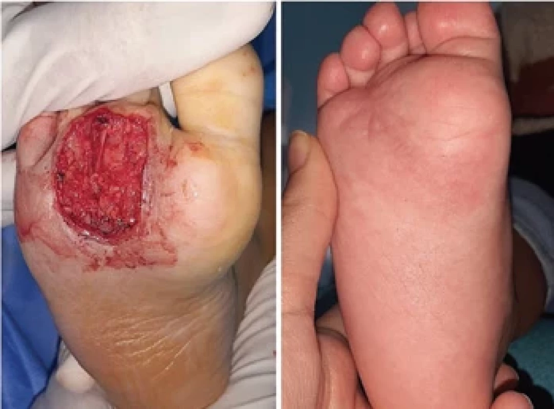
A four-week-old boy presented to the plastic surgery department with a congenital solitary lesion on the right sole that gradually increased in size. The physical examination revealed a 2 cm exophytic, firm, pink, and nodular mass with a central serosanguineous crust (Fig. 1). Biopsy results indicated diffuse sheets of histiocytes in the dermis mixed with lymphocytes, eosinophils, and Touton giant cells, consistent with JXG (Fig. 2). The patient underwent ocular and systemic examinations in the ophthalmology and pediatric departments, revealing no extra-cutaneous lesions. An MRI study showed an ovoid 22 × 15 × 12 mm mass, abutting the plantar aponeurosis with tendinous involvement (Fig. 3). Given that JXG is benign, spontaneously resolving in nature, a watch-and-wait approach was initially chosen. However, at the 12-month follow-up, the nodule neither decreased in size clinically nor radiologically, interfering with weight-bearing and walking due to bleeding. The family was counseled, and after understanding the risks, benefits, and possible complications of surgical intervention, they provided consent for mass excision. On the day of surgery, the patient was brought to the operating room, placed supine on the table, and underwent general anesthesia. An ankle tourniquet was used and inflated prior to incision. The incision was placed around the mass, and dissection was carried out through the plantar adipose layer, just superficial to the tendons. Metzenbaum scissors were used to dissect out the ovoid mass, which communicated deeply with the tendons. The mass, yellowish and distinct from the surrounding fat (Fig. 4), was sent in to pathology for further evaluation. After hemostasis and irrigation, a negative pressure wound dressing was applied for 5 days. Following the development of satisfying granulation tissue, a skin graft was placed to cover the wound. The 6 month follow-up found the area to be healed with no signs of recurrence or toe contracture (Fig. 5).
1. Aspect of exophytic, fi rm, pink and nodular mass with a central serosanguineous crust, measuring 2 cm. 
2. Multiple touton-type giant cells admixed with a predominantly histiocytic infi ltrate (hematoxylin and eosin, original magnifi cation and × 40, respectively). 
3. T2 sagittal view, uniform hypointense subcutaneous soft tissue mass seen in the relatively hyperintense plantar forefoot measuring 2.27 × 1.59 cm. 
4. Yellow macroscopic aspect of the tumor. 
5. From left to right – after excision of the tumor leaving a minor residue, aspect after 6 months follow-up. 
Discussion
Histiocytoses are rare disorders, characterized by the accumulation of cells thought to be derived from dendritic cells (DCs) or macrophages. The first classification of histiocytosis, published in 1987 by the Histiocyte Society (HS) consisted of 3 categories: Langerhans cell (LC) or non-LC-related, and malignant histiocytoses (MH). A new revised classification system appeared in 2016, consisting of 5 groups of diseases: (L) Langerhans-related, (C) cutaneous and mucocutaneous, (M) malignant histiocytoses, (R) Rosai-Dorfman disease, and (H) hemophagocytic lymphohistiocytosis and macrophage activation syndrome [5].
JXG is the most common form of non--Langerhans cell histiocytosis belonging to the C group (cutaneous and mucocutaneous histiocytoses).
JXG is a tumor that occurs predominantly in early childhood or even at birth [6]. In the Kiel pediatric tumor registry analysis, 71.3% of the patients displayed JXG within the first 12 months; in 34.5% of the cases, JXG was present at birth [4]. However, adult cases have also been described, sharing similar histological and immunohistological features [7].
Cutaneous lesions are generally the main symptom in JXG, with extracutaneous involvement being rare [8,9].
JXG is characterized by a wide spectrum of lesions in terms of shape, color, and number [8,10]. In two major studies of XGJ, the majority of patients with JXG presented with a solitary lesion, whereas in the remaining cases, multiple lesions appeared in different regions of the body ranging from 2 or 3 to over 100 [8,9]. The characteristic JXG lesion is a well-circumscribed papule or nodule with a smooth surface. In its initial stages, it is typically erythematous, but it progressively acquires a characteristic yellow-orange color [8,10]. The lesions have a firm consistency. With the exception of certain locations or very large lesions, JXG is usually asymptomatic. The most common location is the head, neck, trunk, and limbs, but lesions may arise at any anatomic site [4]. In the literature, there are about 35 total cases of JXG in the lower extremity; our case report is the 36th case [11–13]. Regression usually occurs over several months to a few years and is usually complete by late childhood [9]. JXGs may resolve without a trace, or may leave behind an atrophic scar, hyperpigmentation, or other minor residua [14].
Ocular JXG is the most common extracutaneous manifestation; its prevalence is estimated at 0.3–0.5% [8,10]. It is usually unilateral and most often affects the iris. The most common presenting symptoms are a unilateral red eye or photophobia, and a tumor in the iris or conjunctiva could also be seen. Ocular lesions, unlike cutaneous lesions, do not resolve spontaneously and may lead to blinding complications such as glaucoma and hyphema. Therefore, it is recommended referring patients with two or more skin lesions or early-onset lesions to go in for an eye examination. Also, parents of the child should be well educated about the ocular signs [15].
Systemic JXG is rare and it usually manifests as multiple cutaneous and/or subcutaneous nodules and involvement of two or more visceral organs (brain, intestines, liver, heart, kidneys, appendix, or lungs) [10]. Depending on the patient’s history and suggestive signs and symptoms, Meyer et al. suggested that all children with two or more cutaneous JXG lesions should undergo in addition to a full skin examination, a laboratory work-up, screening for systemic JXG, including CT or MRI of the brain and chest/abdomen, and Doppler echocardiography if heart involvement is suspected [16].
JXG does not require treatment as it generally resolves spontaneously, as long as it doesn’t have functional impairment [8,9]. Then, lesions can be surgically removed [9]. The challenge in our case was the location of the nodule in the sole causing a delay in the normal development of the child, and the deeply rooted lesion communicating with the tendons. We decided to follow him up until he reached walking age; since the lesion did not resolve, we decided to surgically remove it superficially to the plantar aponeurosis leaving macroscopic residue [17].
Ocular JXG is usually treated by topical and intralesional corticoids, while systemic corticosteroids or surgery may be needed to rapidly treat progressive lesions or complications [13].
Treatment of systemic JXG should be initiated when JXG starts to interfere with vital organ function; it comprises of surgical resection, radiation therapy, and/or chemotherapy [17].
A JXG recurrence has never been observed in literature [4,9]. It is considered that these lesions rarely if ever recur locally even in the presence of positive surgical margins. Therefore, once the pathologic diagnosis of JXG has been established, a re-excision of a tumor with positive margins is seemingly unnecessary [9].
The uniqueness of this case, as mentioned earlier, lies in the challenging decision of whether to proceed with surgery or wait for the resolution of the lesion. Additionally, the tendinous involvement of the mass adds a layer of complexity to the management of the condition.
Conclusion
JXG is generally a benign skin-limited disease that tends to resolve spontaneously, thus not requiring any investigations or specific treatment except in some rare cases. When two or more skin lesions are found, a blood work-up and screening can search for systemic JXG. A regular ocular examination is always advised due to the severity of JXG complications on the eyes.
Roles of the authors
Sarah Saoud, MD and Prof. Ayat Allah OUFKIR conceptualized and designed the study, drafted the initial manuscript, and critically reviewed and revised the manuscript.
Drs. Ghita Belmaati Cherkaoui, and Hanane El Adak collected the data, and critically reviewed and revised the manuscript.
Prof. Nassira Karich and Prof. Fouad Zouaidia collected the data, carried out the initial analyses, and reviewed and revised the manuscript.
All authors approved the final manuscript as submitted and agree to be accountable for all aspects of the work.
Disclosure: The authors have no conflicts of interest to disclose. The authors declare that this study has received no financial support. All procedures performed in this study involving human participants were in accordance with ethical standards of the institutional and/or national research committee and with the Helsinki declaration and its later amendments or comparable ethical standards.
Sources
1. Adamson HG. Society intelligence: the dermatological society of London. Br J Dermatol. 1905, 17 : 222.
2. McDonagh JER. Spontaneous disappearance of an endothelioma (nevo-xanthoma). Proc R Soc Med. 1909, 2 : 142–144.
3. Helwig EB., Hackney VC. Juvenile xanthogranuloma (nevoxanthoendothelioma). Am J Path. 1954, 30 : 625–626.
4. Janssen D., Harms D. Juvenile xanthogranuloma in childhood and adolescence: a clinicopathologic study of 129 patients from the Kiel pediatric tumor registry. Am J Surg Pathol. 2005, 29 (1): 21–28.
5. Emile JF., Abla O., Fraitag S., et al. Revised classification of histiocytoses and neoplasms of the macrophage-dendritic cell lineages. Blood. 2016, 127 (22): 2672–2681.
6. Oza VS., Stringer T., Campbell C., et al. Congenital-type juvenile xanthogranuloma: a case series and literature review. Pediatr Dermatol. 2018, 35 (5): 582–587.
7. Meyer M., Grimes A., Becker E., et al. Systemic juvenile xanthogranuloma: a case report and brief review. Clin Exp Dermatol. 2018, 43 (5): 642–644.
8. Hernández-San Martín MJ., Vargas-Mora P., Aranibar L. Juvenile xanthogranuloma: an entity with a wide clinical spectrum. Actas Dermosifiliogr. 2020, 111 (9): 725–733.
9. Dehner LP. Juvenile xanthogranulomas in the first two decades of life: a clinicopathologic study of 174 cases with cutaneous and extracutaneous manifestations. Am J Surg Pathol. 2003, 27 (5): 579–593.
10. Hernandez-Martin A., Baselga E., Drolet BA., et al. Juvenile xanthogranuloma. J Am Acad Dermatol. 1997, 36 (3 Pt 1): 355–367.
11. Kim JH., Lee SE., Kim SC. Juvenile xanthogranuloma on the sole: dermoscopic findings as a diagnostic clue. J Dermatol. 2011, 38 (1): 84–86.
12. Derner BS., Hoffman K., Storfa A., et al. Isolated forefoot juvenile xanthogranuloma: unique case study and treatment in a pediatric patient. J Foot Ankle Surg. 2020, 59 (6): 1301−1305.
13. Whittam LR., Higgins EH. Juvenile xanthogranuloma on the sole. Pediatr Dermatol. 2000, 17 (6): 460–462.
14. Chang MW. Update on juvenile xanthogranuloma: unusual cutaneous and systemic variants. Semin Cutan Med Surg. 1999, 18 (3): 195–205.
15. Chang MW., Frieden IJ., Good W. The risk of intra-ocular juvenile xanthogranuloma: survey of current practices and assessment of risk. J Am Acad Dermatol. 1996, 34 (3): 445–449.
16. Stover DG., Alapati S., Regueira O., et al. Treatment of juvenile xanthogranuloma. Pediatr Blood Cancer. 2008, 51 (1): 130–133.
17. Kundak S., Çakır Y. Juvenile xanthogranuloma: retrospective analysis of 44 pediatric cases (single tertiary care center experience). Int J Dermatol. 2021, 60 (5): 564–569.
Sarah Saoud, MDDepartment of Burns and Reconstructive SurgeryMohammed VI Oujda HospitalBP 4806 Oujda Universite 60049Moroccoe-mail: sarah.saoud.izem@gmail.comSubmitted: 10. 11. 2023Accepted: 28. 2. 2024Labels
Plastic surgery Orthopaedics Burns medicine Traumatology
Article was published inActa chirurgiae plasticae

2024 Issue 1-
All articles in this issue
- No drains in reduction mammaplasty – a systematic review
- Inset techniques for the DIEP flap – what improves aesthetic outcomes?
- Microsurgical replantation after forehead avulsion – success or failure? A case report
- Nail bed trauma reconstruction and artificial nail replacement – a case report
- Double right triangular shape full-thickness skin grafts technique for short rectangular or square shape donor site defect – original method with a case report
- Congenital isolated juvenile xanthogranuloma of the sole – a unique case report in a newborn
- Reports on vascular catheter-associated thromboembolic events in a burn unit – a gap in the literature?
- The top 10 AI tools for academic surgeons right now
- Editorial
- Acta chirurgiae plasticae
- Journal archive
- Current issue
- Online only
- About the journal
Most read in this issue- No drains in reduction mammaplasty – a systematic review
- Nail bed trauma reconstruction and artificial nail replacement – a case report
- Inset techniques for the DIEP flap – what improves aesthetic outcomes?
- The top 10 AI tools for academic surgeons right now
Login#ADS_BOTTOM_SCRIPTS#Forgotten passwordEnter the email address that you registered with. We will send you instructions on how to set a new password.
- Career

