-
Medical journals
- Career
Rhabdomyosarcoma of the Gluteus Maximus – Case Report, Review of Literature and Emerging Therapeutic Targets
Authors: Gulab Girish Meshram 1; Neeraj Kaur 2; Singh Kanwaljeet Hura 3
Authors‘ workplace: Klinika komplexní onkologické péče, Masarykův onkologický ústav, Brno 1; Ústav psychologie a psychosomatiky, LF MU, Brno 2
Published in: Klin Onkol 2019; 32(3): 201-207
Category: Original Articles
doi: https://doi.org/10.14735/amko2019201Overview
Background: Rhabdomyosarcoma is an uncommon mesodermal cancer, which predominantly affects the young population. Common sites of primary disease include the head and neck region, genitourinary tract and the extremities. Less than 25% of the cases of rhabdomyosarcoma are metastatic at presentation. Embryonal and alveolar are the most common histological subtypes of rhabdomyosarcoma. Literature about rhabdomyosarcoma located in the gluteal region is sparse.
Case: We present a case of a 3-year-old child with alveolar rhabdomyosarcoma arising from the gluteal region. The metastatic workup was negative. We determined the tumour to be of intermediate risk and managed the patient with systemic chemotherapy consisting of cycles of vincristine, actinomycin D and cyclophosphamide, along with local treatment (wide-margin excision and radiotherapy). No recurrence was observed in the follow-up period.
Conclusion: Management of alveolar rhabdomyosarcoma of the gluteus maximus requires a multipronged approach consisting of systemic chemotherapy, local surgery and radiotherapy. Long-term surveillance is imperative in children for early identification and management of relapses and treatment-related adverse effects. Several biological agents and small-molecule inhibitors targeting the signalling and growth pathways of rhabdomyosarcoma are in the pipeline, which hold promise for personalised therapy in the future. However, due to the rarity and molecular heterogenicity of the tumour, integrating these novel agents with the existing therapy would be a challenge.
Keywords:
rhabdomyosarcoma – buttocks
Introduction
Rhabdomyosarcoma is the most common soft-tissue sarcoma afflicting children and adolescents. It is attributed to around 5% of all paediatric cancers [1]. Common affected sites include the head and neck region, genitourinary tract, retroperitonium and, to a lesser extent the extremities [2]. The most common histological variants encountered are the embryonal and alveolar subtypes. Alveolar rhabdomyosarcoma with a size greater than 5 cm has a poor prognosis [3]. In this report we describe a case of alveolar rhabdomyosarcoma affecting the left gluteal region in a 3-year-old child. We also briefly discuss the emerging therapeutic agents in the pipeline.
Case presentation
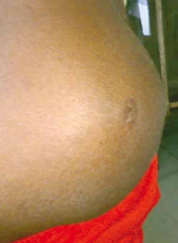
A 3-year-old child presented with a mass in his left gluteal region which had been enlarging gradually for 4 months. History revealed that the swelling was initially painless but had become painful after 2 months for which he had been prescribed painkillers and antibiotics. There was no history of trauma, fever, weight loss or chronic cough. None of the close family members had similar swellings. The general examination was unremarkable. On local examination, a 12 cm firm, tender mass was palpable (Fig. 1). No lymphadenopathy was present. Routine blood investigations did not reveal any abnormalities. Magnetic resonance imaging (MRI) showed a heterogeneous hyperintense infiltrative lesion arising from the gluteus maximus. An incisional biopsy was made. The histological examination was suggestive of the alveolar subtype of rhabdomyosarcoma. High resolution computed tomography (HRCT) of the chest, bilateral bone marrow biopsy and bone scan were negative for metastasis. The patient was referred to a higher cancer centre where he received neoadjuvant chemotherapy consisting of vincristine (V), actinomycin D (A) and cyclophosphamide (C), followed by wide-margin excision of the tumour mass. Radiotherapy was provided before the initiation of chemotherapy and on the 9th week post-chemotherapy. Post-operatively the patient is receiving chemotherapy cycles of VAC. The patient is responding well to chemotherapy without major adverse effects and without relapse/recurrence.
1. Alveolar rhabdomyosarcoma present at the left gluteal region of a 3-year- -old child. 
Discussion
Rhabdomyosarcoma is a relatively rare mesodermal cancer with an estimated annual incidence of 4.3 cases per one million people younger than 20 years of age [1]. Common sites of the primary disease include the head and neck region, genitourinary tract and extremities [2]. Literature about rhabdomyosarcoma located in the gluteal region is sparse. Most cases of rhabdomyosarcoma are sporadic in nature, but there have been associations with familial syndromes such as neurofibromatosis and Li-Fraumeni syndrome [3]. The presence of familial syndromes in our patient was eliminated through detailed history and examination.
As previously mentioned, the two major histological subtypes of rhabdomyosarcoma are embryonal and alveolar. Our patient had been afflicted by the alveolar subtype as histological examination showed the presence of distinct alveolar architecture with aggregates of small round undifferentiated cells separated by dense hyalinized fibrous septa [3,4]. The alveolar subtype accounts for 15–20% of all cases, typically occurs in the second decade of life, has a higher propensity to metastasise and is usually located in the extremities [5].
Around 80% of all cases of the alveolar subtype are caused due to translocations t (2; 13) (q35; q14) or t (1; 13) (p36; q14), which lead to the formation of chimeric transcription factors (PAX3-FKHR or PAX7-FKHR, respectively). These transcription factors cause cell proliferation, angiogenesis and other cancerous changes [3,4]. As our patient did not have enough resources, we could not proceed with mutational analysis for genotype-phenotype correlation.
Most medical centres employ imaging studies such as CT scan or MRI of the primary tumour to determine the size and possible involvement of surrounding vital organ structures [2]. Less than 25% of the cases of rhabdomyosarcoma are metastatic at presentation [5]. The lungs (40–50%), bone marrow (20–30%), bones (10%) and lymph nodes (10–20%) are the most common metastatic sites [2,5]. The metastatic evaluation in our patient was negative.
The prognostic factors which suggested a poor outcome in our patient were the unfavourable site of the primary tumour (buttock), the unfavourable histopathological subtype (alveolar) and the large diameter of the tumour (> 5 cm). The prognostic factors suggesting a positive outcome in our patient were the age (children aged between 1–9 years have a better prognosis), the absence of metastasis/lymph-node involvement and the good response to chemotherapy [2].
As the tumour was invasive in nature with a size greater than 5 cm and without any lymph node or metastasis, it was staged as T2bN0M0 as per TNM (tumour, node, metastasis) classification [2,3]. Our patient was considered to be intermediate-risk and was provided with the VAC regimen, which is the frontline chemotherapy regimen for such patients [3]. The local treatment advised for our patient was radiotherapy and wide-excision of the tumour, following which the VAC regimen was continued.
Incorporating other anticancer agents into the VAC backbone such as ifosfamide, etoposide, carboplatin, irinotecan and vinorelbine in order to improve the outcome rates requires further clinical trials [2]. New molecular targets being explored include inhibiting tyrosine kinase receptors such as epidermal growth factor receptor (erlotinib, geftinib), platelet derived growth factor receptor (olaratumab, dasatinib, pazotinib, sorafenib), vascular endothelial growth factor receptor (sunitinib, apatinib) and anaplastic lymphoma kinase receptor (crizotinib, ceritinib) [6]. Monoclonal antibodies against insulin-like growth factor 1 receptor (cixutumumab, R1507, BMS-754807) have shown promise in early phases of clinical trials [4,6]. Cyclin-dependent kinase inhibitors (palbociclib), and mouse double minute 2 homolog-tumour – tumour protein p53 interaction inhibitor (MI-63) have shown some promise in phase I clinical trials [6]. The role of small GTP-ases family inhibitors (cetuximab, panitumumab, sorafenib), BRAF inhibitors (veumurafenib, dabrafenib) and mitogen activated protein kinase inhibitors (sorafenib, selumetinib, AZD8055) in reducing tumour growth is being explored in preclinical studies [4,7]. Other targets being explored include mammalian target of rapamycin kinase inhibitors (sirolimus, everolimus), phosphadidylinisitol-4,5-bisphosphate 3 kinase inhibitors (temsirolimus, ridaforolimus), programmed cell death-1 receptor inhibitor (pembrolizumab) and polycomb-group protein inhibitors (tazemetostat) [4,5,8]. Drugs targeting the PAX-FKHR fusion proteins (synthetic retinoid fenretinide, fascaplysin) have been isolated, which need further evaluation [5]. Histone deacetylase inhibitors, survivin-responsive conditionally replicating adenovirus and V-ATPase inhibitors (esomeprazole) have been found to reduce cancer growth in preclinical studies [7].
Conclusion
Alveolar rhabdomyosarcoma located in the buttock is an extremely rare entity. Management includes a multimodal approach consisting of systemic chemotherapy, local surgery and radiotherapy. Although several new molecules targeting rhabdomyosarcoma are in the pipeline, integrating them with existing therapy would be a challenge.
This work was supported by the grant of Ministry of Health AZV 15-33590A.
The authors declare they have no potential conflicts of interest concerning drugs, products, or services used in the study.
The Editorial Board declares that the manuscript met the ICMJE recommendation for biomedical papers.
Submitted: 3. 12. 2018
Accepted: 24. 3. 2019
MUDr. Lucie Světláková
Klinika komplexní onkologické péče
Masarykův onkologický ústav
Žlutý kopec 7
65 653 Brno
e-mail: lucie.svetlakova@mou.cz
Sources
1. Hettmer S, Li Z, Billin AN et al. Rhabdomyosarcoma: current challenges and their implications. Cold Spring Harb Presepect Med 2014; 4 (11): a025650. doi: 10.1101/cshperspect.a025650.
2. Panda SP, Chinnaswamy G, Vora T et al. Diagnosis and management of rhabdomyosarcoma in children and adolescents: ICMR consensus document. Indian J Pediatr 2017; 84 (5): 392–402. doi: 10.1007/s12098-017-2315-3.
3. Egas-Bejar D, Huh WW. Rhabdomyosarcoma in adolescent and young adult patients: current perspectives. Adolesc Health Med Ther 2014; 5 : 115–125. doi: 10.2147/AHMT.S44582.
4. Dziuba I, Kurzawa P, Dopierala M et al. Rhabdomyosarcoma in children – current pathologic and molecular classification. Pol J Pathol 2018; 69 (1): 20–32. doi: 10.5114/pjp.2018.75333.
5. Dagher R, Helman L. Rhabdomyosarcoma: an overview. Oncologist 1999; 4 (1): 34–44.
6. Shern JF, Yohe ME, Khan J. Pediatric rhabdomyosarcoma. Crit Rev Oncog 2015; 20 (3–4): 227–243.
7. Sun X, Guo W, Shen JK et al. Rhabdomyosarcoma: advances in molecular and cellular biology. Sarcoma 2015; 2015 : 232010. doi: 10.1155/2015/232010.
8. Hiniker SM, Donaldson SS. Recent advances in understanding and managing rhabdomyosarcoma. F1000Prime Rep 2015; 7 : 59. doi: 10.12703/P7-59.
Labels
Paediatric clinical oncology Surgery Clinical oncology
Article was published inClinical Oncology

2019 Issue 3-
All articles in this issue
- MicroRNAs in Cerebrospinal Fluid as Biomarkers in Brain Tumor Patients
- Prognosis of HPV-Positive and -Negative Oropharyngeal Cancers Depends on the Treatment Modality
- Nanoparticle-Modified Apoferritin Nanotransfer for Targeted Cytostatic Transport
- Prevalence of Anxiety and Depression and Their Impact on the Quality of Life of Cancer Patients Treated with Palliative Antineoplasic Therapy – Results of the PALINT Trial
- Rhabdomyosarcoma of the Gluteus Maximus – Case Report, Review of Literature and Emerging Therapeutic Targets
- Neoadjuvant Hypertermic Isolated Limb Perfusion in Treatment of Undifferentiated Spindle Cell Sarcoma of Lower Limb with Achieved Complete Pathologic Response
- Primary Intracranial Sarcomas, Myxoid Meningeal Sarcoma – a Case Report and Literature Review
- The Loneliness of Patients in the Pre-Terminal and Terminal Stages of Cancer, the Social Dimension of Dying
- Current FIGO Staging for Carcinoma of the Cervix Uteri and Treatment of Particular Stages
- Association of TNF-α -308G>A Polymorphism with Susceptibility to Cervical Cancer and Breast Cancer – a Systematic Review and Meta-analysis
- Clinical Oncology
- Journal archive
- Current issue
- Online only
- About the journal
Most read in this issue- Current FIGO Staging for Carcinoma of the Cervix Uteri and Treatment of Particular Stages
- Rhabdomyosarcoma of the Gluteus Maximus – Case Report, Review of Literature and Emerging Therapeutic Targets
- Prevalence of Anxiety and Depression and Their Impact on the Quality of Life of Cancer Patients Treated with Palliative Antineoplasic Therapy – Results of the PALINT Trial
- The Loneliness of Patients in the Pre-Terminal and Terminal Stages of Cancer, the Social Dimension of Dying
Login#ADS_BOTTOM_SCRIPTS#Forgotten passwordEnter the email address that you registered with. We will send you instructions on how to set a new password.
- Career

