-
Medical journals
- Career
Echocardiography in the Diagnosis of Cardiac Amyloidosis
17. 4. 2023
Cardiac amyloidosis remains a diagnostic challenge that requires great clinical vigilance. The imaging method of choice for evaluating heart failure suspected of amyloidosis is echocardiography. What findings are typical for patients with amyloidosis?
Suspicion of Cardiac Amyloidosis
We should be alerted by patients with unexplained thickening of the left ventricular wall, especially if it appears in older individuals with common cardiac issues such as heart failure with preserved ejection fraction (HFpEF), hypertrophic cardiomyopathy, or severe aortic stenosis.
Typical is heart failure with high levels of NT-proBNP (N-terminal prohormone of brain natriuretic peptide), which do not correspond to the objective findings on echocardiography. Unexplained right heart failure with normal appearing ventricular and valvular function or idiopathic pericardial effusion should also raise suspicion of amyloidosis.
Cardiac amyloidosis may also be accompanied by a set of other extracardiac symptoms (so-called red flags), such as proteinuria, macroglossia, or bilateral carpal tunnel syndrome.
Pathophysiological Processes in Cardiac Tissue
Infiltration of cardiac tissue by amyloid deposits causes thickening and stiffening of ventricular walls, impaired ventricular relaxation and contractility, as well as disturbances in the conduction of electrical signals. Atrial infiltration leads to mechanical dysfunction of the atria and predisposition to cardiac thrombosis and systemic embolism despite sinus rhythm.
Role of Echocardiography in the Diagnostic Process and Typical Findings
The gold standard in the diagnosis of amyloidosis is the demonstration of amyloid deposits in cardiac tissue using Congo red staining of heart biopsy samples. This technique exhibits high sensitivity. However, due to the risks associated with biopsy, it is not suitable for first-line screening. Imaging methods are crucial for diagnosis and prognosis determination of patients.
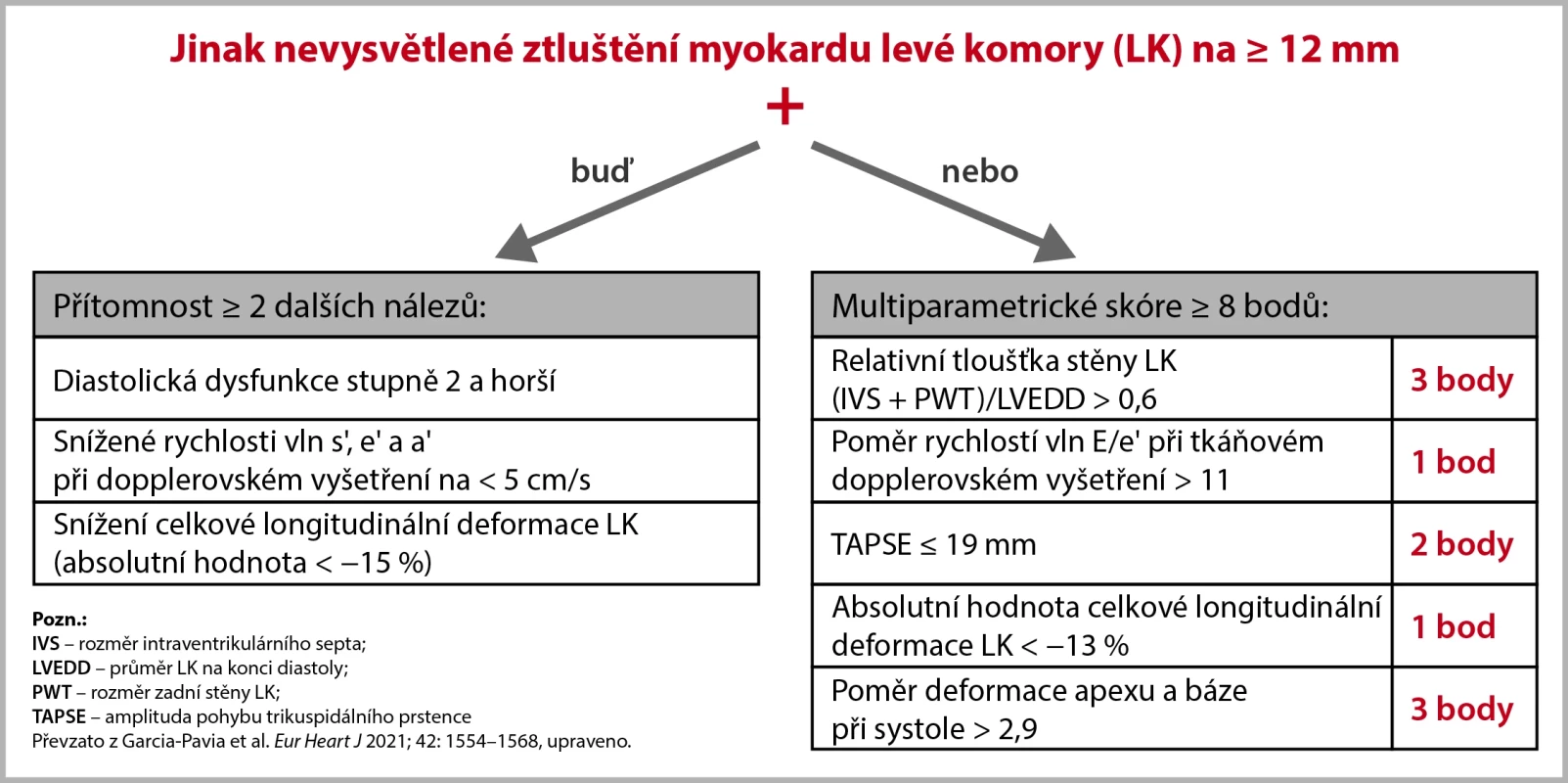
According to the position of the European Society of Cardiology's Working Group on Myocardial and Pericardial Diseases (ESC), the diagnosis of cardiac amyloidosis requires the presence of otherwise unexplained thickening of the left ventricular myocardium to ≥ 12 mm and other findings as per the scheme below.
Image 1. Echocardiographic criteria for non-invasive diagnosis of cardiac amyloidosis. The criteria also apply to cases with amyloidosis proven by extracardiac biopsy. 
Conclusion
The diagnostic accuracy of individual echocardiographic parameters is variable, but their mutual combination increases diagnostic accuracy. The absence of typical echocardiographic features is insufficient to exclude the diagnosis of cardiac amyloidosis, and further examination is warranted if clinical suspicion persists. Echocardiography also cannot differentiate between individual types of amyloidosis. Despite these limitations, it remains the first-choice examination for patients suspected of cardiac amyloidosis.
(este)
Sources:
1. Garcia-Pavia P., Rapezzi C., Adler Y. et al. Diagnosis and treatment of cardiac amyloidosis: a position statement of the ESC Working Group on Myocardial and Pericardial Diseases. Eur Heart J 2021; 42 : 1554–1568, doi: 10.1093/eurheartj/ehab072.
2. Agrawal T., Nagueh S. F. Echocardiographic assessment of cardiac amyloidosis. Heart Fail Rev 2022; 27 (5): 1505–1513, doi: 10.1007/s10741-021-10165-y.
Did you like this article? Would you like to comment on it? Write to us. We are interested in your opinion. We will not publish it, but we will gladly answer you.
Labels
Neurology
Login#ADS_BOTTOM_SCRIPTS#Forgotten passwordEnter the email address that you registered with. We will send you instructions on how to set a new password.
- Career

