-
Medical journals
- Career
Ultrasound in a Patch – Smart System Capable of Monitoring Patient During Regular Activities
25. 4. 2024
A team of scientists from the University of California, San Diego, has developed a miniature autonomous ultrasound system that, when attached to the skin, can visualize tissue located deep beneath the body’s surface. It could be used for continuous monitoring of blood pressure, heart activity, and respiratory parameters in real time, whether during regular activities or intense physical exertion.
Ultrasound Examination Requires Equipment and Knowledge
Ultrasound imaging uses the reflection of high-frequency sound waves from environments with different densities. Echoes captured by the ultrasound probe are then reconstructed into images. Although ultrasound examination is cheaper and safer for the patient compared to other imaging techniques, it requires relatively bulky equipment and a skilled technician or doctor to operate it. The sharpness of the obtained image and the reliability of the results primarily depend on the manipulation of the probe. Traditional ultrasound examination is thus inherently manual and cannot be performed continuously during patient activity.
Several research groups are working on developing wearable ultrasound systems. Their efforts so far have led to the creation of various systems, but these required physical connections to an external power source and data collector or other hardware.
Ultrasound Patch
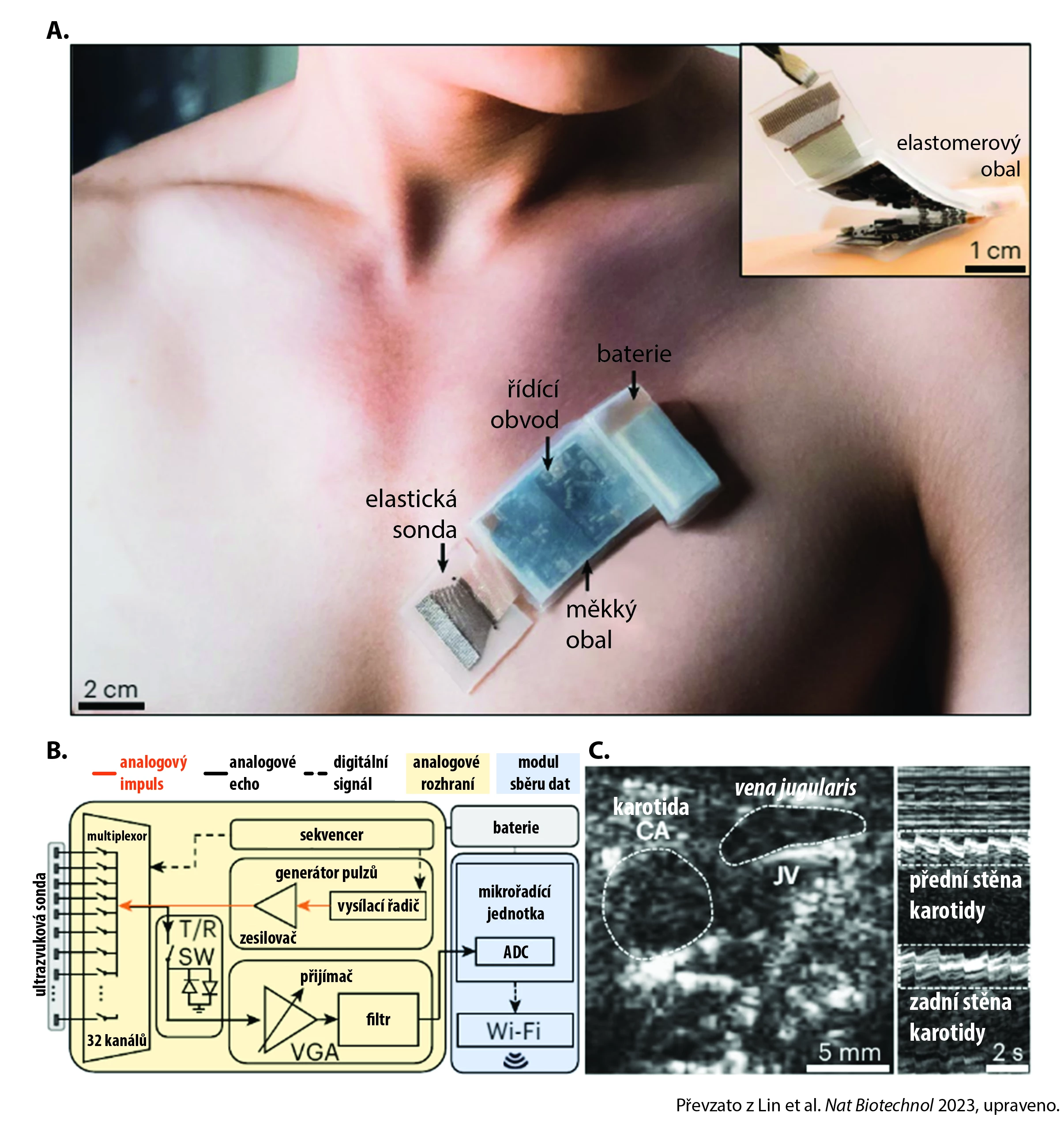
Authors from the University of California, San Diego published their study of a new fully integrated ultrasound heart activity sensor in the journal Nature Biotechnology. The system consists of an elastic ultrasound probe made of a 32-channel array of piezoelectric transducers that convert electrical signals into sound waves and vice versa. The probe is connected to a flexible control circuit using coiled elastic copper wires. The control circuit manages the generation of ultrasound pulses and the collection of echoes, which are then amplified and sent to a microcontroller unit. This unit converts them into digital signals, which are transmitted via Wi-Fi to a computer or smartphone. The system is powered by a standard lithium-polymer battery that lasts up to 12 hours on a single charge. The sensor can detect signals from tissues up to 16 centimeters below the body’s surface.

Image. Fully integrated wireless ultrasound sensor for continuous patient monitoring:
A) Photograph of the device applied to the skin for ultrasound heart activity monitoring using a parasternal approach.
B) Schematic drawing of the device technology.
C) Imaging of the carotid artery (CA) and jugular vein (JV) in B mode during the Valsalva maneuver.Signal Evaluation
For data analysis from the ultrasound patch, the team of scientists developed an algorithm based on machine learning. The wearer’s heart rate and blood pressure are calculated based on the pulsation of the carotid artery and jugular vein. The system is also capable of measuring heart muscle contraction to estimate the ejected blood volume and monitoring diaphragm movements to calculate respiratory parameters. If the patch moves relative to the tissue during the wearer’s activity, the system can select the sensor channels with the best signal in real time.
The device was tested in real-world conditions during both aerobic and anaerobic training. During both activities, the ultrasound patch was able to continuously monitor the carotid artery pulsation, thus tracking blood pressure, heart rate, and heart activity.
And What Practical Uses Are Emerging?
The ultrasound patch does not aim to replace conventional ultrasonography. The quality of the captured signal and the depth of tissue penetration are currently limited. The main advantage of the system is the possibility of continuous real-time monitoring during everyday activities, which can have significant clinical value. For rigorous diagnostics, for instance, the patient's response to intense physical activity is important. Continuous monitoring over a longer period allows for analyzing dynamic changes in the cardiovascular system in response to stressors. These insights could be beneficial for a range of individuals – from athletes optimizing their training plans to patients rehabilitating after cardiac procedures, to high-risk groups needing better stratification and prediction of cardiovascular risk.
(este)
Sources:
1. Lin M., Zhang Z., Gao X. et al. A fully integrated wearable ultrasound system to monitor deep tissues in moving subjects. Nat Biotechnol 2024 Mar; 42 (3): 448−457, doi: 10.1038/s41587-023-01800-0.
2. Patel P. Wearable ultrasound sees deep tissue on the move. IEEE Spectrum, 2023 May 26. Available at: https://spectrum.ieee.org/wearable-ultrasound-wireless
Did you like this article? Would you like to comment on it? Write to us. We are interested in your opinion. We will not publish it, but we will gladly answer you.
Labels
Pharmacy Clinical pharmacology Pharmaceutical assistant
Popular this week- AI Will Help Personalize the Treatment of Atrial Fibrillation
- Czech Gastroenterology Keeps Up with the Times. For 80 Years
- Vaccination Is Entering a New Era Thanks to Innovative Technologies
- Why Endometriosis Requires the Principles of Precision Medicine
- “Bites” from Clinical Research – 2026/4
Recommended for you- Micronutrients in Parenteral Nutrition in Adult Patients – Current Issues and Consensus
- Position of Parenteral Therapy in Patients with Severe COVID-19 Disease
- High Dose of Protein Affects Mortality in Critically Ill Patients if They Do Not Have Sepsis
- The Importance of Adequate Protein Intake in Critical Patients – Analysis of Data from an International Study
- Importance of Home Parenteral Therapy: ESPEN Recommendations from 2020
- Impact of Adequate Protein Intake on Morbidity and Mortality in Critically Ill Patients
Login#ADS_BOTTOM_SCRIPTS#Forgotten passwordEnter the email address that you registered with. We will send you instructions on how to set a new password.
- Career

