-
Medical journals
- Career
Planning Surgery Using 3D Printing in Practice – Case Study
25. 1. 2024
In October 2023, an article by neurosurgeons from Leipzig University was published in the journal 3D Printing in Medicine, focusing on the use of 3D printing in the treatment of complex post-traumatic elbow stiffness. According to the article, the 3D model is a suitable supplement for pre-operative planning, conducting the surgery itself, and post-operative control.
According to the team led by Ronny Grunert, the advantage of 3D printing is the simulation of individual defects and situations for specific patients. The authors of the article present an illustrative case of a 38-year-old man with post-traumatic elbow stiffness and associated bone pathologies – osteophytes and ossifications. The patient suffered a Monteggia lesion – a fracture of the ulna with dislocation of the proximal radius after falling from a height of approximately ten meters.
A year after the surgical osteosynthesis of the olecranon and repositioning of the radial head, the patient's range of motion stopped at an extension/flexion angle of 0/40/135° despite intensive physiotherapy. X-ray and CT examinations showed a properly healed olecranon fracture, but with a large amount of radially and ulnarly protruding osteophytes.
3D model helps to understand relationships and anatomy
Since it was not possible to precisely locate the position of the most obstructive bone growth using CT images, Grunert's team decided to create a 3D model to better analyze key bone structures.
The patient was scanned using multi-spiral CT with a scanning thickness of 1 mm according to the standard hospital protocol. The necessary images were extracted from the tomographic representation under the supervision of an orthopedist and a biomedical engineer (see Fig. 1), from which a 3D mesh for the digital model was calculated (see Fig. 2) and these data were sent to a 3D printer.
Fig. 1 Segmentation of anatomical structures using CT images

Fig. 2 Virtual 3D model in 3D printing software

On the printed model of the patient's elbow made of biocompatible polyamide, doctors subsequently performed a simulated operation (see Fig. 3). They removed individual osteophytes until the free mobility of the joint connection was restored. During the actual surgery, they then knew exactly how to proceed and could determine the optimal and gentlest surgical strategy (see Fig. 4).
Fig. 3 Preoperative simulation of the procedure

Fig. 4 Comparison of the 3D model with the real situation during surgery

The result of the surgery precisely matched the simulation performed on the 3D model. One year after the surgery, the patient achieved an extension/flexion angle of 0/20/130°. In terms of communication with the doctors, the model allowed him to understand the planned surgical procedure and realistically outline its goals.
Fig. 5 Using the patient's 3D printed scapula model for preoperative shaping of the osteosynthetic plate. CT data from the healthy side of the scapula were used to create the model, which was post-processed and mirrored.


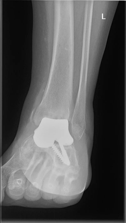
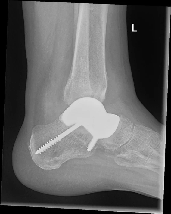
Fig. 6 X-ray images of the ankle joint with a 3D printed bone replacement custom-made for the patient


Improving the quality of performance and patient satisfaction
Using 3D printing and software simulation enables precise planning of complicated surgical procedures. Haptic models offer surgeons the opportunity to reduce preparation time and eliminate intraoperative complications. They can be used to improve theoretical knowledge and practice practical skills, leading to higher quality performance and greater patient satisfaction and cooperation.
Benefits of using 3D modeling:
- preoperative planning
- procedure control during surgery
- postoperative control
- patient consultations
- implantation of custom-made 3D printed titanium implants
The article was created in cooperation with the University Hospital Brno.
(jko)
Sources:
1. Grunert, R., Winkler, D., Frank, F. et al. 3D-printing of the elbow in complex posttraumatic elbow-stiffness for preoperative planning, surgery-simulation and postoperative control. 3D Print Med2023; 9 (1): 28, doi: 10.1186/s41205-023-00191-x.
2. Pešl T., Havránek P. Monteggia lesions in the growing skeleton: principles of therapy. Acta Chir Orthop Traumatol Cech 2010; 77 (1): 32–38, doi: 10.55095/achot2010/005.
3. 3D printing in healthcare: a revolutionary technology surpassing augmented and virtual reality. FN Brno, 30. 10. 2023. Available at: www.fnbrno.cz/3d-tisk-ve-zdravotnictvi-prevratna-technologie-presahujici-rozsirenou-a-virtualni-realitu/t7756
4. Czech Society for 3D Printing in Medicine. Available at: https://3dmedicalprint.cz.
Did you like this article? Would you like to comment on it? Write to us. We are interested in your opinion. We will not publish it, but we will gladly answer you.
Popular this week- AI Will Help Personalize the Treatment of Atrial Fibrillation
- Czech Gastroenterology Keeps Up with the Times. For 80 Years
- Why Endometriosis Requires the Principles of Precision Medicine
- Vaccination Is Entering a New Era Thanks to Innovative Technologies
- “Bites” from Clinical Research – 2026/4
Login#ADS_BOTTOM_SCRIPTS#Forgotten passwordEnter the email address that you registered with. We will send you instructions on how to set a new password.
- Career

