-
Medical journals
- Career
METHODS FOR KINEMATIC ANALYSIS OF HUMAN MOVEMENT IN MILITARY APPLICATIONS: A REVIEW OF CURRENT AND PROSPECTIVE METHODS
Authors: Petr Volf 1; Patrik Kutilek 2; Jan Hejda 2; Slavka Viteckova 3; Pavel Smrcka 3; Karel Hana 3; Zdenek Svoboda 4; Vaclav Krivanek 5
Authors‘ workplace: Department of Biomedical Technology, Faculty of Biomedical Engineering, Czech Technical University in Prague, Kladno, Czech Republic 1; Department of Natural Science, Faculty of Biomedical Engineering, Czech Technical University in Prague, Kladno, Czech Republic 2; Department of Information and Communication Technologies in Medicine, Faculty of Biomedical Engineering, Czech Technical University in Prague, Kladno, Czech Republic 3; Department of Natural Sciences in Kinanthropology, Faculty of Physical Culture, Palacky University of Olomouc, Olomouc, Czech Republic 4; Department of Military Robotics, Faculty of Military Technology, University of Defence, Brno, Czech Republic 5
Published in: Lékař a technika - Clinician and Technology No. 4, 2019, 49, 125-135
Category:
Overview
Expansion of methods employed in the kinematic analysis of human movement for diagnosing of the physical and mental health of subjects can be traced back to the 1990`s when new information technologies and electronic recording systems started their development boom. Evaluation methods of body movement for the diagnostics of physical and mental health expanded significantly in clinical practice. This study presents an overview of these methods with the focus on how applicable the analysis of human movement can be in military practice, where they are currently marginally used. The aim of this study is to offer some recommendations on how particular methods could be utilized in an army context. This article also suggests the most appropriate methods of quantitative evaluation for posture and motion control in the course of standing, gait and other activities carried out in military training and active duty.
Klíčová slova:
chůze – držení těla
Introduction
The expansion of methods of kinematic analysis of the human movement for physical and mental health diagnostics of subjects can be traced back to the 1990’s when new information technologies and electronic recording systems started their development boom [1]. The kinematic analysis of human body movement for the diagnostics of physical and mental health is fre-quently applied in medical observation and treatment within civilian sphere [2]. Kinematic analysis of body motion, which includes evaluation of kinematic data for the quantitative assessment of body motion, can be, however, applicable not only to the civilian sphere but also in some military activities. These might include the developing rehabilitation processes, developing intel-ligent control systems, prosthetics, robotic exoskeletons, or various kinds of customized equipment. This study aims to present an overview and analysis of the recently used methods which evaluate kinematic data on body movement and to refer to how they can be applied in the military sphere. Referring to the research and selected examples, each chapter describes some recently used methods applied in both civilian and military sphere and compares the potential use of their applicability. This article aims to present recommendations for particular methods of evaluation of physical and mental health within specific military areas. It also offers several tools that may help others identify appropriate methods of quantitative assessment of posture and movement control under the conditions of standing, gait and performing various activities within an army context.
This study doesn’t intend to conduct a systematic review of the literature with precise search criteria, but instead to propose a thematic review based the most recent articles and those already known by the authors. Two databases—IEEE Xplore (Institute of Electrical and Electronics Engineers and Institution of Engi-neering and Technology) and ScienceDirect (Elsevier) —were mainly used for the literature search. A combi-nation of keywords, such as movement, soldier, sensors, wearable, kinematics, kinetics, were used as search terms. Publications from 2005–2017 were preferred, however, this range was extended in some cases.
Methods of Quantitative Evalua-tion of Health in Military Personnel Using Kinematic Data
Evaluation of individual kinematic values of move-ment measured by motion capture (MoCap) systems at a particular moment does not seem suitable in diagnostics of physical and mental health since it usually fails to demonstrate the overall complex motion of human body segments. It was, therefore, necessary to develop some alternative methods allowing appropriate processing of the measured kinematic values for motion, the methods which would enable quantitative evaluation of the measured motion as a whole. Although these methods are widely used in civilian medicine, they have a reasonably large potential to find their way into military use. For instance, the measured data allows for evaluation and interpretation of physical and mental health of military personnel or, based on the calculation of numerous factors, they can evaluate the impact of military gear, equipment, and new technology, on soldiers’ movement. Methods of evaluation can differ depending on the kind of movement studied, as well as on the types of MoCap systems themselves. They can be divided into two most widely used areas of clinical practice:
- Evaluation of static position and orientation of body segments (static posture) [3]
- Evaluation of deliberate and active motion of body segments (dynamic posture) [4]
These methods can be further divided into:
- Methods of evaluation of time domain data [5]
- Methods of evaluation of frequency domain data [6]
- Methods of evaluation of the relation between measured variables [7]
- Non-linear methods of data evaluation [8 ̶ 10]
In the following section, this paper will give some examples of how particular methods are applied in the military sphere and the potential application of methods which have not been used in a military context before.
Evaluation of Static Position and Orientation of Body Segments
Data reflecting records of motion control of a static position and orientation of body segments or a body as a whole (static posture) usually take the form of kinematic data developing over a period of time. In the case of force platforms, the record usually includes the development of values of the center of pressure (CoP), i.e. center of mass (CoM) for the whole body. Positions of anatomical points or angles of body segments (or their derivations—velocity and acceleration) are usually recorded by cameras, a gyro-accelerometer or other MoCap systems. A detailed overview of the latest and most frequently used methods of quantitative analysis of the measured data and their relations are documented, for example, in [11 ̶ 14].
Methods of evaluation of time domain data
The most frequent method of evaluation of measured data within the area of civilian medicine (and their prospective application in a military context), is the evaluation of maximum values or of the range of measured values (the difference between the maximum and minimum). This method includes the evaluation of maximum deviations of CoP in the medial-lateral and anterior-posterior direction [15], as measured by e.g. Romberg test. The medians of CoP coordinates were measured as the part of the examination, see Tab. 1. Similarly, data from MoCap systems were assessed by evaluating the acceleration of body segments in medial-lateral and anterior-posterior directions [16] or body CoM [17]. The evaluation of ranges of angles measured by camera systems for individual anatomical planes is nowadays a frequent method of medical examina-tions [18, 19]. Ranges of sway angles for the entire body in particular anatomical axes were evaluated during stance tasks on a dynamic platform [20].
Table 1. Methods of evaluation of static position and orientation of body segments. 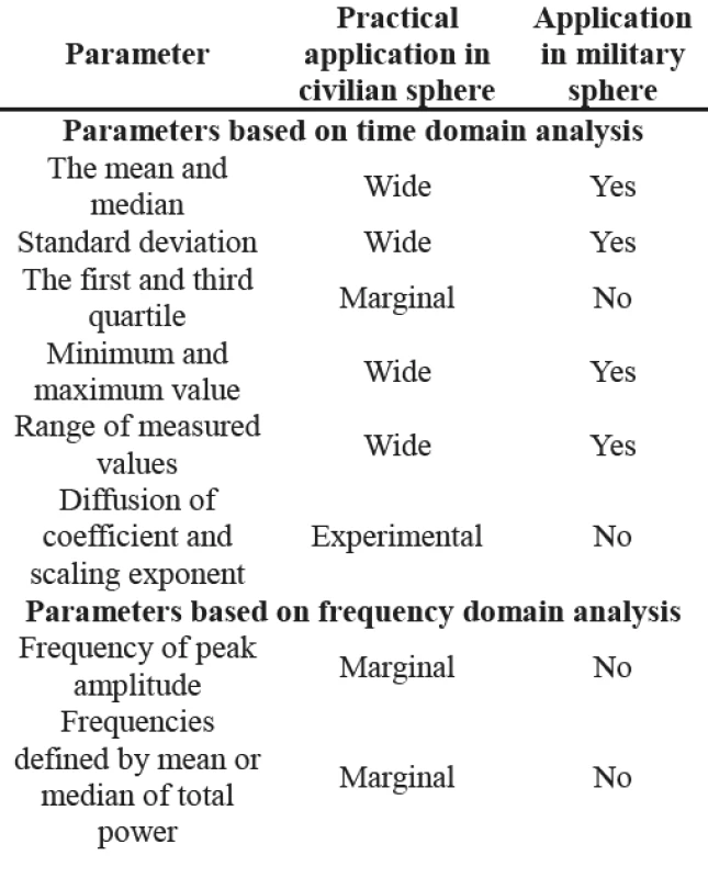
Methods of evaluation of frequency domain data
The evaluation of kinematic data in frequency domain is common in the civilian area. However, data analysis using the amplitude spectrum [21] and the power spectrum [22] is not widely used to evaluate the ability to maintain a static posture when concerning the military sphere. This is due to the fact that these methods are predominantly used in the assessment of neurological disorders, such as hereditary disorders, that can also be identified by evaluation methods using the time domain data. Moreover, they are rarely found among military personnel or war veterans; the diagnosed neurological disorders of war veterans are considered as those in civilians, and were therefore not included in the research of the specific methods used in military medicine. Another reason why frequency domain evaluation is excluded from this research is that when comparing the methods of evaluation for time domain data, their nature—regarding interpretation—is far too complex. In the case of military operations, methods for evaluation of deliberate motion of body segments appear more suitable than evaluation in a static posture.
Methods of evaluation of relationship between measured variables
Although some indicators in quantitative evaluation of kinematic data based on the analysis of relations between measured variables are a standard choice in the civilian sphere (particularly those assessing the static posture of a body segment or the body as a whole [23]), none of these are applicable in a military context. This is due to their computational complexity and intricate interpretation compared to the methods of evaluation of time domain data which has long been used in practice. The vast majority of potentially suitable parameters relies on methods based on an evaluation of postural stability in standing positions, see the overview in Tab. 1 and in [11, 13, 14]. The parameters, such as trajectory length and convex hull enclosing the CoP trajectory, are representations of the measured variables plotted against each other in 2D or 3D diagrams.
Nonlinear methods of data evaluation
Linear evaluation methods stem from the assumption that every behavior pattern in a sequence of repeated tasks is independent of the previous and following behavior. In contrast, nonlinear methods focus on the structure of variability by analyzing fluctuations in behavioral patterns in the course of time and observe how one behavior pattern can affect the other one. Despite the recent rise in the use of nonlinear methods for postural stability and motion assessment, for professionals, they do not represent a preferable choice, especially when compared to previous methods; the main reason is the demanding processing and com-plexity of data interpretation [11]. The nonlinear methods for posturography evaluation in civilian areas, such as CoP analysis [11], are mostly based on Largest Lyapunov Exponent (LLE) [24, 25], wavelet transform (e.g. critical point interval analysis) [26], fractal analysis [27], fluctuation analysis (examining e.g. Hurst
exponent) [28], and calculations of approximate entro-py, sample entropy and multiscale entropy (focusing on e.g. complexity indices) [11]. The investigation of dynamic systems and application of methods for the analysis of dynamic properties of time series and recurring quantification analysis (RQA) presents yet another approach [29, 30]. This method quantifies the number and duration of recurrences of a dynamic system presented by its phase in space trajectory. The applica-tion of RQA has grown significantly in civilian appli-cations in recent years, especially when evaluating CoP motion in subjects with a balance-disorder [9, 31]. Com-puted RQA outcome variables includes e.g. entropy (reflecting complexity of the deterministic structure of the time series) [32], percentage of laminarity (reflecting intermittency) [33], percentage of determinism (reflect-ing predictability, such as a degree of determinism vs. randomness) [30, 33], percentage of recurrence (reflect-ing repetition of data point in phase space) [33], trend (reflecting nonstationarity) [33], maxline (reflecting dynamic stability), etc.Although the RQA method is intended for processing wide ranges of both stationary and nonstationary states of data sets, and is effective with any volume of data, it is mainly used on an experimental basis in the military environment, namely in the area of veterans’ treat-ment [34]. Apparently, the complexity of calculation and the results of interpretation appear to be major drawbacks, similar to previously mentioned methods. Despite no official record of the military using these methods, some effort to introduce them has already been made in the presentations of civilian research published in army journals [24].
Evaluation of Active Motion of Body Segments
Some quantitative methods for the assessment of physical health have already been developed. They are based on the analysis of body segment motion (i.e. dynamic posture) and are recorded by modern MoCap systems. Methods based on the analysis of cyclic and acyclic or symmetric and asymmetric motions are used to evaluate dynamic posture. Rather widely used are methods which evaluate a cyclic motion with a fixed beginning and end to the duration, position, and trajectory; they are usually employed observing the length of trajectory, velocity or frequency of tremor [35, 36]. Methods employed in the assessment of cyclic motion represent another established choice for the evaluation of movement; typical is the focus on repeated movement of the same nature [37] like in gait (walking) or running. Such repeated motion patterns occur in everyday human activities and are therefore often observed and compared. Those analysis are most frequent used in the civilian area for the evaluation of gait, as this is the one of the most typical and important human movement. In clinical practices, various types of cyclic motion are studied applying specifically designed methods; however, standards for quantitative evaluation of particular types of cyclic motion are based on previous studies [5, 31, 44]. In order to make the data comparable across the population, their recording of a particular motion cycle (for example, duration of motion, stride length, motion length, stride time of the gait cycle, etc.), [38] is standardized. Likewise, the measured variables, such as distance, speed, etc. (i.e. recording of motion amplitude) [39] are usually subjected to standardization. The values relating to the basic parameters of particular types of active motions have been carefully examined on healthy subjects, and then compared with the values of parameters observed on research subjects (usually with an impaired physical condition). Tab. 2 illustrates an overview of the basic groups of parameters used in the civilian sphere and the status of their applicability in a military context.
Methods of evaluation of time domain data
In this section, some basic methods used in the civilian sphere and their application in the army will be com-pared. Measurements of kinematic values of transla-tional motion is based on the change of position which allows for the evaluation of velocity and accelera-tion [40]. These values are standardized [39], and are followed by the observation of maximum or minimum values. Observing vertical motion by placing the mark-ers on particular anatomical points during a particular phase of gait cycle is an evaluation example of a kine-matic value [41]. A range of the measured data (the difference between the minimum and the maximum value) such as step length and width derived from CoP displacement [42], is also a frequently observed parame-ter, see Fig. 1. The evaluation of angle variables, usually anatomical angles is, however, more common practice than the evaluation of position parameters. The time derivative of the rotation angle (angular velocity and acceleration) are also observed and analyzed. Similarly to studies focused on the development of the position of body segments coordinates, a range of expected result values from previous research provides a reliable comparison of healthy subjects in rotational motion on major joints. An illustrative example is the develop-ment of the knee or pelvic angle in a gait cycle of healthy subjects [43]. The data record is thus usually standard-ized for motion characteristics, such as the length of one step or stride time of the one gait cycle converted to percentages. Curves of the measured kinematic vari-ables (measured in a particular subject) are captured and then compared to the development values of healthy subjects. Mean values, maximum and minimum values are usually observed as parameters for quantitative evaluation of the body motion.
Image 1. Example of a visualization of the measured data and basic parameters of time domain data, i.e. angle between an upper arm and forearm obtained by process-ing outputs using the gyro-accelerometer system (Xsens MVN system manufactured by Xsens Technologies B.V.); φ – the relative angle at a particular joint, φMAX – the maximum relative angle, φMIN – the maximum relative angle, rRA – the range of the relative angle, tC – the cycle time, tA – the activity duration, ni – the cycle number, s – the sample number. 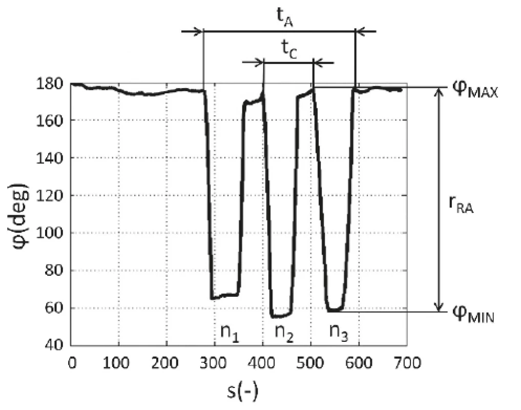
In addition to the above-mentioned traditional methods of analysis of motion data, indices for quanti-tative analysis of quality of a particular type of motion (primarily gait) are also used. The gait indices evaluate the motion of the lower extremities as a whole by calculating the value of the index representing the deviation of the performed gait from the typical gait of healthy subjects.
The most common indices are: Gillette Gait Index (GGI) [44], Gait Deviation Index (GDI) [45], and Gait Profile Score (GPS)/Motion analysis Profile (MAP)
[46]. The Gait Variability Index (GVI) was introduced in 2013 [47]. Calculation of the indices are based on assessing a set of 9 (or 15) measured motion variables focusing on mean values and standard deviations (SD) of spatial-temporal parameters of body motion as a whole or the motion of specific body segments.As for the evaluation of other types of movements, i.e. movements of other body segments (e.g. the upper extremity), diagrams of expected values of angles for respective joints and phases of motion cycle in the healthy population are used to evaluate cyclic mo-tion [48]. Recently, the above indices for the lower extremity motions have been adjusted, and new indices for upper extremity motions have been designed; this can be exemplified on Arm Profile Score (APS) [49], Arm Posture Score (APS) or Arm Posture Index (API)
[50]. A single value of the measured parameter is to assess the motion, while the calculated index is used in the next stage for the evaluation of more measured variables of a complex trunk and arm movement. Naturally, there are other methods evaluating kinematic variables which are used in the evaluation of stability using 2D or 3D cyclograms for state vector (formed by two of three variables) development while differences between trajectories of state vectors (the area defined by trajectories in the defined section plane) are evalu-ated [51]. These novel methods have so far been only used experimentally in the civilian sphere.As for a military application, the most common indices are used for establishing the minimal or maximal values of the measured angles and ranges of angles in a course of the measured movement. They are practi-cally used in war veterans’ rehabilitation programs or in the development of medical devices for veterans (wheel-chairs etc.) [52, 53]. As mentioned before, these indices enable the assessment of maximal values of anatomical angles of the trunk and upper extremities [54, 55], of the kinematic positions of anatomical points (coordinates of anatomical points) [56] and maximum or minimum values of arm accelerations [57]. They are also used for the quantitative assessment of upper extremities’ motion during rehabilitation, focusing on defined ranges of an-gles and displacements (i.e. distances) of segments during specific activities [18].
Cyclic motions studied for military needs is often used for the evaluation of minimum and maximum values of angles of the lower extremity during gait [58], for the assessment of ranges of joint angles of the lower extrem-ity [59 ̶ 61], and for the assesment of trunk motion [62] during gait. These are necessary for the development of new prostheses designed for war veterans or for the design of special implants needed after injuries identi-fied throughout the rehabilitation process [63]. Evalua-tion of the values of angles in particular gait phases
can also be observed during the rehabilitation pro-cess [64, 65]. As already mentioned, minimum and maximum angle values have been defined for respective gait phases in healthy population, and the measured values are compared in the course of rehabilitation. Similarly to the examination of angles, minimum and maximum CoP values for medial-lateral direction and particular gait phase are set and used in rehabilita-tion [66].Development of army equipment (such as back-packs) and its effects on gait quality, is another area where parameters such as step length, step width and ranges of anatomical angles are employed [67]. The same applies to velocities and accelerations during respective gait phases in the development of exoskeletons and smart sensor suits with acceleration sensors [68]. Minimum and maximum values of anatomical angles are key variables in movement activity studies with equipment of different weights, e.g. during jumps [69]. Research has shown that the parameters for quantitative evalua-tion of body motion are already fully used in the military and civilian spheres.
Methods of evaluation of frequency domain data
Methods for evaluating the measured kinematic data in frequency domain, are identical to the methods for evaluating static position and orientation of body segments [70]. This is due to the fact that in most cases, the frequency of deliberate motion does not match the frequency of accidental motions (such as tremors), meaning that accidental motions can be filtered out. Motions suggesting the physical state of subjects can then be studied directly with no regard for deliberate motions.
Similarly to the evaluation of static position and orientation of body segments in frequency domain, the evaluation of active motion of body segments in the frequency domain is only rarely studied and even if it is, it has been only experimental in nature. Rehabilitation practices offer examples of use, such as the power spec-trum of body segments’ motion, to observe responses of the body to the motion of the platform on which the subject was standing [71]. Nevertheless, these represent an isolated example of military-related application. The reasons for their marginal use are identical with those referring to the evaluation of static position and orien-tation of body segments in the frequency domain.
Methods of evaluation of relationship between measured variables
Since MoCap systems usually allow for the recording of more than one variable, mutual development of variables can provide a quantitative parameter of the subject’s condition. In civilian practice, these methods are similar to the methods for the evaluation of static position and orientation of body segments, for example, inclination angles in diagrams for mutual development of two joint angles of the lower limb during cyclic motion [72]. Diagrams representing mutual develop-ment of two or three variables are called cyclograms and are used not only to evaluate the development of joint angles, but also the position and/or orientation of body segments during cyclic gait [73]. Some complex param-eters of quantitative evaluation of cyclogram shape were designed and experimentally used, including its area, circularity, eccentricity, the area of the inertia ellipse, and others [74]. When two kinematic variables during acyclic motion were evaluated, some instances of trajec-tory shape of the mutual dependence of CoP coordi-nates, initializing or terminating a motion phase, were recognized [75]. This example is the transition from a sitting to a standing position. Symmetry indexes (SI) are generally used to assess mutual motions of the
right and left sides of the body. For civilain needs, several types of calculations were designed to define the absolute symmetry of motion [76, 77]. Depending on the type of calculation, the values used are 1 or 0. Based on these indices, a number of other symmetry indices for the evaluation of cyclic motion of segments have been established [78].In 2003, so-called synchronized bilateral cyclograms were introduced to assess the symmetry of motion [79]. This kind of cyclograms stems from the assumption that the development of angles for the left and right joints are identical. The angle of inclination of the diagram is 45º and the area inside the “loop” is zero in the case of abso-lute symmetry [79]. There is a wide range of parameters for the quantitative evaluation of kinematic data intend-ed for the assessment of movement control of body segments or the body as a whole. However, only a minor group of these are used in a military application. Dia-grams for mutual dependencies of kinematic variables were used only for visual demonstration instead of quan-titative parameters calculations. An illustrating example showing how diagrams can be used, is a diagram de-picting the dependency of positions (i.e. coordinates) of anatomical points in space, as used within the testing phase of new medical tools for war veterans (e.g. wheelchairs) [56]. Cyclograms of angular acceleration of cyclic motion (e.g. gait) and various joints of the lower climbs were used in modelling of human postural balance [80] within grant programs for war veterans’ treatment.
Cyclograms were also used for the observation of gait at different speeds and in the development of new bionic prostheses. In the latter case, particularly for describing the dependency of anatomical (or joint) angles on force exerted on the joints (i.e. joint moments) of the lower climbs. The quantitative properties of diagram shapes were, nevertheless, again omitted [58, 66, 81]. In a mili-tary context, there was only one identifiable example when symmetry of motion was assessed. In this case, the symmetry index was used to evaluate gait in the rehabili-tation process of soldiers [82]. Therefore, this method is again used only experimentally.
The above-mentioned nonlinear methods for assess-ing static positions and the orientation of body segments can also be used to assess active movement. These methods include Lyapunov exponents, particularly the largest Lyapunov exponent, which measures the speed at which the nonlinear system diverges from the baseline situation (i.e. perturbation) and evaluates the resistance of cyclic motion (i.e. gait) against small deviations. These were mostly used to analyze cyclic movements and dynamic stability [83, 84]. Another method to evaluate motion data is fractal analysis applied to assess gait data such as in studies of Parkinson’s disease [85]. Floquet analysis represents yet another method for the evaluation of gait stability [86]. The RQA methods in civilian applications have been used to assess kinematic values of translational and rotational motion of individual segments (shank, hand, trunk, head, etc. [87]) of body recorded primarily using accelerometers and gyroscopes. Computed RQA outcome variables are the same as those for evaluation of static position/ orientation of segments [10, 87]. Similarly to the evaluation of static posture of the body, the listed methods which evaluate active body movement are of minor significance in civilian applications, and—apart from some rare exceptions, such as LLE—are used only experimentally. This is mainly due to their relatively new character and short research application history in the field of movement evaluation. A secondary reason is the time-consuming computational algorithm and de-manding interpretation of outcome variables by required by experienced experts. Although military and civilian applications of the mentioned method are closely related, no wider use for them has been found in the army. Fractal analysis is the only method employed in a pilot study of changes with gait variables and oxygen consumption during walking with heavy weight loads [88].Nonlinear methods of data evaluation
Discussion and Recommendations
In the civilian sphere, numerous methods have been designed for monitoring physical and mental state of the subjects. Nevertheless, only a minor portion have been used so far in the military conditions; moreover, even those already used, are fundamental and traditional methods for quantitative evaluation of kinematic data. This stems from the fact that military research is focused on specific areas of application with methods already applied in civilian areas, such as exoskeletons, military assault suits or “smart” prostheses. The opposite ap-proach to developing a method for evaluating the state of physical and mental health under military conditions which could be subsequently used in civilian sphere has not been found. The reason may be the confidential nature of military research which restricts publication in journals, although civilian researchers are permitted to implement their findings in the civilian sphere. How-ever, there is inconclusive data to support this working theory and as emerged from background research into recent scientific works relating to the military.
The parameters of movement evaluation used in the civilian medicine in the early 1990’s were the only parameters quoted in military scientific works. Never-theless, as a result of this research, the new parameters (see Tab. 1 and 2) which have not been used in the army so far, can be recommended for activies such as the evaluation of war veterans’ treatment or for diagnostics of the mental state of soldiers under high-stress condi-tions (such as in the battlefield). Although these methods have not been commonly employed in military appli-cations, their use is not restricted exclusively to medical practice; they can be also used to assess the effects of army equipment or exoskeletons on postural stability and body motion. Another important area for the further use of new methods for the evaluation of body seg-ments’ motion can be found within ergonomics analysis or in the development of control systems and algorithms of motion control for mobile robots or remotely con-trolled cybernetic weapon systems. Most parameters mentioned in the research does not depend on the type of measured values of motion kinematics, i.e. on the type of MoCap system, and therefore can used in various military devices and environments. The parameters of quantitative evaluation can be used with any camera systems (such as remote hospitals in the hinterland), or gyro-accelerometers (during combat). A particular ex-ample of this high potential is, for example, frequency analysis of tremor and its evaluation carried out along with the evaluation of other biological signals (heart rate, temperature, etc.) measured during active combat, as well as during rehabilitation following brain trauma. Methods evaluating the relations between measured variables, which are used in the studies of motion symmetry can reveal the extent of injuries, the progress of rehabilitation, asymmetric the distribution of weight of equipment leading to faster exhaustion, or the impact of exoskeletons on the symmetry of gait.
Table 2. Methods of evaluation of active motion of body segments. 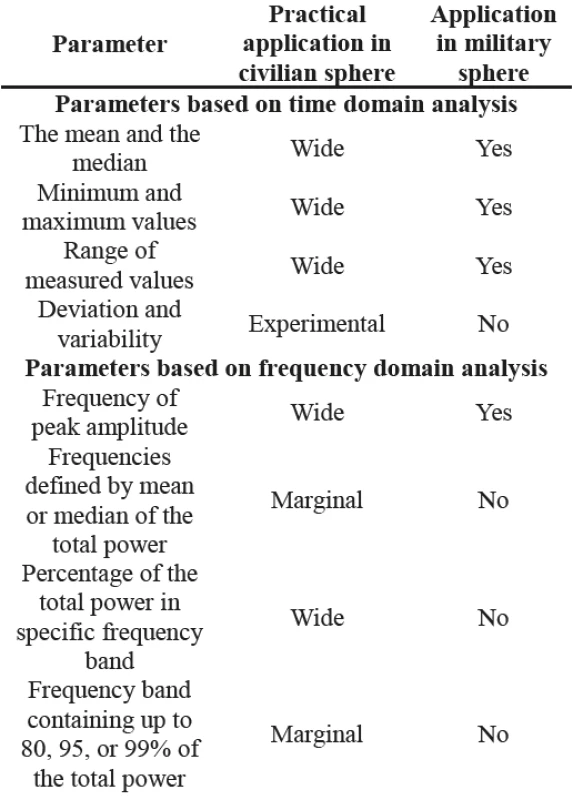
Further prospective for these methods, including their wider usage, are related to further experiments verifying suitability of particular parameters within a required application (evaluation of stress in combat, rehabili-tation efficiency in hospitals, etc). The above mentioned suggests that (MoCap systems and parameters of quanti-tative evaluation of motion) have great potential appli-cations in a military context.
Resulting from the advantages and disadvantages above of individual methods and within the developmet of the FlexiGuard system, designed to monitor the physical and mental health state of soldiers, as well as rescue personnel (eg. for firefighters), the researchers decided to use accelometers [89, 90]. The system was manufactured by the Faculty of Biomedical Engineering of the Czech Technical University in Prague. Based on information from the FlexiGuard system, the physical and mental health condition of a monitored person is assessed. In the case of the motion monitoring, body segment accelerations are measured by accelerometers. Particular sensors are placed on a specific body segment as required. In most cases, it means under a soldier’s uniform on his torso, head, and segments of the upper and lower limbs.
Individual sensors then monitor the motion of those individual body segments. The vector magnitude of the total acceleration is calculated from the measured accel-eration values; three acceleration components are mea-sured at any given point of time on individual axes of the three-axis accelerometer. The value of gravitational acceleration is then subtracted from the total value of acceleration, see Fig. 2.
Image 2. Example of a visualization of the measured magnitude of the acceleration vector in time domain data during gait. It is a record of a triple axis accelerometer located on an individual’s upper arm. 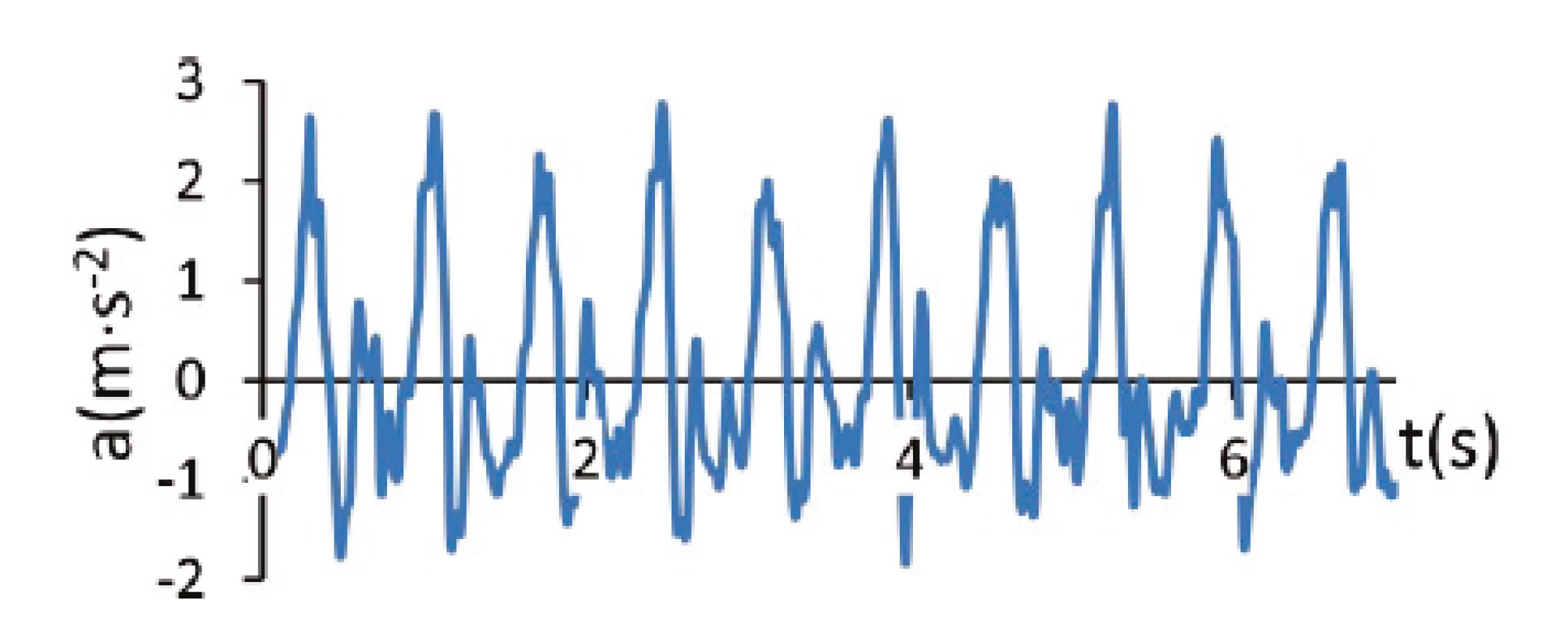
Subsequently, the maximum values and magnitudes of the resulting acceleration vector for specific time slots are determined. This direct calculation of the magnitude of the acceleration vector means that it is not necessary to calibrate the sensor and to transform the data into another (non-anatomical) coordinate system of the seg-ment. The calculated magnitude of the acceleration vector does not depend on the positioning of a sensor located on a particular anatomical point of the segment. This also eliminates the poor positioning of the sensor on the segment, assuming a body segment is rigid or stiff.
Subsequently, the specified data of maximum values and magnitudes of the acceleration vector are analyzed and the types of motions or physical activities are selected. Fundamental parameters and data from the systems located on soldiers’ bodies are compared to threshold values (standards) of the measured properties and provide information about the physical conditions of a proband (e.g. an estimate of energy expenditure). Such information is presented in a command visuali-zation center and can be used immediately, for example, to support decision making or to optimize the profiling of training processes.
The expectation is that other parameters will also be used in the future, such as the frequency of peak amplitude within the data evaluation in frequency domain. The system proposed here allows for the long-term monitoring of a soldiers’ motion during combat deployment in accordance with the results of the review performed. The example illustrates that traditional methods have the potential for the application of other proposed methods.
Conclusions
Based on the analysis of evaluation methods, it is apparent that in most cases the designed methods can be widely utilized in military applications, primarily in the medical field and in the development of new features and components of equipment, including robotic sys-tems.
This applies to the research of physical and mental conditions of soldiers during combat missions or during rehabilitation processes. The output of the study, con-firmed by the researchers’ own experience and testing, is a proposal of methods which can be potentially applied under military conditions.
The authors believe that the outlined methods de-scribed in this article will instigate further use of appro-priate methods for quantitative evaluation of posture and motion control during standing, gait and other activities in the military as well as the civilian sphere.
Acknowledgements
This work was done in Prague in the framework of research project SGS18/201/OHK4/3T/17 of the CTU in Prague. The work presented in this article has also been supported by the Czech Republic Ministry of Defence (University of Defence development program “Re-search of sensor and control systems to achieve battle-field information superiority”). We would also like to thank Anna Gajdosova (Department of Language Studies, The Masaryk Institute of Advanced Studies, Czech Technical University in Prague) for English language check.
Petr Volf
Department of Biomedical Technology
Faculty of Biomedical Engineering
Czech Technical University in Prague
nám. Sítná 3105, CZ-272 01 Kladno
E-mail: petr.volf@fbmi.cvut.cz
Sources
- Mündermann L, Corazza S, Andriacchi TP. Measuring human movement for biomechanical applications using markerless mo-tion capture. Journal of NeuroEngineering and Rehabilitation. 2006;3(1):6. DOI: 10.1186/1743-0003-3-6
- Andriacchi TP, Alexander EJ. Studies of human locomotion: past, present and future. Journal of Biomechanics. 2000 Oct; 33(10):1217–24. DOI: 10.1016/s0021-9290(00)00061-0
- Kutilek P, Socha V, Cakrt O, Svoboda Z. Differences in evalua-tion methods of trunk sway using different MoCap systems. Acta of bioengineering and biomechanics. 2014;16(2):85–94.
- Kutilek P, Socha V, Hana K. Analysis and prediction of upper extremity movements by cyclograms. Open Medicine. 2014 Dec 1;9(6):814–20. DOI: 10.2478/s11536-013-0322-y
- Patterson M, Delahunt E, Sweeney K, Caulfield B. An Ambulatory Method of Identifying Anterior Cruciate Ligament Reconstructed Gait Patterns. Sensors. 2014 Jan 7;14(1):887–99. DOI: 10.3390/s140100887
- Winter DA, Patla AE, Prince F, Ishac M, Gielo-Perczak K. Stiffness Control of Balance in Quiet Standing. Journal of Neurophysiology. 1998 Sep 1;80(3):1211–21.
DOI: 10.1152/jn.1998.80.3.1211 - Kutilek P, Farkasova B. Prediction of lower extremities' movement by angle-angle diagrams and neural networks. Acta of Bioengineering & Biomechanics. 2011;13(1):57–65.
- Bruijn SM, Meijer OG, Beek PJ, van Dieën JH. Assessing the stability of human locomotion: a review of current measures. Journal of The Royal Society Interface. 2013 Jun 6;10(83): 20120999. DOI: 10.1098/rsif.2012.0999
- Mazaheri M, Negahban H, Salavati M, Sanjari MA, Parnianpour M. Reliability of recurrence quantification analysis measures of the center of pressure during standing in individuals with musculoskeletal disorders. Medical Engineering & Physics. 2010 Sep;32(7):808–12. DOI: 10.1016/j.medengphy.2010.04.019
- Sylos Labini F, Meli A, Ivanenko YP, Tufarelli D. Recurrence quantification analysis of gait in normal and hypovestibular subjects. Gait & Posture. 2012 Jan;35(1):48–55.
DOI: 10.1016/j.gaitpost.2011.08.004 - Schubert P, Kirchner M, Schmidtbleicher D, Haas CT. About the structure of posturography: Sampling duration, parametrization, focus of attention (part I). Journal of Biomedical Science and Engineering. 2012;5(9):496–507.
DOI: 10.4236/jbise.2012.59062 - Hernandez ME, Stevenson C, Snider J, Poizner H. Center of pressure velocity autocorrelation as a new measure of postural control during quiet stance. In: 2013 6th International IEEE/ EMBS Conference on Neural Engineering (NER). IEEE; 2013; 1270 : 73. DOI: 10.1109/ner.2013.6696172
- Wollseifen T. Different methods of calculating body sway area. Pharmaceutical Programming. 2011 Dec;4(1–2):91–106. DOI: 10.1179/175709311x13166801334271
- Chow DHK, Leung DSS, Holmes AD. The effects of load car-riage and bracing on the balance of schoolgirls with adolescent idiopathic scoliosis. European Spine Journal. 2007 Mar 6;16(9): 1351–8. DOI: 10.1007/s00586-007-0333-y
- Schiffman JM, Bensel CK, Hasselquist L, Norton K, Piscitelle L. THE EFFECTS OF SOLDIERS’ LOADS ON POSTURAL SWAY. In: Transformational Science and Technology for the Current and Future Force. WORLD SCIENTIFIC; 2006. DOI: 10.1142/9789812772572_0049
- King LA, Horak FB, Mancini M, Pierce D, Priest KC, Chesnutt J, et al. Instrumenting the Balance Error Scoring System for Use With Patients Reporting Persistent Balance Problems After Mild Traumatic Brain Injury. Archives of Physical Medicine and Rehabilitation. 2014 Feb;95(2):353–9.
DOI: 10.1016/j.apmr.2013.10.015 - Nataraj R, Audu ML, Triolo RJ. Center of mass acceleration feedback control of functional neuromuscular stimulation for standing in presence of internal postural perturbations. The Journal of Rehabilitation Research and Development. 2012; 49(6):889-991. DOI: 10.1682/jrrd.2011.07.0127
- Hebert JS, Lewicke J, Williams TR, Vette AH. Normative data for modified Box and Blocks test measuring upper-limb function via motion capture. Journal of Rehabilitation Research and Development. 2014;51(6):918–32.
DOI: 10.1682/jrrd.2013.10.0228 - Triolo RJ, Bailey SN, Miller ME, Lombardo LM, Audu ML. Effects of Stimulating Hip and Trunk Muscles on Seated Stability, Posture, and Reach After Spinal Cord Injury. Archives of Physical Medicine and Rehabilitation. 2013 Sep;94(9):1766–75. DOI: 10.1016/j.apmr.2013.02.023
- Chaudhry H, Findley T, Quigley KS, Bukiet B, Ji Z, Sims T, et al. Measures of postural stability. The Journal of Rehabilitation Research and Development. 2004;41(5):713–720.
DOI: 10.1682/jrrd.2003.09.0140 - Kiyota T, Fujiwara K. Dominant side in single-leg stance sta-bility during floor oscillations at various frequencies. Journal of Physiological Anthropology. 2014 Aug 15;33(1).
DOI: 10.1186/1880-6805-33-25 - Duarte M, Freitas SMSF. Revision of Posturography Based on Force Plate for Balance Evaluation. Brazilian Journal of Physical Therapy. 2010 Jun;14(3):183–92.
DOI: 10.1590/S1413‑35552010000300003 - Prosperini L, Pozzilli C. The Clinical Relevance of Force Platform Measures in Multiple Sclerosis: A Review. Multiple Sclerosis International. 2013;2013 : 1–9.
DOI: 10.1155/2013/756564 - Omid Khayat, M. S. Complex feature analysis of center of pressure signal for age-related subject classification. Annals of Military & Health Sciences Research. 2014;12(1):1-6.
- Lamoth CJC, van Lummel RC, Beek PJ. Athletic skill level is reflected in body sway: A test case for accelometry in combi-nation with stochastic dynamics. Gait & Posture. 2009 Jun;29(4): 546–51. DOI: 10.1016/j.gaitpost.2008.12.006
- Bernard-Demanze L, Dumitrescu M, Jimeno P, Borel L, Lacour M. Age-Related Changes in Posture Control are Differentially Affected by Postural and Cognitive Task Complexity. Current Aging Sciencee. 2009 Jul 1;2(2):135–49.
DOI: 10.2174/1874609810902020135 - Delignières D, Torre K, Bernard P-L. Transition from Persistent to Anti-Persistent Correlations in Postural Sway Indicates Velocity-Based Control. Diedrichsen J, editor. PLoS Compu-tational Biology. 2011 Feb 24;7(2):e1001089.
DOI: 10.1371/journal.pcbi.1001089 - Collins JJ, De Luca CJ. Open-loop and closed-loop control of posture: A random-walk analysis of center-of-pressure trajec-tories. Experimental Brain Research. 1993 Aug;95(2):308–18. DOI: 10.1007/bf00229788
- Pellecchia GL, Shockley K. Application of recurrence quanti-fication analysis: influence of cognitive activity on postural fluctuations. Tutorials in contemporary nonlinear methods for the behavioral sciences. 2005;95–141.
- Ghomashchi H, Esteki A, Nasrabadi AM, Sprott JC, BahrPeyma F. Dynamic patterns of postural fluctuations during quiet standing: A recurrence quantification approach. International Journal of Bifurcation and Chaos. 2011 Apr;21(4):1163–72. DOI: 10.1142/s021812741102891x
- Hasson CJ, Van Emmerik REA, Caldwell GE, Haddad JM, Gagnon JL, Hamill J. Influence of embedding parameters and noise in center of pressure recurrence quantification analysis. Gait & Posture. 2008 Apr;27(3):416–22.
DOI: 10.1016/j.gaitpost.2007.05.010 - Riley M., Balasubramaniam R, Turvey M. Recurrence quanti-fication analysis of postural fluctuations. Gait & Posture. 1999 Mar;9(1):65–78. DOI: 10.1016/s0966-6362(98)00044-7
- Kiefer AW, Cummins-Sebree S, Riley MA, Shockley K, Haas JG. Control of posture in professional level ballet dancers. In Studies in Perception and Action IX: Fourteenth International Conference on Perception and Action. 2010;123.
- Rhea CK, Silver TA, Hong SL, Ryu JH, Studenka BE, Hughes CML, et al. Noise and Complexity in Human Postural Control: Interpreting the Different Estimations of Entropy. Perc M, editor. PLoS ONE. 2011 Mar 17;6(3):e17696.
DOI: 10.1371/journal.pone.0017696 - Flash T, Inzelberg R, Schechtman E, Korczyn AD. Kinematic analysis of upper limb trajectories in Parkinson’s disease. Experimental Neurology. 1992 Nov;118(2):215–26.
DOI: 10.1016/0014-4886(92)90038-r - Majsak M. The reaching movements of patients with Parkinson’s disease under self - determined maximal speed and visually cued conditions. Brain. 1998 Apr 1;121(4):755–66.
DOI: 10.1093/brain/121.4.755 - Wurdeman SR, Myers SA, Jacobsen AL, Stergiou N. Prosthesis preference is related to stride-to-stride fluctuations at the pros-thetic ankle. The Journal of Rehabilitation Research and Devel-opment. 2013;50(5):671–86. DOI: 10.1682/jrrd.2012.06.0104
- Hesse S, Uhlenbrock, D. A mechanized gait trainer for resto-ration of gait. Journal of rehabilitation research and development. 2000;37(6):701–8.
- Stansfield BW, Hillman SJ, Hazlewood ME, Robb JE. Regression analysis of gait parameters with speed in normal children walking at self-selected speeds. Gait & Posture. 2006 Apr;23(3):288–94. DOI: 10.1016/j.gaitpost.2005.03.005
- Farris DJ, Sawicki GS. Human medial gastrocnemius force-velocity behavior shifts with locomotion speed and gait. Proceedings of the National Academy of Sciences. 2012 Jan 4;109(3):977–82. DOI: 10.1073/pnas.1107972109
- Khandoker AH, Palaniswami M, Begg RK. A comparative study on approximate entropy measure and poincaré plot indexes of minimum foot clearance variability in the elderly during walking. Journal of NeuroEngineering and Rehabilitation. 2008 Feb 2; 5(1). DOI: 10.1186/1743-0003-5-4
- Paquette MR, Vallis LA. Age-related kinematic changes in late visual-cueing during obstacle circumvention. Experimental Brain Research. 2010 May 14;203(3):563–74.
DOI: 10.1007/s00221‑010-2263-x - Wu JZ, Chiou SS, Pan CS. Analysis of Musculoskeletal Loadings in Lower Limbs During Stilts Walking in Occupational Activity. Annals of Biomedical Engineering. 2009 Mar 19; 37(6):1177–89. DOI: 10.1007/s10439-009-9674-5
- Wren TAL, Do KP, Hara R, Dorey FJ, Kay RM, Otsuka NY. Gillette Gait Index as a Gait Analysis Summary Measure. Journal of Pediatric Orthopaedics. 2007 Oct;27(7):765–8.
DOI: 10.1097/bpo.0b013e3181558ade - Schwartz MH, Rozumalski A. The gait deviation index: A new comprehensive index of gait pathology. Gait & Posture. 2008 Oct;28(3):351–7. DOI: 10.1016/j.gaitpost.2008.05.001
- Baker R, McGinley JL, Schwartz MH, Beynon S, Rozumalski A, Graham HK, et al. The Gait Profile Score and Movement Analysis Profile. Gait & Posture. 2009 Oct;30(3):265–9. DOI: 10.1016/j.gaitpost.2009.05.020
- Gouelle A, Mégrot F, Presedo A, Husson I, Yelnik A, Penneçot G-F. The Gait Variability Index: A new way to quantify fluctua-tion magnitude of spatiotemporal parameters during gait. Gait & Posture. 2013 Jul;38(3):461–5.
DOI: 10.1016/j.gaitpost.2013.01.013 - Chwala W, Forczek W. Spatial Motions of Head, Trunk and Upper Limbs in Locomotion with Natural Velocity. Studies in Physical Culture and Tourism. 2005, 12(2):47–54.
- Jaspers E, Feys H, Bruyninckx H, Klingels K, Molenaers G, Desloovere K. The Arm Profile Score: A new summary index to assess upper limb movement pathology. Gait & Posture. 2011 Jun;34(2):227–33. DOI: 10.1016/j.gaitpost.2011.05.003
- Riad J, Coleman S, Lundh D, Broström E. Arm posture score and arm movement during walking: A comprehensive assessment in spastic hemiplegic cerebral palsy. Gait & Posture. 2011 Jan; 33(1):48–53. DOI: 10.1016/j.gaitpost.2010.09.022
- Vieten MM, Sehle A, Jensen RL. A Novel Approach to Quantify Time Series Differences of Gait Data Using Attractor Attributes. Perc M, editor. PLoS ONE. 2013 Aug 7;8(8):e71824.
DOI: 10.1371/journal.pone.0071824 - Murphy JO, Audu ML, Lombardo LM, Foglyano KM, Triolo RJ, et al. Feasibility of closed-loop controller for righting seated posture after spinal cord injury. Journal of Rehabilitation Re-search and Development. 2014;51(5):747–60.
DOI: 10.1682/jrrd.2013.09.0200 - Veeger HE, Meershoek LS, van der Woude LH, Langenhoff JM. Wrist motion in hand rim wheelchair propulsion. J Rehabil Res Dev. 1998;35(3):305–13.
- Koontz AM, Lin Y-S, Kankipati P, Boninger ML, Cooper RA. Development of custom measurement system for biomechanical evaluation of independent wheelchair transfers. The Journal of Rehabilitation Research and Development. 2011;48(8):1015–28. DOI: 10.1682/jrrd.2010.09.0169
- Lin J, Hanten WP, Olson SL, Roddey TS, Soto-quijano DA, Lim HK, et al. Shoulder Dysfunction Assessment: Self-report and Impaired Scapular Movements. Physical Therapy. 2006 Aug 1; 86(8):1065–74. DOI: 10.1093/ptj/86.8.1065
- Richter WM, Rodriguez R, Woods KR, Axelson PW. Stroke Pattern and Handrim Biomechanics for Level and Uphill Wheelchair Propulsion at Self-Selected Speeds. Archives of Physical Medicine and Rehabilitation. 2007 Jan;88(1):81–7. DOI: 10.1016/j.apmr.2006.09.017
- Uswatte G, Miltner WHR, Foo B, Varma M, Moran S, Taub E. Objective Measurement of Functional Upper-Extremity Movement Using Accelerometer Recordings Transformed With a Threshold Filter. Stroke. 2000 Mar;31(3):662–7. DOI: 10.1161/01.str.31.3.662
- Eilenberg MF, Geyer H, Herr H. Control of a Powered Ankle–Foot Prosthesis Based on a Neuromuscular Model. IEEE Trans-actions on Neural Systems and Rehabilitation Engineering. 2010 Apr;18(2):164–73. DOI: 10.1109/tnsre.2009.2039620
- DeLisa JA. Gait analysis in the science of rehabilitation. Diane Publishing. 1998;2.
- Hitt J, Merlo J, Johnston J, Holgate M, Boehler A, Hollander K., Sugar T. Bionic running for unilateral transtibial military ampu-tees. Military Academy West Point NY. 2010.
- Ventura JD, Klute GK, Neptune RR. The effects of prosthetic ankle dorsiflexion and energy return on below-knee amputee leg loading. Clinical Biomechanics. 2011 Mar;26(3):298–303. DOI: 10.1016/j.clinbiomech.2010.10.003
- To CS, Kobetic R, Bulea TC, Audu ML, Schnellenberger JR, Pinault G, et al. Sensor-based hip control with hybrid neuro-prosthesis for walking in paraplegia. Journal of Rehabilitation Research and Development. 2014;51(2):229–44.
DOI: 10.1682/jrrd.2012.10.0190 - Hardin E, Kobetic R, Murray L, Corado-Ahmed M, Pinault G, Sakai J, et al. Walking after incomplete spinal cord injury using an implanted FES system: A case report. The Journal of Reha-bilitation Research and Development. 2007;44(3):333–46. DOI: 10.1682/jrrd.2007.03.0333
- Schnall BL, Baum BS, Andrews AM. Gait Characteristics of
a Soldier With a Traumatic Hip Disarticulation. Physical Therapy. 2008 Dec 1;88(12):1568–77.
DOI: 10.2522/ptj.20070337 - Dillon MP, Fatone S, Hansen AH. Effect of prosthetic design on center of pressure excursion in partial foot prostheses. The Journal of Rehabilitation Research and Development. 2011; 48(2):161–78. DOI: 10.1682/jrrd.2010.09.0167
- Hansen AH, Childress DS, Meier MR. A simple method for determination of gait events. Journal of Biomechanics. 2002 Jan;35(1):135–8. DOI: 10.1016/s0021-9290(01)00174-9
- Qu X, Yeo JC. Effects of load carriage and fatigue on gait characteristics. Journal of Biomechanics. 2011 Apr;44(7):1259–63. DOI: 10.1016/j.jbiomech.2011.02.016
- Martin T, Lockhart T, Jones M, Edmison J. Electronic textiles for in situ biomechanical measurements. Alexandria: Springfield VA, U.S. Department of Commerce, National Technical Infor-mation Service. 2004.
- Sell TC, Chu Y, Abt JP, Nagai T, Deluzio J, McGrail MA, et al. Minimal Additional Weight of Combat Equipment Alters Air Assault Soldiers’ Landing Biomechanics. Military Medicine. 2010 Jan;175(1):41–7. DOI: 10.7205/milmed-d-09-00066
- Hess CW, Pullman SL. Tremor: clinical phenomenology and assessment techniques. Tremor and other hyperkinetic move-ments. 2012;2.
- Audu ML, Murphy JO, Triolo RJ. Trunk stability after spinal cord injury. In Banff, Alberta, Canada: 17th Annual Meeting of the International Functional Electrical Stimulation Society (IFESS). 2012.
- Kutilek P, Hejda J, Svoboda Z. Kinematic Quantification of Gait Asymmetry in Patients with Elastic Ankle Wrap Based on Cyclograms. In: IFMBE Proceedings. Springer International Publishing; 2014. p. 117–20.
DOI: 10.1007/978-3-319-00846-2_29 - Orendurff MS, Segal AD, Klute GK, Berge JS, Rohr ES, Kadel NJ. The effect of walking speed on center of mass displacement. Journal of Rehabilitation Research & Development. 2004;41(6): 829–34.
- Goswami A. A new gait parameterization technique by means of cyclogram moments: Application to human slope walking. Gait & Posture. 1998 Aug;8(1):15–36.
DOI: 10.1016/s0966-6362(98)00014-9 - Winter D. Human balance and posture control during standing and walking. Gait & Posture. 1995 Dec;3(4):193–214. DOI: 10.1016/0966-6362(96)82849-9
- Horváth M, Tihanyi T, Tihanyi J. Kinematic and kinetic analyses of gait patterns in hemiplegic patients. Facta universitatis-series: Physical Education and Sport. 2001;1(8):25–35.
- Ancillao A, Camerota F, Castori M, Albertini G, Galli M, Celletti C. Temporomandibular joint mobility in adult females with Ehlers-Danlos syndrome, hypermobility type (also known as joint hypermobility syndrome). Journal of Cranio-Maxillary Diseases. 2012;1(2):88–94. DOI: 10.4103/2278-9588.105697
- Chester VL, Calhoun M. Gait Symmetry in Children with Autism. Autism Research and Treatment. 2012;2012 : 1–5. DOI: 10.1155/2012/576478
- Goswami A. Kinematic Quantification of Gait Asymmetry Based on Bilateral Cyclograms. U.S. patent No 7,503,900, 2009.
- Kuo AD. An optimal control model for analyzing human postural balance. IEEE Transactions on Biomedical Engineering. 1995; 42(1):87–101. DOI: 10.1109/10.362914
- Herr HM, Grabowski AM. Bionic ankle–foot prosthesis nor-malizes walking gait for persons with leg amputation. Proceedings of the Royal Society B: Biological Sciences. 2011 Jul 13;279(1728):457–64. DOI: 10.1098/rspb.2011.1194
- Meijer R, Plotnik M, Zwaaftink E, van Lummel RC, Ainsworth E, Martina JD, et al. Markedly impaired bilateral coordination of gait in post-stroke patients: Is this deficit distinct from asym-metry? A cohort study. Journal of NeuroEngineering and Rehabilitation. 2011;8(1):23. DOI: 10.1186/1743-0003-8-23
- England SA, Granata KP. The influence of gait speed on local dynamic stability of walking. Gait & Posture. 2007 Feb;25(2): 172–8. DOI: 10.1016/j.gaitpost.2006.03.003
- Kang HG, Dingwell JB. Dynamic stability of superior vs. inferior segments during walking in young and older adults. Gait & Pos-ture. 2009 Aug;30(2):260–3.
DOI: 10.1016/j.gaitpost.2009.05.003 - Kirchner M, Schubert P, Liebherr M, Haas CT. Detrended Fluctuation Analysis and Adaptive Fractal Analysis of Stride Time Data in Parkinson’s Disease: Stitching Together Short Gait Trials. Hernandez-Lemus E, editor. PLoS ONE. 2014 Jan 23; 9(1):e85787. DOI: 10.1371/journal.pone.0085787
- Bisi M, Riva F, Stagni R. Measures of gait stability: performance on adults and toddlers at the beginning of independent walking. Journal of NeuroEngineering and Rehabilitation. 2014;11(1): 131. DOI: 10.1186/1743-0003-11-131
- Josiński H, Michalczuk A, Świtoński A, Szczęsna A, Wojciechowski K. Recurrence plots and recurrence quanti-fication analysis of human motion data. AIP Conference Proceedings. AIP Publishing. 2016. DOI: 10.1063/1.4951961
- Schiffman JM, Chelidze D, Adams A, Segala DB, Hasselquist L. Nonlinear analysis of gait kinematics to track changes in oxygen consumption in prolonged load carriage walking: A pilot study. Journal of Biomechanics. 2009 Sep;42(13):2196–9. DOI: 10.1016/j.jbiomech.2009.06.011
- Schlenker J, Socha V, Smrcka P, Hana K, Begera V, Kutilek P, et al. FlexiGuard: Modular biotelemetry system for military applications. In: International Conference on Military Tech-nologies (ICMT) 2015. IEEE; 2015.
DOI: 10.1109/miltechs.2015.7153712 - Hon Z, Smrcka P, Hana K, Kaspar J, Muzik J, Fiala R, Viteznik M, Vesely T, Kucera L, Kuttler T, Kliment R, Navratil V. A surveillance system for enhancing the safety of rescue teams. Communications-Scientific letters of the University of Zilina. 2015;17(1):81–6.
Labels
Biomedicine
Article was published inThe Clinician and Technology Journal
2019 Issue 4-
All articles in this issue
- METHODS FOR KINEMATIC ANALYSIS OF HUMAN MOVEMENT IN MILITARY APPLICATIONS: A REVIEW OF CURRENT AND PROSPECTIVE METHODS
- THE EFFECT OF FRAME RATE AND CALIBRATION ON LUNG MONITORING WITH ELECTRICAL IMPEDANCE TOMOGRAPHY
- THE EFFECT OF SPEED ON GAIT ASYMMETRY IN SUBJECT WITH CONGENITAL TIBIAL DEFICIENCY: A CASE STUDY
- THE ADOPTION OF AUTOMATED FiO2 CONTROL INTO POLISH NICUS: 2012–2019
- The Clinician and Technology Journal
- Journal archive
- Current issue
- Online only
- About the journal
Most read in this issue- METHODS FOR KINEMATIC ANALYSIS OF HUMAN MOVEMENT IN MILITARY APPLICATIONS: A REVIEW OF CURRENT AND PROSPECTIVE METHODS
- THE EFFECT OF FRAME RATE AND CALIBRATION ON LUNG MONITORING WITH ELECTRICAL IMPEDANCE TOMOGRAPHY
- THE ADOPTION OF AUTOMATED FiO2 CONTROL INTO POLISH NICUS: 2012–2019
- THE EFFECT OF SPEED ON GAIT ASYMMETRY IN SUBJECT WITH CONGENITAL TIBIAL DEFICIENCY: A CASE STUDY
Login#ADS_BOTTOM_SCRIPTS#Forgotten passwordEnter the email address that you registered with. We will send you instructions on how to set a new password.
- Career


