-
Články
- Vzdělávání
- Časopisy
Top články
Nové číslo
- Témata
- Kongresy
- Videa
- Podcasty
Nové podcasty
Reklama- Kariéra
Doporučené pozice
Reklama- Praxe
Reprogramming of from Virulent to Persistent Mode Revealed by Complex RNA-seq Analysis
To establish infection and colonize within a host, infecting pathogens have to cope with a variety of destructive surroundings. The food-borne pathogen Y. pseudotuberculosis can cause persistent infection in mice. Upon infection, Y. pseudotuberculosis passes the anti-microbial gastrointestinal milieu and finally remains associated with lymphoid follicles in cecal tissue surrounded by polymorphonuclear leukocytes, indicating that the bacteria are exposed to multiple environmental cues. We performed complex RNA-seq of small cecal biopsies of infected mice to reveal Y. pseudotuberculosis gene expression in vivo. We found that Y. pseudotuberculosis underwent reprogramming from a virulent phenotype, expressing virulence genes during early infection, to an adapted phenotype capable of persisting in the harsh cecal environment. Persistence was characterized by a novel expression pattern with down-regulation of virulence genes and up-regulation of genes involved in anaerobiosis, chemotaxis, and protection against oxidative and acidic stress. Mutagenesis of selected genes revealed that the regulator rovA was critical for the establishment of infection, and that arcA, fnr, frdA, and wrbA play critical roles in maintaining infection for long periods of time. Our study shows the power of RNA deep sequencing, which can be used to reveal the in vivo expression patterns of small amounts of bacteria in complex intestinal environments.
Published in the journal: . PLoS Pathog 11(1): e32767. doi:10.1371/journal.ppat.1004600
Category: Research Article
doi: https://doi.org/10.1371/journal.ppat.1004600Summary
To establish infection and colonize within a host, infecting pathogens have to cope with a variety of destructive surroundings. The food-borne pathogen Y. pseudotuberculosis can cause persistent infection in mice. Upon infection, Y. pseudotuberculosis passes the anti-microbial gastrointestinal milieu and finally remains associated with lymphoid follicles in cecal tissue surrounded by polymorphonuclear leukocytes, indicating that the bacteria are exposed to multiple environmental cues. We performed complex RNA-seq of small cecal biopsies of infected mice to reveal Y. pseudotuberculosis gene expression in vivo. We found that Y. pseudotuberculosis underwent reprogramming from a virulent phenotype, expressing virulence genes during early infection, to an adapted phenotype capable of persisting in the harsh cecal environment. Persistence was characterized by a novel expression pattern with down-regulation of virulence genes and up-regulation of genes involved in anaerobiosis, chemotaxis, and protection against oxidative and acidic stress. Mutagenesis of selected genes revealed that the regulator rovA was critical for the establishment of infection, and that arcA, fnr, frdA, and wrbA play critical roles in maintaining infection for long periods of time. Our study shows the power of RNA deep sequencing, which can be used to reveal the in vivo expression patterns of small amounts of bacteria in complex intestinal environments.
Introduction
Yersinia pseudotuberculosis is a food borne pathogen that can penetrate the intestinal epithelium and cause gastroenteritis. This enteropathogen invades lymphoid follicles of Peyer’s patches and cecum, where it survives extracellularly before breaking the barrier and becoming systemic [1,2]. All pathogenic Yersinia species are capable of inhibiting important host immune mechanisms in local lymph nodes, and this essential virulence property is dependent on the plasmid-encoded Yersinia outer proteins (Yops) YopE, YopH, YopJ, YopM, YopT, YpkA, and YopK. Upon intimate contact with a target host cell, the Yops are delivered into the host cell via the Yersinia type three secretion system (T3SS) [3]. Inside the target cell, the Yop effectors interfere with several key mechanisms of the host immune defense; for example, YopH and YopE inhibit phagocytosis and YopJ interferes with the production of pro-inflammatory signaling molecules [4]. Polymorphonuclear neutrophils (PMNs), which are rapidly recruited to infection sites, are the main target cells for Y. pseudotuberculosis T3SS-mediated Yop translocation during infection [2,5]. Current knowledge of Y. pseudotuberculosis virulence mechanisms is based, to a great extent, on studies using the acute mouse infection model in which infection results in systemic infection.
We recently found that the enteric pathogen Y. pseudotuberculosis can cause persistent infection in mice, where it persists associated with the lymphoid follicles of the cecum [6]. In this model, low dose oral infection (106–107 colony-forming units (CFUs)) leads to asymptomatic infection in 20–30% of infected mice with an observed infection duration as long as 115 days. Even if no signs of disease are present, Y. pseudotuberculosis persistence is associated with an immune response in which the Y. pseudotuberculosis foci are surrounded by PMNs and bacteria are shed in feces [6]. Yersinia, Salmonella, and Campylobacter have all been reported to infect and affect the ileocecal area in humans [7], suggesting that the cecum is a beneficial niche for bacterial persistence.
Many pathogenic bacteria are capable of maintaining infection in mammalian hosts, giving rise to persistent infections [8]. Diagnosis of a persistent infection can be difficult, as symptoms are not always obvious. Prolonged persistent infections can cause chronic inflammation, which can lead to complications, or even precipitation of certain diseases in susceptible hosts [9]. In addition, persistent bacterial infections are a major cause of the overuse of antibiotics in both humans and animal husbandry. Increasing evidence indicates that persistence contributes to the development and spread of antibiotic resistance [10]. Therefore, the identification of bacterial mechanisms involved in the development of persistent infections is of great interest. One well-established model of persistence is Salmonella typhimurium; upon infection of Nramp1-expressing mice, this intracellular pathogen can persist inside phagocytic cells in classical granuloma lesions in the spleen, liver, and mesenteric lymph nodes [11,12]. Another model of S. typhimurium is the infection of antibiotic-treated DBA/2 and 129Sv/Ev mice, which results in colitis and chronic cholangitis [13]. Similar to the Y. pseudotuberculosis persistence model, the colitis phase is associated with PMN infiltration into the cecum and bacterial shedding in the feces. Studies of S. typhimurium persistence using these models have shown a variety of factors that contribute to persistent infection [14], including effectors encoded by the pathogenicity islands SPI1 and SPI2, which are required for initial invasion and intracellular growth. Two SPI2 factors, Sse1 and SseK2, have been implicated as being important for later stages of infection, with Sse1 affecting host cell adhesion and migration. Different adhesive proteins and factors that protect against host-derived antimicrobial peptides and factors that aid in coping with oxidative and nitrosative stress have been found important for sustained colonization of S. typhimurium in the gastrointestinal tract, as well as systemic persistence [14].
The mechanisms enabling Y. pseudotuberculosis to persist in cecal tissue in the presence of immune cells for a prolonged period of time are largely unknown. Our previous study showed that the T3SS effectors YopH and YopE contributed to Y. pseudotuberculosis persistence in the cecum, likely by enabling initial colonization in the presence of phagocytic cells [6]. One way to understand mechanisms and metabolic traits that are important for Y. pseudotuberculosis persistence in the cecum is to identify the genes involved. Several methods have been used to identify genes induced in vivo during infection, such as in vivo expression technology (IVET) [15], signature-tagged mutagenesis (STM) [16], cDNA microarray analysis [17,18], and the recently developed RNA sequencing technology with massively parallel cDNA sequencing (RNA-seq) [19]. IVET and STM, which are both based on infections with bacterial libraries, are not suitable for in vivo studies of enteropathogenic Yersinia due to restricted clonal invasion of the intestinal tissue by this pathogen [20,21]. Furthermore, because the intestinal tract is colonized by the intestinal microflora and harbors many commensal bacteria, the reliability of DNA microarray is greatly diminished due to cross-reactions between species-specific probes on the microarray chips. In contrast, RNA-seq provides a promising approach for monitoring gene expression in a specific organism in the presence or absence of others. This method is sensitive and allows accurate discrimination between similar RNAs originating from different species, offering an excellent opportunity to reveal the gene expression patterns of pathogens within host tissues, even in heavily colonized environments, such as the intestine, stomach, and cecum.
In this study, we performed RNA-seq on Y. pseudotuberculosis YPIII in small cecal tissue biopsies from mice at early and persistent stages of infection to reveal mechanisms of importance for persistent infection. We found that the bacteria underwent substantial transcriptional reprogramming. Initially, genes encoded on the virulence plasmid, including T3SS and associated effectors, were highly up-regulated. At the persistent stage these genes were found to be down-regulated, and other genes were up-regulated, including those encoding for anaerobic growth, motility, protection against acidic and oxidative stress, and genes indicating envelope perturbation, suggesting adaptation to the harsh environment in cecal tissue.
Results
Heterogeneous RNA populations of Y. pseudotuberculosis-infected cecal tissues sequenced by RNA-seq
To identify mechanisms promoting Y. pseudotuberculosis persistence, RNA-seq was employed to determine the differential gene expression profiles of bacteria in the cecum during the early phase of infection and during persistence. To obtain infected tissue for the isolation of Y. pseudotuberculosis RNA, FVB/N mice were infected orally with bioluminescent wild-type (wt) bacteria at an infection dose of ∼2×10⁷ CFUs. The infection was monitored in real time by an in vivo imaging system (IVIS) at certain intervals for 42 days. In agreement with that reported earlier [6], we found bacterial foci associated with the cecal lymphoid tissue (Fig. 1A–B), massive infiltration of PMNs surrounding the bacterial foci (Fig. 1C–D) as well as superficial destruction of the epithelial lining and mixed inflammatory infiltrates.
Fig. 1. Persistent Y. pseudotuberculosis resides in cecal tissue in the presence of an immune response. 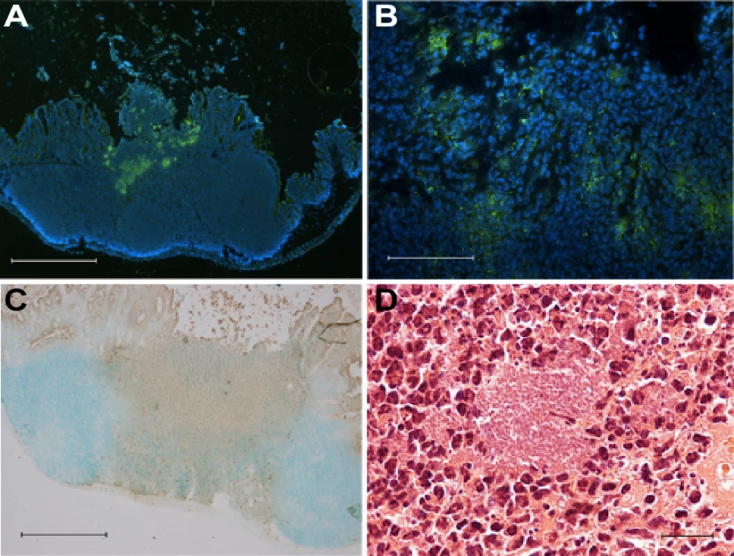
(A-B) Immunofluorescent staining of Y. pseudotuberculosis in cecum from a mouse with persistent asymptomatic infection (35 dpi) using anti-Yersiniae rabbit polyclonal serum detected by anti-rabbit Al488 (green). Nuclei were stained with DAPI (blue); (A) 4× magnification, scale bar 500 μm, (B) 40× magnification, scale bar 50 μm. (C) Immunohistochemical staining of PMNs with anti-Ly6G6C in cecal tissue from a persistently infected asymptomatic mouse (35 dpi). Positive cells are brown (DAB) and the background is green. (methyl green). 4× magnification, scale bar 500 μm. (D) Hematoxylin-eosin staining of persistently infected cecal tissue (42 dpi). 60× magnification, scale bar 20 μm. For sample preparation, isolated cecal tissues from 2 and 42 days post-infection (dpi) were analyzed by IVIS to verify that they contained Yersinia. The bioluminescent signal from Y. pseudotuberculosis allowed identification of the precise location of the bacteria in the tissue. Small biopsies (3 mm Ø) of cecal tissue containing bioluminescent bacteria were isolated using a hole punch. Total RNAs were extracted from biopsies from two mice infected for 2 days and two asymptomatic mice infected for 42 days. As a control, we extracted total RNAs from the cecal tissue of two un-infected mice. We also included RNA samples from bacteria grown in Luria broth (LB) in vitro at 26°C and in Ca2+-depleted LB at 37°C, a condition known to induce T3SS [22], hereafter referred to as T3SS-inducing conditions. The quality and quantity of all RNA samples were determined using an Agilent Bioanalyzer 2100, and all total RNA preparations had RIN values >7. This analysis revealed pure bacterial RNA in the in vitro samples (16S and 23S rRNAs) and mouse RNA (18S and 26S rRNAs) in samples from un-infected cecal tissue. As expected, both eukaryotic and prokaryotic RNAs were detected in the samples from infected cecal tissue and appeared as four distinct bands: 16S, 18S, 23S, and 26S rRNAs (Fig. 2A). However, as we previously recovered only 1×10⁵ to 2×10⁶ CFUs Y. pseudotuberculosis from cecal tissues [6], the amount of prokaryotic RNA was unexpectedly high in the infected tissues. Therefore, we performed qPCR to determine the Yersinia RNA abundance in infected tissues by comparing ymoA expression as an indication of Yersinia RNAs and GAPDH expression as an indication of host RNAs, finding ∼0.2% Yersinia RNA in the total RNA preparations. Therefore, the remarkably higher amount of prokaryotic RNA compared to that predicted for Yersinia RNA was assumed to reflect the presence of other microbial inhabitants in the samples.
Fig. 2. Y. pseudotuberculosis infection alters the bacterial composition of the cecum. 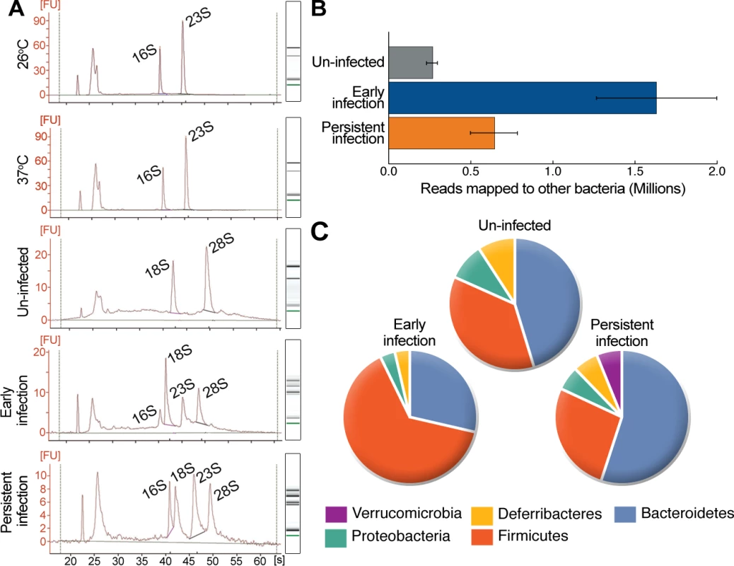
(A) Representative Bioanalyzer 2100 electrographs and associated gel pictures for replicates of in vitro-derived RNA samples (grown at 26°C and 37°C), in vivo-derived samples of early (isolated from mouse cecal tissue 2 dpi) and persistent infection (isolated from mouse cecal tissue 42 dpi), and uninfected samples (isolated from uninfected mouse cecal tissue). (B) The number of reads mapping to 16S rRNA from different bacteria in non-depleted in vivo-derived samples. Data represent the mean ± SD of the two replicates for each sample group. (C) Relative abundance of different bacterial phyla in samples according to reads mapped to the 16SMicrobial database. The proportions are given as the percent of bacterial phyla identified in specific samples. Y. pseudotuberculosis infection alters the bacterial composition of the cecum
To identify other bacteria in the cecum during the two different phases of infection, the in vivo-derived total RNA samples were analyzed by RNA-seq. The sequencing reads from these samples were mapped initially to the NCBI 16SMicrobial database with a full alignment parameter in order to map only unique reads to each 16S rRNA sequence. Next, matched 16S rRNA sequences were filtered with at least 80% coverage due to conserved regions in the 16S rRNA sequences. The number of reads mapped to each sample was normalized to the depth of sequencing in order to estimate the relative bacterial load in the tissue samples. Compared to un-infected samples, the bacterial RNA content of samples from early infection was 5.2-fold higher, and from persistent infection 3.7-fold higher, according to the normalized ratio of mapped reads for each tissue sample (Fig. 2B and S1 Table). The presence of other bacteria in uninfected samples likely reflects luminal bacteria, whereas the greater amount of bacteria in infected cecum samples may indicate that the infection leads to dysregulation of the luminal microbiota and/or that luminal bacteria gained access to the tissue. We identified 11 species in uninfected samples, 30 in early infection samples, and 11 in persistent infection samples (S1 Table). The identified species were grouped by phylum using RDP Classifier [23]. The abundance and composition of bacterial phyla in uninfected samples were similar to previous reports [24,25] but differed in samples from infected cecums. Samples from early infection (Firmicutes 60%, Bacteroidetes 27%, Proteobacteria 3.3%) differed from samples from persistent infection (Firmicutes 27%, Bacteroidetes 55%, Proteobacteria 9%, Verrucomicrobia 9%; Fig. 2C. The two independent replicates of the persistent sample contained similar species, and the strictly anaerobic Gram-negative bacterium Akkermansia muciniphila [26] was present in high abundance (75% of its transcriptome was revealed by RNA-seq; S1 Fig.).
Mapping Y. pseudotuberculosis-specific reads in mixed populations of cDNAs
Given the relatively small amount of Y. pseudotuberculosis in the cecal tissue, samples were enriched for bacterial mRNA by depleting fractions of poly(A)-tagged RNAs, rRNAs, and tRNAs prior to RNA-seq. The enrichment procedure was performed with both in vivo and in vitro total RNA samples.
Rigorous validations on mapped reads were required due to the presence of RNAs from other microbial inhabitants and homologous mRNAs that did not represent true Y. pseudotuberculosis transcripts. The genomes available for bacterial species found in the in vivo samples (42 genomes) were used as reference to optimize the alignment parameters for sequencing reads. Eventually, the optimization trials ended with a strict criterion of 95% alignment specificity to retrieve reads uniquely mapped to Y. pseudotuberculosis in the presence of other bacterial genomes. Finally, the uniqueness of these reads was double-checked with probabilistic variant detection, which searches for single nucleotide polymorphisms (SNPs) using CLC Genomic Workbench. With sample enrichment, optimized alignment parameters, and high sequencing depth, we revealed ∼92% of the metatranscriptome, which was composed of mouse and 42 other bacterial species in addition to Y. pseudotuberculosis. The reads mapped to Y. pseudotuberculosis were used in the subsequent RNA-seq analysis in order to calculate the expression of each open reading frame (ORF). The range of reads per ORF for in vitro and in vivo-derived samples was up to more than 200,000 and 490, respectively. Therefore, fewer ORFs were mapped (1551 ORFs, 36% transcriptome coverage) in vivo than in vitro, which had complete coverage (Table 1). Because we reached full coverage of the mouse transcriptome, and in some cases up to 75% coverage of other bacterial transcriptomes in the in vivo-derived samples (S1 Fig.), the relatively low coverage of Y. pseudotuberculosis was due to the very low abundance of its transcripts. Nevertheless, the RNA-seq results for Y. pseudotuberculosis showed very high correlation (Pearson and Spearman correlation R-values ≥0.98) of normalized RPKMO values (i.e., reads per kilobase pairs of a gene per million reads aligning to annotated ORFs) between biological replicates of all samples, which verifies the robustness of the analysis (Table 1). In addition, transcriptionally active regions were found to be the same for both in vitro and in vivo-derived samples (Fig. 3A, highlighted with gray borders). Even though the number of reads varied (53 to 111,817 reads) between different in vivo and in vitro samples, the distributions of the reads were similar for active regions (Fig. 3B). As qPCR requires more starting material than RNA-seq, we tested the differential expression of 9 genes that were highly expressed during persistent infection. The qPCR analysis confirmed the differential expression of all tested genes (Fig. 3C). Thus, the RNA-seq results for the in vivo samples provided valid information about differentially expressed genes in Y. pseudotuberculosis during early versus persistent infection in mice.
Tab. 1. Summary of RNA-seq reads. 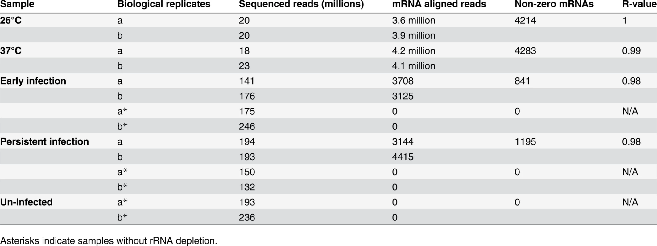
Asterisks indicate samples without rRNA depletion. Fig. 3. In vivo Y. pseudotuberculosis gene expression revealed by RNA-seq. 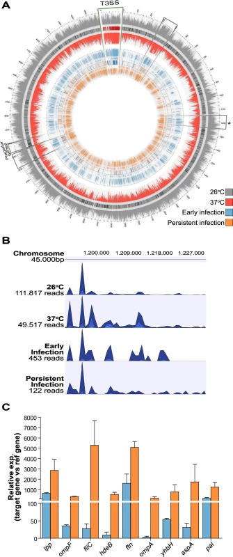
(A) Gene expression data for Y. pseudotuberculosis from two biological replicates of in vitro and in vivo-derived samples. From outside to inside, the 10 circles in the plot correspond to: (1) a histogram of RPKMO values for each gene expressed at 26°C; (2) genes expressed only at 26°C under in vitro conditions; (3) heat map (combined gray and red Brewer palettes) of the log2 difference in RPKMO values for genes expressed at both 26°C and 37°C; (4) genes expressed only at 37°C under in vitro conditions; (5) histogram of RPKMO values for each gene expressed at 37°C; (6) histogram of RPKMO values for each gene expressed during the early phase of infection (2 dpi) in FVB/N cecum; (7) genes expressed only during the early phase of infection in FVB/N cecum; (8) heat map (combined blue and orange Brewer palettes) of the log2 difference in RPKMO values for genes expressed during both the early phase of infection and persistent infection (42 dpi) in the cecum; (9) genes expressed only during persistent infection in the FVB/N cecum; and (10) a histogram of RPKMO values for each gene expressed during persistent infection in FVB/N cecum. Regions outlined with green borders indicate the locations of genes encoding T3SS components and effectors of the virulence plasmid and genes involved in flagellar assembly on the chromosome. Regions outlined with gray borders indicate the locations of transcriptionally active regions. The asterisk indicates a chromosomal region shown in Fig. 3B. The plots were created using Circos [62]. (B) Distributions of reads mapped to a specific transcriptionally active region on the Y. pseudotuberculosis YPIII chromosome (from 1,191 Mb to 1,230 Mb) in one replicate of each sample group. The height of each peak corresponds to the relative number of reads mapped to the region. The tracks were created using CLC Genomic Workbench. (C). Expression of indicated genes during early (blue; 2 dpi) and persistent infections (orange; 42 dpi) determined by qPCR of cDNAs from two biological and three technical replicates for each gene. lpp, ompF, fliC, hdeB, ftn, ompA, yhbH, aspA and pal gene expressions were 1.1, 6.8, 9.2, 4.8, 9.7, 1.7, 2.3, 1.1 and 1.1 log2fold upregulated respectively during persistent infection in RNA-seq analysis. In vivo-based RNA-seq disclosed transcriptional reprogramming with repression of T3SS and induction of motility during persistence
RNA-seq analysis of the in vitro samples revealed that 665 genes were differentially expressed (log2 fold change ≥0.7, p <0.05) under the different in vitro growth conditions. A total of 146 genes were up-regulated at 37°C (T3SS-inducing conditions), 55 of which were located on the 70-kb virulence plasmid that encodes the T3SS components; 519 genes, including 29 flagellar genes, were up-regulated at 26°C. This confirmation of T3SS induction by the temperature shift to 37°C, combined with Ca2+-depletion [22] and the motile phenotype of Y. pseudotuberculosis at 26°C [27], verifies the reliability of the RNA-seq analysis. The global expression patterns of cultured bacteria at 26°C and 37°C are shown on a histogram and heat map in Fig. 3A (see also S2 Table).
A total of 1288 genes were found to be differentially expressed (log2 fold change ≥0.7) in vivo. RPKMO values detected by RNA-seq are shown in a histogram and heat map in Fig. 3A to highlight the differences in individual ORFs during early and persistent infection (see also S3 Table). Surprisingly, the T3SS components encoded on the virulence plasmid that were highly expressed during the early stage of infection were distinctly down-regulated during persistent infection. Another conspicuous finding was the up-regulation of flagella and chemotaxis genes. T3SS is known to be induced at 37°C, but flagella are down-regulated at this temperature; in vitro, flagella are expressed only at 26°C (confirmed by qPCR in the same samples used in RNA-seq; Fig. 4A–B). In analogy, the T3SS master regulator lcrF was up-regulated during early infection and down-regulated during persistence (S3 Table), and the flagellar regulator flhCD was down-regulated during early infection and up-regulated during persistence (Fig. 4B). In addition, up-regulation of the gene encoding the adhesion protein invA, which is co-regulated with flagella [28], and its positive regulator rovA [29] suggested that other genes that are only expressed at 26°C in vitro could be up-regulated during persistence. Accordingly, a comparison of the expression patterns of in vivo and in vitro-derived samples showed that, during the early phase of infection, bacteria have an expression pattern similar to that seen in vitro at 37°C, whereas the expression pattern of persistent bacteria was much more similar to that of bacteria grown in vitro at 26°C (Fig. 5A and S4 Table). These results clearly indicate that, though increased temperature triggers T3SS and associated virulence genes during initial infection, other environmental cues are responsible for the observed transcriptional reprogramming of Y. pseudotuberculosis during prolonged infection.
Fig. 4. T3SS genes and flagellar genes are differentially regulated during persistent infection. 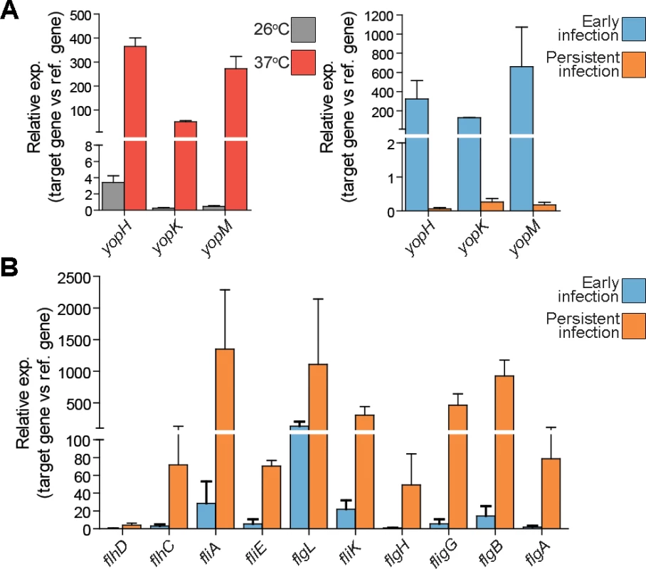
(A) Expression of indicated yop effectors in vitro (left) at 26°C (gray) and at 37°C inducing conditions (red), and in vivo (right) during early (2 dpi; blue) and persistent infections (42 dpi; orange) as determined by qPCR of cDNAs from two biological and three technical replicates for each gene. (B) Expression of indicated flagellar genes in vivo during early (2 dpi; blue) and persistent infection (42 dpi; orange) as determined by qPCR. All qPCR analyses were performed with cDNAs from two biological and three technical replicates. flhDC, fliA, fliE, flgL, fliK, flgH, flgG, flgB, and flgA genes expressions were 5.2, 8.3, 9.9, 3.6, 2.3, 3.8, 3.1, 5.2 and 3.6 log2fold upregulated respectively during persistent infection according to RNA-seq analysis. Fig. 5. Y. pseudotuberculosis undergoes transcriptional reprogramming for adaption to persistence. 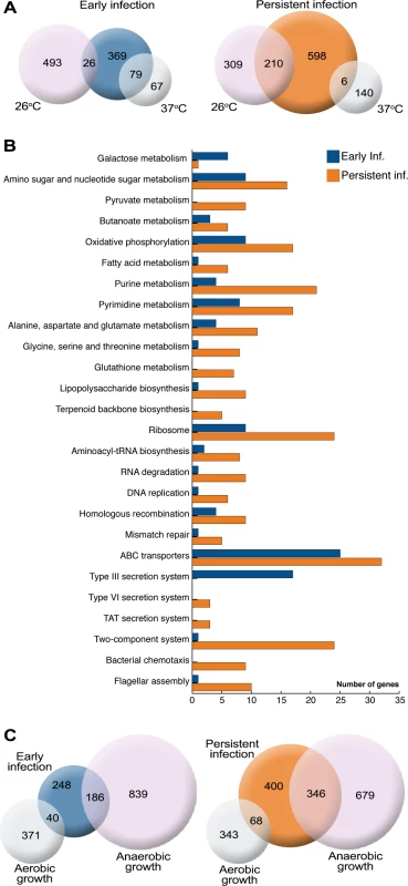
(A) Comparison of genes up-regulated in Y. pseudotuberculosis in vitro at 26°C and 37°C compared to in vivo during early (2 dpi) and persistent (42 dpi) stages of infection. Similarities are shown with the number of genes up-regulated in both groups. (B) Functional annotation of Y. pseudotuberculosis genes up-regulated during early and persistent infection (KEGG pathway mapping tool). (C) Comparison of the in vivo gene expression profiles and the expression profiles of bacteria grown under anaerobic conditions in vitro. The analysis included genes up-regulated (>1.8-fold) during anaerobic or aerobic growth in both the exponential and stationary growth phase compared to genes up-regulated during early and persistent infection. Similarities are shown with the number of genes up-regulated in both groups. Flagellar genes are known to be down-regulated at 37°C; therefore, the up-regulation of flagellar genes at later time points of infection at this temperature suggests that motility may be important for certain stages of persistence. However, Y. pseudotuberculosis is expected to remain flagellated for some time after the temperature shift and therefore flagella have also been assumed to participate in initial infection. To investigate this possibility, Y. pseudotuberculosis was grown at 26°C and then shifted to 37°C, followed by sampling at different time points for the detection of flagellated bacteria using atomic force microscopy. This analysis showed that the flagella remained for at least 2 hours after shifting temperature (S2 Fig.).
Functional clustering of differentially expressed genes uncovers environmental cues that promote reprogramming
To obtain an overview of the diversity of metabolic pathways and other functional systems utilized by persistent bacteria, functional clustering was performed using KEGG pathway mapping [30] for the 1288 genes differentially expressed in vivo (Fig. 5B). Down-regulation of T3SS and up-regulation of motility genes during persistence were verified. Induction of genes involved in ribosome biogenesis, amino-acyl tRNA biosynthesis, and RNA degradation suggests an active metabolic state during persistent infection. Induction of DNA replication and repair, as well as purine and pyrimidine biosynthesis, indicates proliferation of persistent bacteria and correlates well with constant bacterial shedding from the tissue to luminal sites during infection. The induction of TAT secretion components, which are involved in the transport of proteins synthesized mostly under anaerobic conditions [31], and induction of genes indicative of oxidative phosphorylation reflect an anaerobic/microaerophilic environment. Moreover, up-regulation of genes associated with two-component signal transduction systems, type VI secretion system, and chemotaxis indicates the presence of various external stimuli within the cecal environment. In addition to the functional annotations, the up-regulation of other genes indicates that the bacteria are influenced by different environmental conditions, such as acidic, oxidative, and other forms of stress (Table 2). The up-regulation of genes encoding proteins involved in envelope biogenesis, including a variety of inner and outer membrane proteins, and lipopolysaccharide biosynthesis also suggests an environmental influence. Therefore, the expression pattern of persistent bacteria suggests adaptation to an environment with limited oxygen and oxidative and acidic stress, a need for motility/chemotaxis, and modulation of the bacterial surface (Table 2).
Tab. 2. Up-regulated genes indicative of different environmental cues. 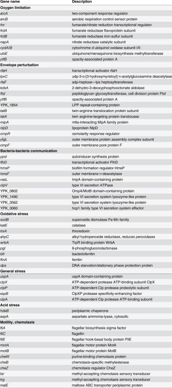
The cecal microaerophilic environment influences the expression pattern of Y. pseudotuberculosis
The up-regulation of several genes encoding proteins involved in oxidative phosphorylation in persistent bacteria (Fig. 5B) raised questions about energy metabolism and nutrient utilization. The cecal environment is expected to be anaerobic, and this was indicated by the sequencing data. The induction of genes involved in switching from aerobic to anaerobic respiration, such as the two-component system genes arcA-arcB, the fumarate nitrate reductase gene fnr encoding a global regulator of anaerobic growth, and other functional genes encoding proteins involved in microaerophilic/anaerobic respiration (Table 2), prompted us to investigate the influence of the anaerobic environment. We compared the expression profiles of Y. pseudotuberculosis grown in vitro under aerobic and anaerobic conditions at 26°C during the exponential and stationary phases using microarrays. Comparison of the differentially expressed genes identified in persistent bacteria by RNA-seq with the genes identified by microarray to be differentially expressed during anaerobiosis showed high similarity between persistent infection and in vitro anaerobic growth (Fig. 5C). Up to 42.5% of the genes up-regulated during persistence were also up-regulated during anaerobic growth (Fig. 5C and S4–S5 Tables). We found no obvious bias towards genes differentially expressed in the logarithmic or stationary phase (S3 Fig.). Thus, a substantial part of the expression profile of persistent bacteria is due to limited oxygen availability. The induction of some genes associated with anaerobic growth was also evident in samples from early infection (Fig. 5C), suggesting adaptation to the new environment at this stage.
Crp/CsrA/RovA regulatory cascades influence the expression pattern of persistent bacteria
The observed up-regulation of invA and its positive regulator rovA suggests that the RovA regulatory cascade contributes to the expression pattern of persistent Y. pseudotuberculosis. RovA is a regulator of the MarR/SlyA family, which controls different physiological processes [32]. Expression of rovA is controlled by the global regulators CsrA and Crp [33], which were both up-regulated during persistence. PhoP, which positively regulates rovA in Y. pestis and some Y. pseudotuberculosis strains, is not functional in the YPIII strain, where instead the Csr system via differential regulation of Csr RNAs influences production of RovA [34]. The RovA regulon of the Y. pseudotuberculosis YPIII strain (grown in vitro at 26°C) was recently revealed by microarray analysis [35]. A comparison of the gene expression patterns of persistent Y. pseudotuberculosis and the reported regulon [35] revealed 27.7% of the RovA regulon with 62 activated (lpp, fliC, and ftn verified by qPCR; Fig. 3C) and 26 suppressed genes in persistent bacteria (S4 Fig. and S4 Table). A comparison of the Y. pseudotuberculosis in vivo transcriptome and the Crp and CsrA regulons [35] revealed that more than 20% of the respective regulons represented genes identified as being differentially expressed during persistence. Some of the identified genes were shared and others were unique for Crp or CsrA (S4 Fig. and S4 Table). Notably, the fractions of Crp, CsrA, and RovA regulons observed in persistent bacteria appear to be relatively high with regard to the low coverage of the Y. pseudotuberculosis in vivo transcriptome.
Establishment of persistent infection requires arcA, fnr, frdA, and wrbA
To determine the importance of potential persistence genes identified in the RNA-seq analysis, we constructed a set of single gene deletion mutants to test in the mouse infection model of persistent infection. We selected genes implicated in different environmental responses (i.e., rovA, arcA, fnr, hdeB, uspA, napA, frdA, motB, cheW, and wrbA). Infection was achieved and monitored with IVIS for up to 42 dpi. Among the mice infected with the wt strain 31.6% had persistent infection, 31.6% cleared the infection, and 36.8% succumbed to severe disease (Fig. 6A), and this distribution is in accordance with that reported earlier [6]. The ΔrovA strain was completely attenuated and did not establish infection, indicating that it is indispensable for initiation of infection. Three of the mutants lacking genes involved in anaerobic respiration (arcA and fnr) or oxidative stress (wrbA) had a reduced capacity to establish persistence (Δfnr, 15.4%; ΔarcA, 7.7%; and ΔwrbA, 6.7%). These strains also gave less and later onset of severe disease than the wt strain (Δfnr 15.4%; ΔarcA, 7.7%; and ΔwrbA, 14.2%; Fig. 6A and S5 Fig.). A similar but less dramatic phenotype was observed for the ΔfrdA mutant, which had a reduced capacity to establish persistence (7.7%) but still caused severe disease (38.5%), with even earlier onset than the wt strain (Fig. 6A and S6 Fig.). ΔarcA, Δfnr, ΔfrdA, and ΔwrbA, established infections as wt, with almost similar levels of clearance during the first 7–14 days. However, after this, there were significant differences between these mutants and the wild-type strain with higher degrees of clearance, suggesting that these gene products are important during the persistent state (Fig. 6B). Interestingly, ΔhdeB was cleared to almost the same extent as the wt strain but was unable to establish persistent infection; this strain also appeared to be more aggressive, with infection resulting in more frequent and faster onset severe disease (68.8% vs. 31.6% for the wt strain; Fig. 6A and S5 Fig.). The ΔuspA mutant had a higher frequency of persistence than the wt strain (55.6% vs. 31.6%). The other mutants, ΔnapA, ΔmotB, and ΔcheW, had infection profiles similar to the wt strain (Fig. 6A).
Fig. 6. Establishment of persistent infection requires arcA, fnr, frdA, and wrbA. 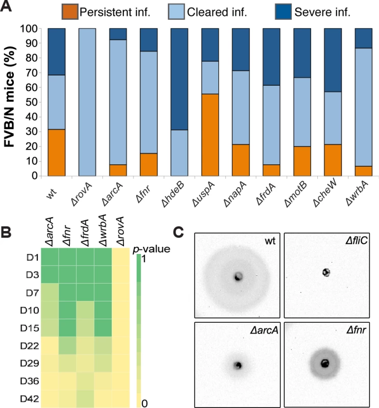
(A) Infection profile 42 dpi for FVB/N mice infected orally with107 CFUs of wt Y. pseudotuberculosis (n = 20) and indicated mutant strains (each group n = 16). The infections were monitored by IVIS at certain intervals up to 42 dpi. (B) Heatmap showing differences in clearance (by p-value) between wt and indicated mutant strains at different time points during the 42 day infection period. Heatmap color scale, from green to yellow, was adjusted according to p-values from 1 to 0. p-values were calculated with 2×2 contingency table by Fisher’s Exact Test, see also S6 Table. (C) Motility profile of wt Y. pseudotuberculosis and indicated mutant strains under anaerobic conditions at 26°C. Images were captured by the ChemiDoc XRS System (Bio-Rad), showing the bioluminescent signal produced by Y. pseudotuberculosis YPIII/pIBX. Microaerophilic and acidic environments influence T3SS and motility
The decreased virulence and persistence of the mutants related to anaerobic growth (ΔarcA, Δfnr, and ΔfrdA), combined with the down-regulation of T3SS and up-regulation of motility genes during persistent infection, prompted us to analyze T3SS secretion and motility in vitro in the presence and absence of oxygen. Secretion of T3SS effectors was reduced under anaerobic conditions for wt and all tested mutants (S6A Fig.). The reduction in T3SS effectors was also confirmed by qPCR (S6B Fig.). Moreover, to evaluate the contributions of the selected genes to bacterial motility, strains with mutations affecting the establishment of persistent infection were subjected to a motility test at 26°C and under T3SS-inducing conditions at 37°C in the presence and absence of oxygen. Neither the wt nor any of the mutants were motile at 37°C, independent of oxygen, but all strains except a ΔfliC mutant were motile at 26°C in the presence of oxygen. Interestingly, in the absence of oxygen at 26°C, the motility of Δfnr and ΔarcA mutants was significantly reduced (Fig. 6C).
Next, we investigated the influence of acid on T3SS and motility. Similar to what was observed for low oxygen, acidic conditions (pH 5.2) inhibited the induction of T3SS under inducing conditions at 37°C (S6A–S6B Fig.). Inhibition of T3SS under acidic conditions in Y. pseudotuberculosis was reported previously and suggested to involve pH-dependent inhibition of YscU proteolysis [36]. Furthermore, acidic conditions completely repressed the motility of the wt strain and all mutants tested in this study. Thus, both low oxygen and acidic conditions have a negative effect on T3SS induction and represent environmental cues that can contribute to the observed reprogramming of Y. pseudotuberculosis from a virulent to adaptive state.
Discussion
In this study, we applied in vitro and in vivo-based RNA-seq to determine key players that enable Y. pseudotuberculosis to establish a persistent infection. We found that the bacterium undergoes reprogramming from a virulent phenotype, massively expressing T3SS components in the early invasive phase of infection, to an adapted phenotype capable of persisting in a microaerophilic and hostile environment. This finding has clear impact on future rationales to identify bacterial targets for new antibiotics.
As shown here, RNA-seq is a powerful method for retrieving robust information about bacterial gene expression profiles during in vivo infection. We show that pathogen gene induction can be detected even if the amount of infecting bacteria in the isolated tissue is very low and mixed with other bacterial species. Thorough work with varied optimization steps allowed us to discriminate Yersinia-specific reads at the resolution of a single base with partial coverage from in vivo-derived samples and full coverage from in vitro-derived samples. We found that very strict read mapping parameters should be used to discriminate Y. pseudotuberculosis-specific reads for in vivo data. This strategy, which was established and optimized in this study, provides a controlled solution for discriminating between species-specific transcripts in complex RNA populations. Using this methodology we obtained robust data, revealing 36% of the Y. pseudotuberculosis in vivo transcriptome and providing novel information about bacterial gene expression during infection.
A discrepancy in the level of coverage between in vitro and in vivo-derived bacteria was expected based on previous studies of Vibrio cholerae [19] and Campylobacter jejuni [37]. The starting materials in those studies were cecal or intestinal contents, not infected tissue, and contained 100–300 times more bacteria. In addition, the overall coverage in those studies was higher than what we obtained with our tissue biopsy approach. However, the overall coverage for the metatranscriptome was >92%, and the ratio of Y. pseudotuberculosis total RNA was ∼0.2%. Full coverage of a bacterial transcriptome in such a complex population with low abundance is estimated to require a sequencing depth of at least 1.5–2 billions reads [38]. This estimate was calculated based on only the host and Y. pseudotuberculosis RNA being present in the sample, whereas additional RNAs from many bacterial species were present in our samples. Therefore, the depth of sequencing for such biopsy samples may need to be several times higher than 1.5–2 billion reads.
The massive expression of T3SS virulence genes during the early phase of infection is likely necessary to break the epithelial barrier and defend against innate immune cells. This assumption is supported by previous data showing that yopH or yopE mutants are defective in establishing the initial infection and less able to cause persistent infection [6]. Later, the bacteria become persistent with a novel expression profile, suggesting substantial transcriptional reprogramming. At this stage, the T3SS components are down-regulated. Thus, the bacteria prefer to use other genetic resources to adapt to the environment instead of producing massive amounts of invasive T3SS components.
Taking the host temperature into consideration, the repertoire of up-regulated genes in persistent bacteria was remarkably different from the repertoire of bacteria grown in vitro at 37°C. One striking observation was the up-regulation of flagella at 37°C, as achieving the induction of flagella is impossible at this temperature in vitro. Therefore, the situation in the animal greatly differs from laboratory settings. The surprising finding that the expression pattern seen during persistence is similar to the pattern seen at 26°C in vitro, indicates that the pathogens during infection encounters multiple environmental cues, other than the temperature that substantially influences its gene expression. Consequently, the reprogramming likely enables bacteria to persist in the harsh environment in cecum lymphoid follicles, where the tissue-associated bacteria are surrounded by PMNs. The functional annotation analysis revealed genes indicative of a microaerophilic environment with acidic and oxidative stress factors. We found similarities between the repertoire of up-regulated genes in bacteria grown in vitro under anaerobic conditions and the repertoire of persistent bacteria in the cecums of infected mice. Given the presence of PMNs in the cecum during persistence, it is not surprising that adaption requires protection against acidic and oxidative stress, or that it involves modulation of the bacterial surface for protection. Reprogramming probably occurs after initial colonization that requires T3SS, and later on certain environmental cues force the pathogen to reprogram its transcriptome where the induced gene products aid in maintaining long-term survival in this particular niche. At this late stage of infection the pathogen resists elimination by PMNs with very low expression of its T3SS-associated virulence arsenal, which is puzzling. However, the pathogen may be less recognized by innate immune cells due to its adapted phenotype with altered surface, and maybe also secretion of protective factors. In addition, we cannot exclude that presence of other microbial inhabitant(s) contributes. The observed up-regulation of type VI secretion genes previously reported to participate in bacteria-bacteria communication [39], as well as up-regulation of genes involved in biofilm formation and quorum sensing, may reflect interactions with other bacteria.
Up-regulation of genes involved in DNA replication and repair, RNA degradation, tRNA biosynthesis, and ribosome biogenesis suggests a metabolically active state for persistent bacteria, but it is not a direct indication of whether persistent bacteria are in a dormant or replicative form. We hypothesize that bacteria have a restricted replicative form in order to maintain bacterial load with consistent bacterial shedding into the feces. Motility may also be required for efficient shedding and spread of the bacteria within a restricted host environment, as shown by reduce bacterial shedding into the cecal lumen in a flagellar mutant of avian pathogenic Escherichia coli in a chick persistence model [40].
We show that regulators of anaerobic growth, as well as genes involved in oxidative/acidic stress, are important for the establishment of persistent infection with Y. pseudotuberculosis. Both arcA and fnr mutants demonstrated a markedly reduced ability to establish severe or persistent infection in mice, demonstrating the importance of reprogramming to anaerobic respiration. The function and importance of arcA and fnr have not been studied extensively in Yersinia. Here, we show that these gene products control motility, which has also been shown for arcA in Salmonella enterica sv. Typhimurium and E. coli [41,42]. In an earlier study, arcA was reported to be dispensable for acute Y. pseudotuberculosis infection upon intragastric inoculation in BALB/c mice [43], whereas another more recent study showed that a Y. pseudotuberculosis arcA mutant had attenuated virulence [35]. Whether the former study is contradictory to the latter and our results or just reflects different requirements of arcA depending on infection dose or intestinal delivery of bacteria remains to be elucidated. However, in agreement with our data, arcA has been implicated in virulence for a variety of bacteria [44,45]–[46–48]. Importantly, our data also show that a reduced ability to cause persistent infection is not directly coupled to decreased virulence in general, as increased virulence and an absence or reduced level of persistence was observed for the hdeB and frdA mutants. However, the mechanisms responsible for the virulence phenotypes of the hdeB and uspA mutants, with the former resulting in increased severe disease and the latter increased persistence, is not obvious and requires further investigation.
The regulatory pathways responsible for the switch, where T3SS is down-regulated is a central question in this context. We show that T3SS cannot be induced under low oxygen or acidic conditions. Therefore, regulatory circuits mediating T3SS repression are active under these conditions. Notably, FNR indirectly represses the expression of T3SS effectors under anaerobic conditions in Shigella flexneri [49]. However, we showed here that FNR per se has no effect on T3SS secretion in Y. pseudotuberculosis, as T3SS was repressed at the same level in the fnr mutant and wt strains under anaerobic conditions. The same was observed with the arcA mutant.
We found that many of the genes (20%) that were differentially expressed during persistence overlapped with the Crp/CsrA/RovA regulons, indicating that this regulatory circuit contributes to persistence in the host. The global regulatory systems of Crp and CsrA involves energy metabolism, but also control of certain virulence functions [33]. The interplay between these regulators and RovA is delicate and incorporates a series of complex regulatory loops that can be influenced by other regulators including UvrY and Hfq (all up-regulated during persistence). CsrA can control RovA via RovM, and independently of RovA, positively regulate flagella/motility genes as well as arcA [33,35]. Induction of flagella/motility genes is also influenced by Crp, which can control CsrA and promote induction of rovA. We hypothesize that genes induced by the Crp/CsrA/RovA regulatory cascades, which are mainly down-regulated during the early phase when T3SS is on, participate in reprogramming of Yersinia physiology by promoting expression of genes necessary for persisting in the cecal environment. RovA has been shown to be critical for virulence in Y. enterocolitica and Y. pestis [29,50]. By analogy, we found that RovA was required for virulence upon low dose infection. Interestingly, in contrast to the arcA, fnr, frdA, and wrbA mutants that initially infected cecum, but thereafter were efficiently cleared, the rovA mutant did not establish infection at all upon oral infection of mice. Hence, not only is RovA required for the positive regulation of many genes expressed at 26°C in vitro and during persistence, but is important also for initial infection where the gene expression pattern actually resembles that seen at 37°C in vitro with expression of T3SS. This suggests that RovA contributes to adaption to the host environment also at early stages of infection. Regulators of the Mar/SlyA family have been implicated in the regulation of genes involved in coping with diverse environmental stresses [32]. As such, RovA could contribute to the initial infection by regulating genes important for resistance to low pH in the stomach and to reactive oxygen metabolites produced by innate immune cells in cecum.
The persistence route may reflect the life cycle of this enteropathogen. In such a cycle we hypothesize that, during the initiation of infection, Y. pseudotuberculosis still has flagella and expresses T3SS virulence genes for breaking the epithelial barrier (Fig. 7). Flagella expression is supported by in vitro data, which showed flagellated bacteria up to 2 hours after shifting the temperature to 37°C. T3SS components are expected to be instrumental for resisting the attack from arriving PMNs during the early phase. In later stages, the bacterium adapts to the environment by reducing the expression of T3SS components and increasing the expression of genes important for survival in the cecum lymphoid compartment, from where it can spread to other hosts by fecal shedding, possibly through motility. In this context, Y. pseudotuberculosis has been found in the colon of wild mice with hyperemic cecal membranes [51], suggesting that this compartment is a potential reservoir for this pathogen.
Fig. 7. Hypothetical model of Y. pseudotuberculosis reprogramming for persistent infection in cecum. 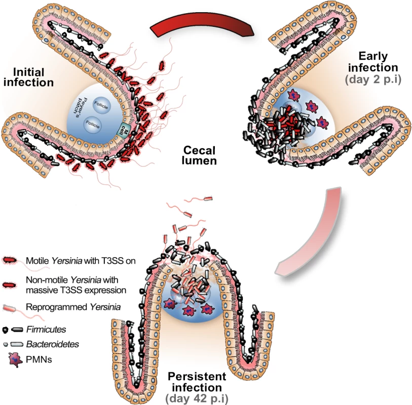
Upon initial infection, Y. pseudotuberculosis is still flagellated and expresses T3SS virulence genes. At the early stage of infection (2 dpi) the T3SS is important for colonization of tissue, including breaking the epithelial barrier and resisting the attack from arriving PMNs. At the persistent stage of infection (42 dpi), Y. pseudotuberculosis had reprogrammed its transcriptome by reducing the expression of T3SS components and increasing the expression of genes important for survival in the cecal lymphoid compartment. At this stage the bacteria are flagellated and can spread to other hosts by shedding into the feces, possibly through motility. Materials and Methods
Strains and growth conditions
YPIII/pIBX, a kanamycin-resistant bioluminescent Y. pseudotuberculosis strain (S6 Table), was used in this study. The YPIII strain represents a well established model for Y. pseudotuberculosis that has been used for decades to elucidate various aspects of Y. pseudotuberculosis pathogenesis, including identification of Yops [2,4–6,33,36,52]. For in vitro total RNA preparation, the strain was cultured at 26°C or 37°C in brain heart infusion (BHI) broth or LB at acidic (pH 5.2) or normal pH supplemented with 50 μg/ml kanamycin, 5 mM EGTA, and 20 mM MgCl2 for T3SS induction at 37°C. For microarray analysis, wt Y. pseudotuberculosis YPIII was grown at 25°C in LB medium supplemented with 10 g/l glucose and 0.2 M HEPES buffer under aeration or under anaerobic growth conditions (in a nitrogen atmosphere). Additional glucose was added to maximize energy production and the growth rate under anaerobic growth conditions. The absence of oxygen in the culture medium was tested by a gas chromatograph (GC-WLD, Carlo Erba Vega Series 6000) coupled with a detector and integrator (Spectra-Physics, SP4270) using a Poropak QS (100–120 Mesh) column and helium (Westalen 4.6) at 300 kPa. See Supplemental Experimental Procedure for the mutant strains used in this study.
Mutant construction
In order to generate an in-frame deletion mutant of the gene of interest, approximately 200 nucleotides from the 5’ and 3’ flanking regions of the gene were amplified by PCR and ligated together into SalI and BglII (New England Biolabs, Inc) linearized pDM4 [53] using the In-Fusion HD Cloning Kit (Clontech Laboratories, Inc) according to the manufacturer’s instructions. The plasmid was transformed into E. coli DH5αλpir and selected on a Cml (25 μg/ml)-containing agar plate. Positive colonies were confirmed by colony PCR. Plasmids purified from positive clones were sequenced to confirm insertion. Confirmed plasmid constructs were transformed into E. coli conjugation strain S17-1λpir for conjugal mating with Y. pseudotuberculosis YPIII-Xen04. Positive allelic exchange was selected as described previously [53]. Finally, in-frame deletion mutants of arcA, fnr, hdeB, uspA, cheW, frdA, and motB were confirmed by sequencing (S7 Table).
Ethics statement
Mice were housed in accordance with the Swedish National Board for Laboratory Animals guidelines. All animal procedures were approved by the Animal Ethics Committee of Umeå University (Dnr A108-10). Mice were allowed to acclimate to the new environment for one week before the experiments.
Mouse infection and bioluminescent imaging
Eight-week-old female FVB/N (Taconic Farms, Inc) mice were deprived of food and water for 16 hours prior to oral infection with ∼107 CFUs of wt or mutant Y. pseudotuberculosis YPIII-Xen04 strains, which were supplied in their drinking water for 6 hours. Bacteria were subcultured on LB agar plates supplemented with kanamycin (50 μg/ml). For infection, the bacteria were grown overnight in LB at 26°C and concentrations estimated by absorbance at OD600nm. Cultures were re-suspended to 107 CFUs/ml in sterilized tap water supplemented with 150 mM NaCl. The infection dose was determined by viable count and drinking volume. Mice were inspected frequently for signs of infection and to ensure that infected mice showing prominent clinical signs were euthanized promptly to prevent suffering. The infections were monitored using IVIS Spectrum (Caliper LifeSciences, Inc.) every third day after infection to 15 dpi, and then every week up to 42 dpi. Prior to imaging, the mice were anesthetized using the XGI-8 gas anesthesia system (Caliper LifeSciences, Inc), which allowed control over the duration of anesthesia. Oxygen mixed with 2.5% IsoFloVet (Orion Pharma, Abbott Laboratories Ltd, Great Britain) was used for the initial anesthesia, and 0.5% isoflurane in oxygen was used during imaging. To analyze bacterial localization within organs, mice were euthanized, the intestine, mesenteric lymph nodes, liver, and spleen removed, and the organs imaged by bioluminescent imaging (BLI). Acquisition and analysis were performed using Living Image software, version 3.1 (Caliper LifeSciences, Inc.).
Immunohistochemistry
Cecal tissue was fresh frozen in isopentane pre-chilled with liquid nitrogen and kept at −80°C. For detection of Yersinia in the tissue, 10-μm cryosections were fixed and stained with α-Yersinia serum and for immunohistochemistry sections were stained with rat-α-mouse Gr-1 (clone RB6-8C5, BD Biosciences Pharmingen) as described previously [6]. Cecal sections were also stained with hematoxylin-eosin using standard methods. Analysis was performed using a NIKON Eclipse 90i microscope and images captured with a Hamamatsu Orcha C4742-95 camera or NIKON DSFi1 camera and NIS-Elements AR 3.2 software (Nikon Instruments).
Bacterial total RNA isolation from mouse cecal tissue and in vitro bacterial cultures
Total RNA was isolated as described previously [54,55] with small modifications. Dissected cecums were emptied by flushing the luminal contents several times with 1× PBS using a sterile syringe. Parts of the cecal tissue associated with Y. pseudotuberculosis-Xen4 (bioluminescent) was cut out using a sterile 3 mm hole punch and immediately transferred to RNAlater (Ambion) for overnight incubation at 4°C after IVIS confirmation of bacterial in the isolated tissue. The RNAlater solution was removed the next day and the tissue samples stored at −80°C. The tissue was homogenized using Dispomix Drive (Medic Tools AG, Switzerland) and all steps performed at 4°C. The samples were transferred to previously cooled Dispomix homogenization tubes (Medic Tools AG, Switzerland) containing 1 ml of Solution D and homogenized twice using homogenization program 9. Tissue lysates were spun down with a quick spin. Each sample was aliquoted (0.5 ml) into separate 2 ml bead beater tubes containing small (0.1 mm) and big (1 mm) glass beads and treated with Mini-Beadbeater (Biospec Products Inc, USA) at a fixed speed for 1 min. Samples were cooled on ice for 1 min and the following added sequentially: 50 μl 2M sodium acetate (pH 4.0), 500 μl water-saturated phenol (Invitrogen, CA, USA), and 100 μl chloroform:isoamyl alcohol (49 : 1). The samples were inverted vigorously by hand. Suspensions were centrifuged for 20 min at 10,000g after cooling on ice for 15 min. The upper aqueous phase was transferred to RNase-free 1.5 ml tubes and 1 ml isopropanol added to precipitate the RNA. Samples were incubated at −20°C for 2 hours and centrifuged for 20 min at 10,000g. The RNA pellet was dissolved in 0.3 ml Solution D, 0.3 ml isopropanol added, and the resulting aliquots for each sample combined in one tube. The final suspensions were incubated for 30 min at −20°C and centrifuged for 20 min at 10,000g. The RNA pellets were suspended in 50 μl RNase-free water. DNA contamination was removed using the Qiagen DNase Kit according to the manufacturer’s instructions. The same procedure was applied to bacterial cultures grown in vitro except for the homogenization step with the Dispomix Drive homogenizer. The quality and concentration of total RNA isolated from the cecum and in vitro cultured bacteria was assessed by microcapillary electrophoresis using an Agilent 2100 Bioanalyzer (Agilent Technologies, Palo Alto, CA). All preparations used in this study had an RIN value >7.0.
Depletion of rRNA and poly(A)-tagged RNA from total RNA preparations
The MICROBEnrich Kit (Ambion) was used to enrich bacterial mRNAs in total RNA samples from cecums by removing 18S and 26S rRNAs and polyadenylated mRNAs according to the manufacturer’s instructions. To deplete the bacterial rRNA and tRNA in total RNAs from in vivo and in vitro samples, we used a MICROBExpress Kit (Ambion) according to the manufacturer’s instructions.
Illumina TruSeq RNA library preparation
RNA libraries for sequencing were prepared using TruSeq RNA kits (Illumina, CA, USA) according to the manufacturer’s instructions, but with the following changes. The RNA samples were EtOH precipitated and subsequent protocols (starting from cDNA synthesis in the Illumina provided protocol) were automated using an MBS 1200 pipetting station (Nordiag AB, Sweden). All purification steps and gel-cuts were replaced by the magnetic bead clean-up methods described previously [56].
RNA-seq
Quality and base trimming (5 nt from 5’ end and 5 nt from 3’ end of each read) were performed on 100-nt-long paired-end Illumina 2000 Hiseq reads from in vivo and in vitro sample libraries. Trimmed reads for each in vivo library were mapped to the NCBI 16SMicrobial database to determine the bacterial species in the cecal tissue biopsies with 100% identity. Identified bacterial species with available reference genomes (42 annotated bacterial genomes), Y. pseudotuberculosis YPIII, and the pYV plasmid from NCBI were used as reference genomes for mapping. A variety of mapping tests were performed by loosening or strengthening the alignment settings to optimize the filtration of non-Y. pseudotuberculosis YPIII-specific reads with SNP calling using Probabilistic Variant Detection in CLC Genomics Workbench after each mapping attempt. The rRNA and tRNA annotations were removed from the reference genomes prior to RNA-seq in order to avoid bias from the rRNA depletion procedures. The mRNA expression level and RPKMO value was calculated for each gene in in vivo and in vitro samples using annotated NC_010465 and NC_006153 as reference genomes in CLC Genomic Workbench for RNA-seq. Analysis of differentially expressed genes were performed on normalized RPKMO values by CLC Genomic Workbench for RNA-seq. The zero read values for the ORFs in in vivo samples were normalized by adding 0.01 pseudocounts in order to avoid dividing by zero [57–59]. As many of the reads detected for individual ORFs were only present in samples from either early or persistent infection, proper p-values could not be calculated for the in vivo data set. For in vitro data, the ORFs with less than 10 reads in all four replicates and p >0.05 were removed from the analysis to filter out false-positive values. The raw RNA-seq data have been deposited in NCBI’s Gene Expression Omnibus and are accessible through GEO Series accession number GSE55292.
cDNA preparation and qPCR
Total pure bacterial RNA isolated from bacterial cultures grown in BHI/LB medium at pH 7.2 at 26°C/37°C (in some experiments at acidic pH or under anaerobic conditions at 37°C) and heterogenous RNA isolated from infected cecums were used as templates for cDNA synthesis with the RevertAid H Minus First Strand cDNA Synthesis Kit (Fermentas). The qPCR reactions were performed in triplicate for each condition using the Quantimix Easy Syg Kit (Biotools) or KAPA SYBR FAST qPCR Kit (Kapabiosystems) and Bio-Rad i5 Light Cycler. After optimization experiments (S7 Fig.) gyrB was selected as internal control to calculate the relative expression of tested genes.
Microarray and data analysis
The design and analysis of the microarray (Agilent, 8×15K format) for the transcriptome analysis of Y. pseudotuberculosis YPIII were described previously [33]. YPIII was grown aerobically and anaerobically in four independent cultures at 25°C to the exponential and stationary phase. Bacteria were pelleted, mixed with 0.2 volumes of stop solution (5% water-saturated phenol), and snap frozen in liquid nitrogen. After thawing on ice, total RNA was prepared using the SV Total RNA Purification Kit (Promega) and remaining genomic DNA removed by rDNAse (Macherey-Nagel) digestion as described by the manufacturer. RNA concentration and quality were determined by measuring A260 and A280 with an Agilent 2100 Bioanalyzer using the Nano 6000 kit, and the absence of DNA was excluded by PCR of intergenic regions. Total RNA from the independent cultures was labeled using the ULS™ Fluorescent Labeling Kit for Agilent Arrays (Kreatech) as follows: 1 μg total RNA was used for RNA-labeling with Cy5 (for wt RNA) and Cy3 (for mutant RNA). Non-incorporated Cy5/Cy3 was removed using KREApure purification columns from the ULS™ Fluorescent Labeling Kit as suggested by the manufacturer, and the degree of labeling was analyzed with a Nanodrop (Peqlab). Subsequently, 300 ng Cy5-labelled RNA and 300 ng Cy3-labelled RNA were mixed, fragmented, and hybridized for 17 h at 65°C to custom-made Agilent microarray slides using the Agilent Gene Expression Hybridization Kit as described by the manufacturer. Four replicates were utilized in each experiment. After washing and drying the microarray slide, the data were scanned using an Axon GenePix Personal 4100A scanner and array images captured using the software package GenePix Pro 6.015. The microarray data was processed using the software package R (www.r-project.org) in combination with the “Bioconductor” software framework [60] as described previously [33]. The overall fold-changes of a gene represented by at least three probes are given as median values for all probes. The set of resulting differentially expressed genes (fold-change ≥ 2) was analyzed by the topGO package for Gene Ontology (GO) term enrichment [61]. MIAME compliant array data were deposited in the Gene Expression Omnibus (GEO) database and are available via the following accession numbers: GSE56475 and GSE56475.
Motility assay
Bacteria from overnight cultures were inoculated into LB and grown to exponential phase. A 5 μl aliquot of each culture was spotted on LB (pH 7.4 or pH 5) with 0.25% agar. Plates were incubated at 26°C or 37°C under aerobic or anaerobic conditions for 48 hours. The bioluminescent signal from bacteria on the plates was monitored using a ChemiDoc XRS System (Bio-Rad).
Analysis of protein secretion by Y. pseudotuberculosis
Overnight bacterial cultures were diluted 25-times in LB media and allowed to grow for 2 hours at 26°C. The medium was changed to T3SS-inducing conditions (5 mM EGTA, 20 mM MgCl2, pH 7.4 or 5) and incubated for 4 hours at 37°C under aerobic and anaerobic conditions. The culture supernatant was collected by centrifugation at 4000 rpm for 10 min and filtered using 0.45-μm filters. The secreted proteins were concentrated by TCA precipitation. Samples were loaded onto 12% SDS-PAGE according to the cultures’ OD600 values after 4 hours incubation.
Visualization of flagella by atomic force microscopy
Bacterial cultures grown overnight were diluted 25-times in LB media and allowed to grow for 2 hours at 26°C to OD600 = 0.2. The growth conditions were then changed to T3SS-inducing conditions at 37°C. One milliliter was taken from the bacterial cultures every hour for analysis with atomic force microscopy. Each sample was centrifuged for 4 min at 1500 rpm, washed once with 2 mM MgCl2, and re-suspended in 50–200 μl of the same solution. Ten microliters of each sample was placed on freshly cleaved ruby red mica (Goodfellow Cambridge Ltd, Cambridge), incubated 5 min at room temperature, and blotted dry before being placed into a desiccator for a minimum of 2 hours. Images were collected by a Nanoscope V AFM (Bruker software) using ScanAsyst in air with ScanAsyst cantilevers at a scan rate of approximately 0.9–1 Hz. The final images were flattened and/or plane-fitted in both axes using Bruker software and presented in amplitude (error) mode.
Statistical analysis
Windows Microsoft Excel 2011 and CLC Genomic Workbench were utilized for statistical tests and linear regression analysis of RNA-seq and infection data. Multiple RNA-seq were compared by the paired t-test on Gaussian data. Bonferroni correction was employed for multiple comparison analysis. Similarities between replicates were determined by Spearman and Pearson’s R value. One-tailed test were conducted to calculate p-values for difference in mutant infections clearance with 2×2 contingency table by Fisher’s exact test.
Accession numbers
Kyoto Encyclopedia of Genes an Genomes (KEGG) accession numbers for the genes mentioned in this study are as follow; arcA (ypy:YPK_3606), fnr (ypy:YPK_1944), rovA (ypy:YPK_2381), frdA (ypy:YPK_3813), hdeB (ypy:YPK_1140), uspA (ypy:YPK_0120), napA (ypy:YPK_1387), wrbA (ypy:YPK_2363), motB (ypy:YPK_0802), cheW (ypy:YPK_1750), csrA (ypy:YPK_3372), crp (ypy:YPK_0248), dnaG (ypy:YPK_0635), gyrB (ypy:YPK_0004), mdh (ypy:YPK_3761), rpoC (ypy:YPK_0341), fliC (ypy:YPK_2381), lpp (ypy:YPK_1854), ompF (ypy:YPK_2649), ompA (ypy:YPK_2630), ftn (ypy:YPK_2438), aspA (ypy:YPK_3825), yhbH (ypy:YPK_3353), pal (ypy:YPK_2955), flhC (ypy:YPK_1746), flhD (ypy:YPK_1745), fliA (ypy:YPK_2380), fliE (ypy:YPK_2390), fliK (ypy:YPK_2396), flgL (ypy:YPK_2415), flgH (ypy:YPK_2419), flgG (ypy:YPK_2420), flgB (ypy:YPK_2425), flgA (ypy:YPK_2426), invA (ypy:YPK_2429), uvrY (ypy:YPK_2326), hfq (ypy:YPK_3799), yopB (pYV0055), yopD (pYV0054), yopH (pYV0094), yopE (pYV0025), yopK (pYV0040), yopM (pYV0047), lcrF (pYV0076).
Supporting Information
Zdroje
1. Simonet M, Richard S, Berche P (1990) Electron microscopic evidence for in vivo extracellular localization of Yersinia pseudotuberculosis harboring the pYV plasmid. Infect Immun 58 : 841–845. 2307522
2. Westermark L, Fahlgren A, Fallman M (2014) Yersinia pseudotuberculosis efficiently escapes polymorphonuclear neutrophils during early infection. Infect Immun 82 : 1181–1191. doi: 10.1128/IAI.01634-13 24379291
3. Galan JE, Wolf-Watz H (2006) Protein delivery into eukaryotic cells by type III secretion machines. Nature 444 : 567–573. doi: 10.1038/nature05272 17136086
4. Viboud GI, Bliska JB (2005) Yersinia outer proteins: role in modulation of host cell signaling responses and pathogenesis. Annu Rev Microbiol 59 : 69–89. doi: 10.1146/annurev.micro.59.030804.121320 15847602
5. Durand EA, Maldonado-Arocho FJ, Castillo C, Walsh RL, Mecsas J (2010) The presence of professional phagocytes dictates the number of host cells targeted for Yop translocation during infection. Cellular microbiology 12 : 1064–1082. doi: 10.1111/j.1462-5822.2010.01451.x 20148898
6. Fahlgren A, Avican K, Westermark L, Nordfelth R, Fallman M (2014) Colonization of cecum is important for development of persistent infection by Yersinia pseudotuberculosis. Infect Immun 82 : 3471–3482. doi: 10.1128/IAI.01793-14 24891107
7. Puylaert JB, Van der Zant FM, Mutsaers JA (1997) Infectious ileocecitis caused by Yersinia, Campylobacter, and Salmonella: clinical, radiological and US findings. European radiology 7 : 3–9. doi: 10.1007/s003300050098 9000386
8. Monack DM, Mueller A, Falkow S (2004) Persistent bacterial infections: the interface of the pathogen and the host immune system. Nat Rev Microbiol 2 : 747–765. doi: 10.1038/nrmicro955 15372085
9. Ternhag A, Torner A, Svensson A, Ekdahl K, Giesecke J (2008) Short - and long-term effects of bacterial gastrointestinal infections. Emerg Infect Dis 14 : 143–148. doi: 10.3201/eid1401.070524 18258094
10. Cohen NR, Lobritz MA, Collins JJ (2013) Microbial persistence and the road to drug resistance. Cell host & microbe 13 : 632–642. doi: 10.1016/j.chom.2013.05.009 23768488
11. Monack DM, Bouley DM, Falkow S (2004) Salmonella typhimurium persists within macrophages in the mesenteric lymph nodes of chronically infected Nramp1+/+ mice and can be reactivated by IFNgamma neutralization. J Exp Med 199 : 231–241. doi: 10.1084/jem.20031319 14734525
12. Monack DM (2012) Salmonella persistence and transmission strategies. Curr Opin Microbiol 15 : 100–107. doi: 10.1016/j.mib.2011.10.013 22137596
13. Stecher B, Paesold G, Barthel M, Kremer M, Jantsch J, et al. (2006) Chronic Salmonella enterica serovar Typhimurium-induced colitis and cholangitis in streptomycin-pretreated Nramp1+/+ mice. Infect Immun 74 : 5047–5057. doi: 10.1128/IAI.00072-06 16926396
14. Monack DM (2013) Helicobacter and salmonella persistent infection strategies. Cold Spring Harbor perspectives in medicine 3: a010348. doi: 10.1101/cshperspect.a010348 24296347
15. Angelichio MJ, Camilli A (2002) In vivo expression technology. Infect Immun 70 : 6518–6523. doi: 10.1128/IAI.70.12.6518-6523.2002 12438320
16. Saenz HL, Dehio C (2005) Signature-tagged mutagenesis: technical advances in a negative selection method for virulence gene identification. Curr Opin Microbiol 8 : 612–619. doi: 10.1016/j.mib.2005.08.013 16126452
17. Fukuto HS, Svetlanov A, Palmer LE, Karzai AW, Bliska JB (2010) Global gene expression profiling of Yersinia pestis replicating inside macrophages reveals the roles of a putative stress-induced operon in regulating type III secretion and intracellular cell division. Infect Immun 78 : 3700–3715. doi: 10.1128/IAI.00062-10 20566693
18. Lathem WW, Crosby SD, Miller VL, Goldman WE (2005) Progression of primary pneumonic plague: a mouse model of infection, pathology, and bacterial transcriptional activity. Proc Natl Acad Sci U S A 102 : 17786–17791. doi: 10.1073/pnas.0506840102 16306265
19. Mandlik A, Livny J, Robins WP, Ritchie JM, Mekalanos JJ, et al. (2011) RNA-Seq-based monitoring of infection-linked changes in Vibrio cholerae gene expression. Cell host & microbe 10 : 165–174. doi: 10.1016/j.chom.2011.07.007 21843873
20. Oellerich MF, Jacobi CA, Freund S, Niedung K, Bach A, et al. (2007) Yersinia enterocolitica infection of mice reveals clonal invasion and abscess formation. Infect Immun 75 : 3802–3811. doi: 10.1128/IAI.00419-07 17562774
21. Handley SA, Newberry RD, Miller VL (2005) Yersinia enterocolitica invasin-dependent and invasin-independent mechanisms of systemic dissemination. Infect Immun 73 : 8453–8455. doi: 10.1128/IAI.73.12.8453-8455.2005 16299350
22. Brubaker RR, Surgalla MJ (1964) The Effect of Ca++ and Mg++ on Lysis, Growth, and Production of Virulence Antigens by Pasteurella Pestis. J Infect Dis 114 : 13–25. doi: 10.1093/infdis/114.1.13 14118042
23. Wang Q, Garrity GM, Tiedje JM, Cole JR (2007) Naive Bayesian classifier for rapid assignment of rRNA sequences into the new bacterial taxonomy. Appl Environ Microbiol 73 : 5261–5267. doi: 10.1128/AEM.00062-07 17586664
24. Vijay-Kumar M, Aitken JD, Carvalho FA, Cullender TC, Mwangi S, et al. (2010) Metabolic syndrome and altered gut microbiota in mice lacking Toll-like receptor 5. Science 328 : 228–231. doi: 10.1126/science.1179721 20203013
25. Cullender TC, Chassaing B, Janzon A, Kumar K, Muller CE, et al. (2013) Innate and adaptive immunity interact to quench microbiome flagellar motility in the gut. Cell Host Microbe 14 : 571–581. doi: 10.1016/j.chom.2013.10.009 24237702
26. Derrien M, Vaughan EE, Plugge CM, de Vos WM (2004) Akkermansia muciniphila gen. nov., sp. nov., a human intestinal mucin-degrading bacterium. Int J Syst Evol Microbiol 54 : 1469–1476. doi: 10.1099/ijs.0.02873-0 15388697
27. Kapatral V, Olson JW, Pepe JC, Miller VL, Minnich SA (1996) Temperature-dependent regulation of Yersinia enterocolitica Class III flagellar genes. Molecular microbiology 19 : 1061–1071. doi: 10.1046/j.1365-2958.1996.452978.x 8830263
28. Young GM, Badger JL, Miller VL (2000) Motility is required to initiate host cell invasion by Yersinia enterocolitica. Infect Immun 68 : 4323–4326. doi: 10.1128/IAI.68.7.4323-4326.2000 10858252
29. Revell PA, Miller VL (2000) A chromosomally encoded regulator is required for expression of the Yersinia enterocolitica inv gene and for virulence. Molecular microbiology 35 : 677–685. doi: 10.1046/j.1365-2958.2000.01740.x 10672189
30. Kanehisa M, Goto S, Sato Y, Furumichi M, Tanabe M (2012) KEGG for integration and interpretation of large-scale molecular data sets. Nucleic Acids Res 40: D109–114. doi: 10.1093/nar/gkr988 22080510
31. Jack RL, Sargent F, Berks BC, Sawers G, Palmer T (2001) Constitutive expression of Escherichia coli tat genes indicates an important role for the twin-arginine translocase during aerobic and anaerobic growth. J Bacteriol 183 : 1801–1804. doi: 10.1128/JB.183.5.1801-1804.2001 11160116
32. Ellison DW, Miller VL (2006) Regulation of virulence by members of the MarR/SlyA family. Curr Opin Microbiol 9 : 153–159. doi: 10.1016/j.mib.2006.02.003 16529980
33. Heroven AK, Sest M, Pisano F, Scheb-Wetzel M, Steinmann R, et al. (2012) Crp induces switching of the CsrB and CsrC RNAs in Yersinia pseudotuberculosis and links nutritional status to virulence. Front Cell Infect Microbiol 2 : 158. doi: 10.3389/fcimb.2012.00158 23251905
34. Nuss AM, Schuster F, Kathrin Heroven A, Heine W, Pisano F, et al. (2014) A direct link between the global regulator PhoP and the Csr regulon in Y. pseudotuberculosis through the small regulatory RNA CsrC. RNA biology 11 : 580–593. 24786463
35. Bucker R, Heroven AK, Becker J, Dersch P, Wittmann C (2014) The Pyruvate—Tricarboxylic Acid Cycle Node: a Focal Point of Virulence Control in the Enteric Pathogen Yersinia pseudotuberculosis. J Biol Chem doi: 10.1074/jbc.M114.581348 25164818
36. Frost S, Ho O, Login FH, Weise CF, Wolf-Watz H, et al. (2012) Autoproteolysis and intramolecular dissociation of Yersinia YscU precedes secretion of its C-terminal polypeptide YscU(CC). PLoS One 7: e49349. doi: 10.1371/journal.pone.0049349 23185318
37. Taveirne ME, Theriot CM, Livny J, DiRita VJ (2013) The complete Campylobacter jejuni transcriptome during colonization of a natural host determined by RNAseq. PLoS One 8: e73586. doi: 10.1371/journal.pone.0073586 23991199
38. Westermann AJ, Gorski SA, Vogel J (2012) Dual RNA-seq of pathogen and host. Nature reviews Microbiology 10 : 618–630. doi: 10.1038/nrmicro2852 22890146
39. Schwarz S, West TE, Boyer F, Chiang WC, Carl MA, et al. (2010) Burkholderia Type VI Secretion Systems Have Distinct Roles in Eukaryotic and Bacterial Cell Interactions. PLoS Pathog 6. doi: 10.1371/journal.ppat.1001068 20865170
40. La Ragione RM, Sayers AR, Woodward MJ (2000) The role of fimbriae and flagella in the colonization, invasion and persistence of Escherichia coli O78:K80 in the day-old-chick model. Epidemiol Infect 124 : 351–363. doi: 10.1017/S0950268899004045 10982058
41. Evans MR, Fink RC, Vazquez-Torres A, Porwollik S, Jones-Carson J, et al. (2011) Analysis of the ArcA regulon in anaerobically grown Salmonella enterica sv. Typhimurium. BMC Microbiol 11 : 58. doi: 10.1186/1471-2180-11-58 21418628
42. Kato Y, Sugiura M, Mizuno T, Aiba H (2007) Effect of the arcA mutation on the expression of flagella genes in Escherichia coli. Biosci Biotechnol Biochem 71 : 77–83. doi: 10.1271/bbb.60375 17213678
43. Flamez C, Ricard I, Arafah S, Simonet M, Marceau M (2008) Phenotypic analysis of Yersinia pseudotuberculosis 32777 response regulator mutants: new insights into two-component system regulon plasticity in bacteria. Int J Med Microbiol 298 : 193–207. doi: 10.1016/j.ijmm.2007.05.005 17765656
44. Parish T, Smith DA, Roberts G, Betts J, Stoker NG (2003) The senX3-regX3 two-component regulatory system of Mycobacterium tuberculosis is required for virulence. Microbiology 149 : 1423–1435. doi: 10.1099/mic.0.26245-0 12777483
45. Rickman L, Saldanha JW, Hunt DM, Hoar DN, Colston MJ, et al. (2004) A two-component signal transduction system with a PAS domain-containing sensor is required for virulence of Mycobacterium tuberculosis in mice. Biochem Biophys Res Commun 314 : 259–267. doi: 10.1016/j.bbrc.2003.12.082 14715274
46. De Souza-Hart JA, Blackstock W, Di Modugno V, Holland IB, Kok M (2003) Two-component systems in Haemophilus influenzae: a regulatory role for ArcA in serum resistance. Infect Immun 71 : 163–172. doi: 10.1128/IAI.71.1.163-172.2003 12496162
47. Wong SM, Alugupalli KR, Ram S, Akerley BJ (2007) The ArcA regulon and oxidative stress resistance in Haemophilus influenzae. Mol Microbiol 64 : 1375–1390. doi: 10.1111/j.1365-2958.2007.05747.x 17542927
48. Sengupta N, Paul K, Chowdhury R (2003) The global regulator ArcA modulates expression of virulence factors in Vibrio cholerae. Infect Immun 71 : 5583–5589. doi: 10.1128/IAI.71.10.5583-5589.2003 14500477
49. Marteyn B, West NP, Browning DF, Cole JA, Shaw JG, et al. (2010) Modulation of Shigella virulence in response to available oxygen in vivo. Nature 465 : 355–358. doi: 10.1038/nature08970 20436458
50. Cathelyn JS, Crosby SD, Lathem WW, Goldman WE, Miller VL (2006) RovA, a global regulator of Yersinia pestis, specifically required for bubonic plague. Proceedings of the National Academy of Sciences of the United States of America 103 : 13514–13519. doi: 10.1073/pnas.0603456103 16938880
51. Backhans A, Fellstrom C, Lambertz ST (2011) Occurrence of pathogenic Yersinia enterocolitica and Yersinia pseudotuberculosis in small wild rodents. Epidemiol Infect 139 : 1230–1238. doi: 10.1017/S0950268810002463 21073763
52. Bolin I, Portnoy DA, Wolf-Watz H (1985) Expression of the temperature-inducible outer membrane proteins of yersiniae. Infect Immun 48 : 234–240. 3980086
53. Milton DL, Norqvist A, Wolf-Watz H (1992) Cloning of a metalloprotease gene involved in the virulence mechanism of Vibrio anguillarum. J Bacteriol 174 : 7235–7244. 1429449
54. Chomczynski P, Sacchi N (2006) The single-step method of RNA isolation by acid guanidinium thiocyanate-phenol-chloroform extraction: twenty-something years on. Nat Protoc 1 : 581–585. doi: 10.1038/nprot.2006.83 17406285
55. Zoetendal EG, Booijink CC, Klaassens ES, Heilig HG, Kleerebezem M, et al. (2006) Isolation of RNA from bacterial samples of the human gastrointestinal tract. Nat Protoc 1 : 954–959. doi: 10.1038/nprot.2006.143 17406329
56. Borgstrom E, Lundin S, Lundeberg J (2011) Large scale library generation for high throughput sequencing. PLoS One 6: e19119. doi: 10.1371/journal.pone.0019119 21589638
57. Cai G, Li H, Lu Y, Huang X, Lee J, et al. (2012) Accuracy of RNA-Seq and its dependence on sequencing depth. BMC bioinformatics 13 Suppl 13: S5. doi: 10.1186/1471-2105-13-S13-S5 23320920
58. Friedman J, Alm EJ (2012) Inferring correlation networks from genomic survey data. PLoS Comput Biol 8: e1002687. doi: 10.1371/journal.pcbi.1002687 23028285
59. Gan Q, Schones DE, Ho Eun S, Wei G, Cui K, et al. (2010) Monovalent and unpoised status of most genes in undifferentiated cell-enriched Drosophila testis. Genome biology 11: R42. doi: 10.1186/gb-2010-11-4-r42 20398323
60. Gentleman RC, Carey VJ, Bates DM, Bolstad B, Dettling M, et al. (2004) Bioconductor: open software development for computational biology and bioinformatics. Genome Biol 5: R80. doi: 10.1186/gb-2004-5-10-r80 15461798
61. Alexa A, Rahnenfuhrer J, Lengauer T (2006) Improved scoring of functional groups from gene expression data by decorrelating GO graph structure. Bioinformatics 22 : 1600–1607. doi: 10.1093/bioinformatics/btl140 16606683
62. Krzywinski M, Schein J, Birol I, Connors J, Gascoyne R, et al. (2009) Circos: an information aesthetic for comparative genomics. Genome Res 19 : 1639–1645. doi: 10.1101/gr.092759.109 19541911
Štítky
Hygiena a epidemiologie Infekční lékařství Laboratoř
Článek Differential Reliance on Autophagy for Protection from HSV Encephalitis between Newborns and AdultsČlánek The Molecular Basis for Control of ETEC Enterotoxin Expression in Response to Environment and HostČlánek Different Infectivity of HIV-1 Strains Is Linked to Number of Envelope Trimers Required for EntryČlánek Preferential Use of Central Metabolism Reveals a Nutritional Basis for Polymicrobial Infection
Článek vyšel v časopisePLOS Pathogens
Nejčtenější tento týden
2015 Číslo 1- Stillova choroba: vzácné a závažné systémové onemocnění
- Diagnostika virových hepatitid v kostce – zorientujte se (nejen) v sérologii
- Perorální antivirotika jako vysoce efektivní nástroj prevence hospitalizací kvůli COVID-19 − otázky a odpovědi pro praxi
- Choroby jater v ordinaci praktického lékaře – význam jaterních testů
- Diagnostický algoritmus při podezření na syndrom periodické horečky
-
Všechny články tohoto čísla
- The Importance of Pathogen Load
- Implication of Gut Microbiota in Nonalcoholic Fatty Liver Disease
- Infections in Humans and Animals: Pathophysiology, Detection, and Treatment
- Helminth-Induced Immune Regulation: Implications for Immune Responses to Tuberculosis
- The M3 Muscarinic Receptor Is Required for Optimal Adaptive Immunity to Helminth and Bacterial Infection
- An Iron-Mimicking, Trojan Horse-Entering Fungi—Has the Time Come for Molecular Imaging of Fungal Infections?
- Modulates the Unfolded Protein Response in during Infection
- Differential Reliance on Autophagy for Protection from HSV Encephalitis between Newborns and Adults
- Identification of HNRNPK as Regulator of Hepatitis C Virus Particle Production
- Parasite Biomass-Related Inflammation, Endothelial Activation, Microvascular Dysfunction and Disease Severity in Vivax Malaria
- : Trypanosomatids Adapted to Plant Environments
- Early Virus-Host Interactions Dictate the Course of a Persistent Infection
- TLR3 Signaling in Macrophages Is Indispensable for the Protective Immunity of Invariant Natural Killer T Cells against Enterovirus 71 Infection
- The Epstein-Barr Virus Encoded BART miRNAs Potentiate Tumor Growth
- Macrophage-Derived Human Resistin Is Induced in Multiple Helminth Infections and Promotes Inflammatory Monocytes and Increased Parasite Burden
- Dissemination of a Highly Virulent Pathogen: Tracking The Early Events That Define Infection
- Variability in Tuberculosis Granuloma T Cell Responses Exists, but a Balance of Pro- and Anti-inflammatory Cytokines Is Associated with Sterilization
- The Shear Stress of Host Cell Invasion: Exploring the Role of Biomolecular Complexes
- The Molecular Basis for Control of ETEC Enterotoxin Expression in Response to Environment and Host
- Different Infectivity of HIV-1 Strains Is Linked to Number of Envelope Trimers Required for Entry
- Secreted Herpes Simplex Virus-2 Glycoprotein G Modifies NGF-TrkA Signaling to Attract Free Nerve Endings to the Site of Infection
- Preferential Use of Central Metabolism Reveals a Nutritional Basis for Polymicrobial Infection
- A New Family of Secreted Toxins in Pathogenic Neisseria Species
- A Human Type 5 Adenovirus-Based Therapeutic Vaccine Re-programs Immune Response and Reverses Chronic Cardiomyopathy
- Regulation of Oncogene Expression in T-DNA-Transformed Host Plant Cells
- GITR Intrinsically Sustains Early Type 1 and Late Follicular Helper CD4 T Cell Accumulation to Control a Chronic Viral Infection
- Cell Cycle-Independent Phospho-Regulation of Fkh2 during Hyphal Growth Regulates Pathogenesis
- Virus-Induced NETs – Critical Component of Host Defense or Pathogenic Mediator?
- Environmental Drivers of the Spatiotemporal Dynamics of Respiratory Syncytial Virus in the United States
- Protective Efficacy of Centralized and Polyvalent Envelope Immunogens in an Attenuated Equine Lentivirus Vaccine
- Transmitted Virus Fitness and Host T Cell Responses Collectively Define Divergent Infection Outcomes in Two HIV-1 Recipients
- Systemic Expression of Kaposi Sarcoma Herpesvirus (KSHV) Vflip in Endothelial Cells Leads to a Profound Proinflammatory Phenotype and Myeloid Lineage Remodeling
- Dengue Virus RNA Structure Specialization Facilitates Host Adaptation
- DNA Is an Antimicrobial Component of Neutrophil Extracellular Traps
- Uropathogenic Superinfection Enhances the Severity of Mouse Bladder Infection
- Well-Ordered Trimeric HIV-1 Subtype B and C Soluble Spike Mimetics Generated by Negative Selection Display Native-like Properties
- The Phylogenetically-Related Pattern Recognition Receptors EFR and XA21 Recruit Similar Immune Signaling Components in Monocots and Dicots
- Reprogramming of from Virulent to Persistent Mode Revealed by Complex RNA-seq Analysis
- Compartment-Specific and Sequential Role of MyD88 and CARD9 in Chemokine Induction and Innate Defense during Respiratory Fungal Infection
- Bacterial Flagella: Twist and Stick, or Dodge across the Kingdoms
- Elucidation of the RamA Regulon in Reveals a Role in LPS Regulation
- IL-1α Signaling Is Critical for Leukocyte Recruitment after Pulmonary Challenge
- Chronic Filarial Infection Provides Protection against Bacterial Sepsis by Functionally Reprogramming Macrophages
- Specificity and Dynamics of Effector and Memory CD8 T Cell Responses in Human Tick-Borne Encephalitis Virus Infection
- Promiscuous RNA Binding Ensures Effective Encapsidation of APOBEC3 Proteins by HIV-1
- Viral Activation of MK2-hsp27-p115RhoGEF-RhoA Signaling Axis Causes Cytoskeletal Rearrangements, P-body Disruption and ARE-mRNA Stabilization
- PLOS Pathogens
- Archiv čísel
- Aktuální číslo
- Informace o časopisu
Nejčtenější v tomto čísle- Infections in Humans and Animals: Pathophysiology, Detection, and Treatment
- : Trypanosomatids Adapted to Plant Environments
- Environmental Drivers of the Spatiotemporal Dynamics of Respiratory Syncytial Virus in the United States
- Dengue Virus RNA Structure Specialization Facilitates Host Adaptation
Kurzy
Zvyšte si kvalifikaci online z pohodlí domova
Autoři: prof. MUDr. Vladimír Palička, CSc., Dr.h.c., doc. MUDr. Václav Vyskočil, Ph.D., MUDr. Petr Kasalický, CSc., MUDr. Jan Rosa, Ing. Pavel Havlík, Ing. Jan Adam, Hana Hejnová, DiS., Jana Křenková
Autoři: MUDr. Irena Krčmová, CSc.
Autoři: MDDr. Eleonóra Ivančová, PhD., MHA
Autoři: prof. MUDr. Eva Kubala Havrdová, DrSc.
Všechny kurzyPřihlášení#ADS_BOTTOM_SCRIPTS#Zapomenuté hesloZadejte e-mailovou adresu, se kterou jste vytvářel(a) účet, budou Vám na ni zaslány informace k nastavení nového hesla.
- Vzdělávání


