-
Články
- Vzdělávání
- Časopisy
Top články
Nové číslo
- Témata
- Kongresy
- Videa
- Podcasty
Nové podcasty
Reklama- Kariéra
Doporučené pozice
Reklama- Praxe
Acute kidney injury requiring renal replacement therapy due to Clostridioides difficile infection in a 15-year-old boy
Akutní poškození ledvin s nutností eliminační léčby jako důsledek infekce Clostridioides difficile u 15letého chlapce
Akutní gastroenteritida se běžně vyskytuje v klinické praxi pediatrů. Většinou se jedná o tzv. self-limited onemocnění s nekomplikovaným průběhem. Autoři uvádějí případ adolescenta s gastroenteritidou vedoucí k těžkému akutnímu poškození ledvin. Pro významný pokles glomerulární filtrace musela být zahájena eliminační terapie. V intermitentní hemodialýze jsme pokračovali sedm dní až do dostatečného zlepšení renálních funkcí. Clostridioides difficile byl identifikován jako příčina zvracení, krvavých průjmů a následné dehydratace. Pokud je nám známo, jedná se o první případ infekce C. difficile doprovázeného akutním poškozením ledvin vyžadující eliminační léčbu u dítěte.
Klíčová slova:
průjem – dítě – hemodialýza – Clostridioides difficile – akutní poškození ledvin
Authors: J. Papež 1; J. Štarha 1; Z. Doležel 1; L. Homola 2; P. Jabandžiev 1
Authors place of work: Department of Paediatrics, University Hospital Brno and Faculty of Medicine, Masaryk University, Brno, Czech Republic 1; Department of Children’s Infectious Diseases, University Hospital Brno and Faculty of Medicine, Masaryk University, Brno, Czech Republic 2
Published in the journal: Epidemiol. Mikrobiol. Imunol. 71, 2022, č. 4, s. 208-211
Category: Krátké sdělení
Summary
Acute gastroenteritis is commonly seen in pediatric clinical practice. It is a largely self-limited disease with a benign course. We present a case of teenager with gastroenteritis resulting in severe acute kidney injury. The decline in glomerular filtration was so significant that renal replacement therapy had to be initiated. We had to continue in intermitent hemodialysis for seven days until sufficient improvement in renal function. Clostridioides difficile was identified as a cause of vomiting, bloody diarrhea and subsequent dehydration. To our knowledge, this is the first reported case of C. difficile-associated diarrhea accompanied by acute kidney injury requiring renal replacement therapy in a child.
Keywords:
Diarrhea – Clostridioides difficile – child – acute kidney injury – hemodialysis
INTRODUCTION
Acute gastroenteritis (AGE) is a frequent reason for presentation to general practice or emergency departments. It can affect people of all ages, but is particularly common in young children and remains one of the major causes of morbidity and mortality in this group worldwide. The most important and dangerous complications associate with AGE are dehydration, electrolyte disturbances, and metabolic acidosis [1]. Children, especially infants, are more susceptible than adults to dehydration because of their greater basal fluid and electrolyte requirements per kilogram. Acute kidney injury (AKI) resulting from kidney hypoperfusion is one of the leading causes of acute renal failure in children [2]. Although in a majority of individuals laboratory signs of AKI are transient, there may in very rare cases be such a reduction in glomerular filtration rate that renal replacement therapy (RRT) is required.
C. difficile is a Gram-positive, anaerobic, spore-forming, toxin-producing bacillus. The clinical picture of C. difficile infection ranges from mild diarrhea to severe colon inflammation. Life-threatening complications are pseudomembranous colitis, toxic megacolon, and intestinal perforation [3]. Additional complications of severe colitis include dehydration, electrolyte disturbances, hypotension, renal failure, and sepsis [4]. Although severe C. difficile infection (CDI) is reported among children [5], complications related to CDI are not common [6].
Herein, we report the case of a 15-year-old boy with C. difficile-associated diarrhea accompanied by severe AKI requiring RRT.
CASE REPORT
The boy was born at gestational age 40 weeks to nonconsanguineous parents with no adverse perinatal events. He had normal growth and age-appropriate psychomotor development. He was not followed up in any specialist outpatient clinic and had never been hospitalized before. The family history was unremarkable. At 15 years of ages, he was admitted to our hospital with 3-day history of vomiting and bloody diarrhea. These complaints began after he had eaten a beef burger, he had prepared the day before and left it lying in the sunshine. The day prior to admission, he had vomited 5 times and had bloody diarrhea approximately once in every 2 hours. He urinated only once in the morning and drank only a single cup of tea throughout the day.
Upon physical examination, the boy presented clinical signs of severe dehydration. His blood pressure was low (97/58 mmHg) and heart rate was 98/min. Laboratory findings revealed severe azotemia and ion disturbances; creatinine: 1094 μmol/L (59–104 μmol/L), blood urea nitrogen (BUN): 62.4 mmol/L (2.8–8.1 mmol/L), uric acid: 838 μmol/L (102–417 μmol/L), potassium: 9.3 mmol/L (3.5–5.1 mmol/L), sodium: 121 mmol/L (136 – 145 mmol/L), chloride: 85 mmol/L (98–107 mmol/L). Leukocytosis (17.5 . 109/L) was the only pathology indicated by a complete blood count test. C-reactive protein was slightly elevated at 12.2 mg/L. Investigation of acid-base balance revealed mild metabolic acidosis (pH 7.26, PaCO2 3.7 kPa, PaO2 9.9 kPa, bicarbonate 12.9 mmol/l, BD -12.2). Urinalysis showed microscopic hematuria, mild proteinuria, and sterile pyuria. In order to replace dehydration losses normal saline solution was administered.
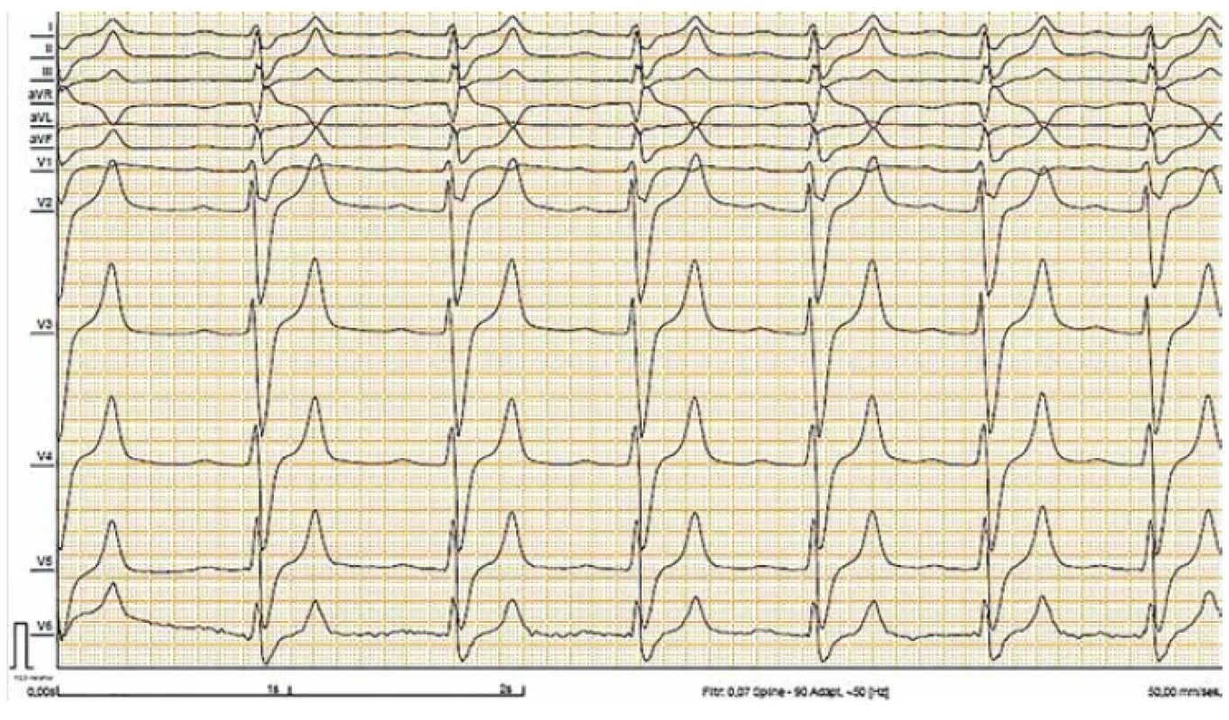
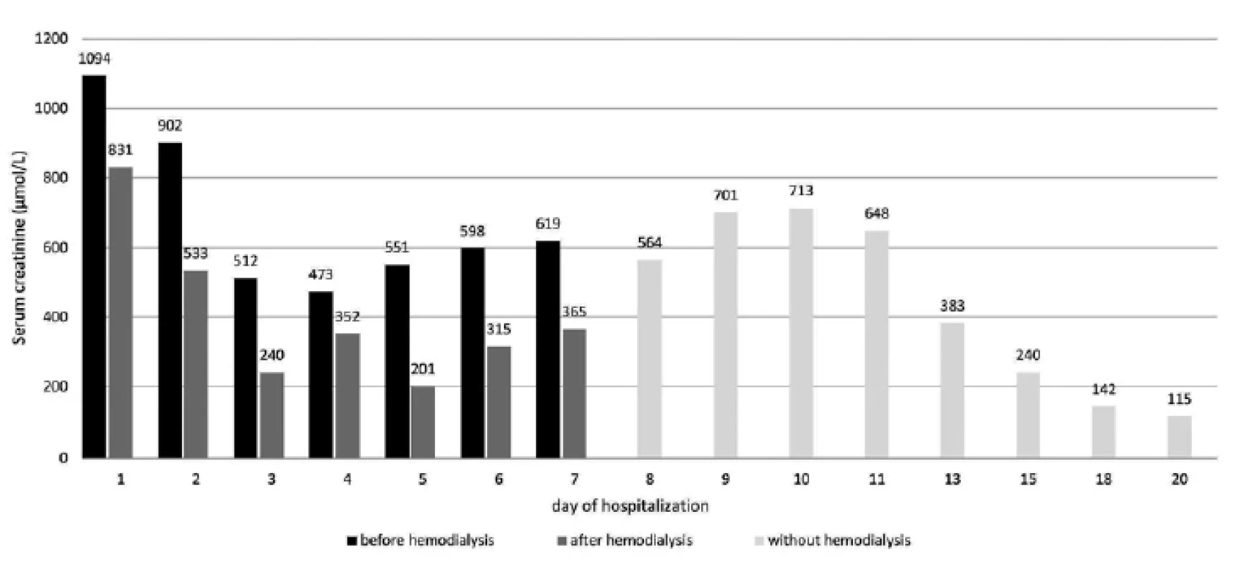
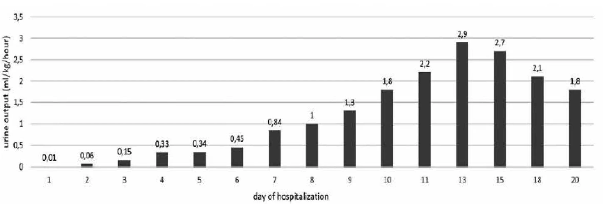
An electrocardiogram performed immediately after admission revealed changes typical of severe hyperkalemia (tall, peaked T waves and widened QRS complexes) (Figure 1). To treat hyperkalemia we used calcium gluconate, inhaled β-agonist salbutamol, and intravenous sodium bicarbonate. This approach dropped the blood serum potassium to 5.9 mmol/L. A permanent urinary catheter was inserted to accurately measure urine output. Abdomen ultrasound revealed slightly increased echogenicity of the kidney parenchyma. After insertion of a central venous catheter, intermittent hemodialysis (IHD) was initiated. Enzyme immunoassay (EIA) of stool sample was positive for C. difficile antigen but negative for toxins. However, genes for toxins A and B of C. difficile were the only positives on the gastrointestinal (GIT) panel of patient’s stool sample (a polymerase chain reaction test for detecting 22 common gastrointestinal pathogens). According to this finding, we indicated antibiotic treatment with fidaxomicin, a bactericidal macrolide, for 10 days. We continued IHD for the next 7 days. Azotemia was improving and urine output was gradually increasing. Development of serum creatinine levels and urine output are shown in Figure 2 and Figure 3. IHD was well tolerated in the patient and no side effects occurred. We started oral nutrition slowly as soon as blood disappeared from the stool. It was possible to discontinue the IHD after 1 week. After rising transiently, renal parameters started to decline spontaneously. The boy was discharged after 20 days of hospitalization. At 6-month follow-up, the kidneys were functioning normally and he is without any complaints.
Fig. 1. ECG changes in severe hyperkalemia 
Fig. 2. Development of serum creatinine level during hospitalization 
Fig. 3. Development of urine output during hospitalization 
DISCUSSION
AGE is defined by three or more diarrheal stools within 24 h. This common disease of childhood is usually caused by a viral, bacterial, or parasitic infection. Typical signs are rapid onset of complaints, nausea, vomiting, fever, abdominal pain, and diarrhea. Even though in the vast majority of cases the course is self-limited [7], complications can occur. Dehydration is the main sign of AGE complications. It is caused by a loss of water and essential salts and minerals that potentially lead to AKI. A study by Marzuillo et al. showed that duration of symptoms prior to hospitalization as anamnestic, dehydration > 5% as clinical, and serum bicarbonate levels as biochemical parameters were significant and independent predictors for AKI in children hospitalized for AGE [8]. The very likely cause of AKI and severe course in our patient’s case was volume depletion (prerenal AKI) due to prolonged diarrhea and highly decreased fluid intake. Following severe azotemia, hyperkalemia and anuria were the indications for IHD to be started. We had to continue with IHD for 7 days until kidney function recovery.
One of the less common etiological agents of AGE in children is C. difficile. This Gram-positive, spore-forming, and toxin-producing bacterium is a common cause of health care-associated infectious gastroenteritis in adults. Deshpande et al. found an increasing trend also in the incidence of pediatric CDI [9]. These data suggest greater effort should be made to prevent and treat this infection in children.
The ability of C. difficile to cause colitis depends on a range of virulence factors. The pathogenetic effects are mainly secondary to the production of toxin A and toxin B [10]. These exotoxins primarily target intestinal epithelial cells in the intestinal tract. Disruption in the actin cytoskeleton and loss of epithelial barrier integrity leads to apoptosis and tissue damage resulting in significant inflammation [11]. The production of toxins was not detected in our patient. We only identified the genes for toxins by the GIT PCR panel. These findings raise the question whatever CDI was the cause of the condition. We have to admit that C. difficile colonisation could be the possible answer and we just failed in detection of the accurate pathogen. In addition, C. difficile is not a typical food-borne disease.
Testing for C. difficile should not be routinely performed in children younger than 2 years due to high rates (9%–37%) of asymptomatic colonization [12]. In contrast, testing is recommended for older children with prolonged or worsening diarrhea and risk factors (e.g. immunocompromising conditions, underlying inflammatory bowel disease) or relevant exposures (e.g. contact with the healthcare system or recent antibiotics) [13]. We think that consumed a burger was the source of C. difficile in our patient who had no specific predispositions to CDI.
According to current recommendations we used fidaxomicin for the treatment of CDI in our patient [14]. This antibiotic treatment was effective and well tolerated.
Even though the boy is without any complaints and the kidneys are working normally, he remains in our follow-up. It has been proven that episodes of AKI requiring RRT in the setting of an intensive care unit are associated with increased mortality 1 year after discharge and an increased requirement for renal replacement even 18 months after discharge [15].
CONCLUSION
Our pediatric patient presented with severe AKI following C. difficile-associated diarrhea. He was successfully treated with specific antibiotics and hemodialysis. The patient’s general condition improved, azotemia gradually resolved, and the patient could be discharged after full recovery 20 days following admission. With increasing incidence of CDI, we should remember the possible complications of this potentially life-threatening disease.
Do redakce došlo dne 27. 2. 2022.
Adresa pro korespondenci:
MUDr. Jan Papež
Pediatrická klinika FN Brno
Jihlavská 20
625 00 Brno
e-mail: papez.jan@fnbrno.czEpidemiol Mikrobiol Imunol, 2022; 71(4): 208–211
Zdroje
1. Elliott EJ. Acute gastroenteritis in children. BMJ, 2007;334(7583): 35–40.
2. Whyte DA, Fine RN. Acute renal failure in children. Pediatr Rev., 2008;29(9):299–306; quiz –7.
3. Rupnik M, Wilcox MH, Gerding DN. Clostridium difficile infection: new developments in epidemiology and pathogenesis. Nat Rev Microbiol., 2009;7(7):526–536.
4. Cohen SH, Gerding DN, Johnson S, et al. Clinical practice guidelines for Clostridium difficile infection in adults: 2010 update by the society for healthcare epidemiology of America (SHEA) and the infectious diseases society of America (IDSA). Infect Control Hosp Epidemiol., 2010;31(5):431–455.
5. Kim J, Shaklee JF, Smathers S, et al. Risk factors and outcomes associated with severe clostridium difficile infection in children. Pediatr Infect Dis J., 2012;31(2):134–138.
6. Spivack JG, Eppes SC, Klein JD. Clostridium difficile-associated diarrhea in a pediatric hospital. Clin Pediatr (Phila), 2003;42(4):347–352.
7. Musil V, Homola L, Vrba M, et al. Clinical and microbiological characteristics of Clostridium difficile infection in children hospitalized at the Departement of Paediatric Infectious Diseases in Brno between 2013 and 2017. Epidemiol Mikrobiol Imunol., 2019;68(1):15–22.
8. Marzuillo P, Baldascino M, Guarino S, et al. Acute kidney injury in children hospitalized for acute gastroenteritis: prevalence and risk factors. Pediatr Nephrol., 2021;36(6):1627–1635.
9. Deshpande A, Pant C, Anderson MP, et al. Clostridium difficile infection in the hospitalized pediatric population: increasing trend in disease incidence. Pediatr Infect Dis J., 2013;32(10):1138–1140.
10. Leffler DA, Lamont JT. Clostridium difficile Infection. N Engl J Med., 2015;373(3):287–288.
11. Aktories K, Schwan C, Jank T. Clostridium difficile Toxin Biology. Annu Rev Microbiol., 2017;71 : 281–307.
12. Jangi S, Lamont JT. Asymptomatic colonization by Clostridium difficile in infants: implications for disease in later life. J Pediatr Gastroenterol Nutr., 2010;51(1):2–7.
13. McDonald LC, Gerding DN, Johnson S, et al. Clinical Practice Guidelines for Clostridium difficile Infection in Adults and Children: 2017 Update by the Infectious Diseases Society of America (IDSA) and Society for Healthcare Epidemiology of America (SHEA). Clin Infect Dis., 2018;66(7):987–994.
14. Wolf J, Kalocsai K, Fortuny C, et al. Safety and Efficacy of Fidaxomicin and Vancomycin in Children and Adolescents with Clostridioides (Clostridium) difficile Infection: A Phase 3, Multicenter, Randomized, Single-blind Clinical Trial (SUNSHINE). Clin Infect Dis., 2020;71(10):2581–2588.
15. Mizera L, Dürr MM, Rath D, et al. Long-term outcome after dialysis - dependent renal failure on the intensive care unit. Med Klin Intensivmed Notfmed., 2021;116(7):570–577.
Štítky
Hygiena a epidemiologie Infekční lékařství Mikrobiologie
Článek Rejstřík
Článek vyšel v časopiseEpidemiologie, mikrobiologie, imunologie
Nejčtenější tento týden
2022 Číslo 4- Stillova choroba: vzácné a závažné systémové onemocnění
- Jak souvisí postcovidový syndrom s poškozením mozku?
- Perorální antivirotika jako vysoce efektivní nástroj prevence hospitalizací kvůli COVID-19 − otázky a odpovědi pro praxi
-
Všechny články tohoto čísla
- Carriage of Neisseria meningitidis in newly enlisted young military professionals during the COVID-19 pandemic
- Comparing the epidemiological situation of selected sexually transmitted infection in three Czech regions between 2006 and 2013
- Molecular genotyping of Streptococcus agalactiae isolates with non-typeable serotype, Czech Republic, 2008–2020
- Acute kidney injury requiring renal replacement therapy due to Clostridioides difficile infection in a 15-year-old boy
- Pečenkovy epidemiologické dny, Plzeň 2022
- 100 let od narození RNDr. Evy Aldové, CSc.
- 100 let od narození doc. MUDr. Josefa Pečenky, DrSc.
- Rejstřík
- Epidemiologie, mikrobiologie, imunologie
- Archiv čísel
- Aktuální číslo
- Informace o časopisu
Nejčtenější v tomto čísle- Carriage of Neisseria meningitidis in newly enlisted young military professionals during the COVID-19 pandemic
- Acute kidney injury requiring renal replacement therapy due to Clostridioides difficile infection in a 15-year-old boy
- Molecular genotyping of Streptococcus agalactiae isolates with non-typeable serotype, Czech Republic, 2008–2020
- Comparing the epidemiological situation of selected sexually transmitted infection in three Czech regions between 2006 and 2013
Kurzy
Zvyšte si kvalifikaci online z pohodlí domova
Autoři: prof. MUDr. Vladimír Palička, CSc., Dr.h.c., doc. MUDr. Václav Vyskočil, Ph.D., MUDr. Petr Kasalický, CSc., MUDr. Jan Rosa, Ing. Pavel Havlík, Ing. Jan Adam, Hana Hejnová, DiS., Jana Křenková
Autoři: MUDr. Irena Krčmová, CSc.
Autoři: MDDr. Eleonóra Ivančová, PhD., MHA
Autoři: prof. MUDr. Eva Kubala Havrdová, DrSc.
Všechny kurzyPřihlášení#ADS_BOTTOM_SCRIPTS#Zapomenuté hesloZadejte e-mailovou adresu, se kterou jste vytvářel(a) účet, budou Vám na ni zaslány informace k nastavení nového hesla.
- Vzdělávání


