-
Články
- Vzdělávání
- Časopisy
Top články
Nové číslo
- Témata
- Kongresy
- Videa
- Podcasty
Nové podcasty
Reklama- Kariéra
Doporučené pozice
Reklama- Praxe
Submucosal calcifying fibrous tumor of the stomach: A case report
Submukózny kalcifikujúci fibrózny tumor žalúdka – kazuistika
Kalcifikujúci fibrózny tumor žalúdka je zriedkavý benígny fibrózny tumor, ktorý vzniká v podkožných alebo hlbokých mäkkých tkanivách u detí a mladých dospelých, avšak možno sa s ním stretnúť aj u starších ľudí, najmä v oblasti pleury alebo peritoneálnej dutiny. Z literatúry sú známe len ojedinelé prípady výskytu v gastrointestinálnom trakte, respektíve v žalúdku. V práci prezentujeme našu skúsenosť s touto neobvyklou léziou objavenou u 68 ročnej ženy. Klinicky bol zistený pendulujúci, submukózne uložený tumor malej kurvatúry žalúdka. Histologicky je kalcifikujúci fibrózny tumor jedinečná lézia, ktorú tvoria denzné hyalinizované kolagénové vlákna, nevýrazné roztrúsené fibroblasty, rôzne množstvo psamomatóznych teliesok alebo dystrofických kalcifikátov a ložiská lymfoplazmocytárnej infiltrácie. Naša práca je zameraná na krátky prehľad a diferenciálnu diagnostiku kalcifikujúceho fibrózneho tumoru a iných známejších alebo naopak nezvyčajných vretenobunkových lézií žalúdka.
Kľúčové slová:
kalcifikujúci fibrózny tumor – vretenobunkové lézie – žalúdok
Authors: Magdaléna Štofíková 1,2; Boris Rychlý 1; Jozef Bocko 1,2; Dušan Daniš 1,2
Authors place of work: Cytopathos, Ltd., Bratislava, Slovakia 1; Department of Pathological Anatomy, Faculty of Medicine, Slovak Medical University, Bratislava, Slovakia 2
Published in the journal: Čes.-slov. Patol., 52, 2016, No. 3, p. 164-167
Category: Původní práce
Summary
The calcifying fibrous tumor is a rare benign fibrous tumor which occurs in subcutaneous or deep soft tissues in children and young adults, but also is frequently seen in pleural and intraabdominal locations in older people. Gastric involvement has been only sporadically reported in the literature. We present here our experience with this unusual lesion discovered in a 68-year-old woman. Clinically, the tumor was described as a pendulating, submucosally located mass, in the body of the stomach on a lesser curvature. The calcifying fibrous tumor is a histologically distinct lesion composed of dense hyalinized collagen fibers, inconspicuous scattered fibroblasts, a varying amount of psammoma bodies or dystrophic calcifications and foci of lymphoplasmacytic infiltration. In this report we will focus on a brief review and differential diagnosis of this tumor and other more common or not widely known gastric spindle cell lesions.
Keywords:
calcifying fibrous tumor – spindle cell lesion – stomach
The calcifying fibrous tumor (CFT) is a rare benign fibrous tumor which has a wide anatomical distribution. Originally thought to represent soft tissue lesions in children and young adults, it is also not infrequently seen in pleural or intraabdominal locations in older people (1-5). Gastric involvement has been only sporadically reported in the literature - tumors have been described under the serosal surface or as intrinsic visceral lesions (3-4,6-21). Clinical symptoms and imaging findings of gastric CFT are nonspecific (22). The tumor is usually found incidentally, in the form of a polyp or it can manifest with an ulceration (6-21). Final diagnosis is made by pathologist. Histologically, CFT is composed of dense hyalinized collagen fibers, inconspicuous scattered fibroblasts, a varying amount of psammoma bodies or dystrophic calcifications and foci of lymphoplasmacytic infiltration. Despite its characteristic microscopic appearance, occurrence of CFT in unusual anatomic sites, as in the gastric submucosa, it requires careful assessment and differentiation from other more common or not widely known spindle cell lesions.
CASE REPORT
We present our experience with this unusual lesion discovered in a 68-year-old woman. A CT scan of the abdomen showed a polypoid submucosally located mass lying in the junction between the gastric antrum and body along the lesser curvature of the stomach. The tumor measured 3 cm in diameter and exhibited contrast enhancement. Furthermore, gastric wall infiltration was not evident (Fig. 1).
Fig. 1. Contrast enhanced CT scan of the abdomen demonstrating a submucosal mass (white arrow) in the junction between the gastric antrum and body along the lesser curvature of the stomach. 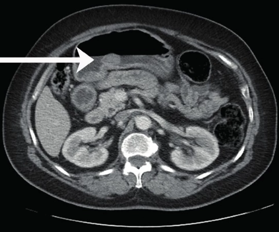
During gastrotomy, the tumor was found to be a pendulating nodule and treatment consisted of local tumor excision together with overlying mucosa. Unfortunately, no other clinical data are available. For the histological examination we received an elipsoid mass with smooth borders, partially covered with gastric mucosa, measuring 3.2 cm in the greatest dimension, on the cut surface whitish to grey, firm and homogenous. The whole specimen was routinely processed and stained with hematoxylin-eosin (HE). Immunohistochemistry and DNA analysis of the c-kit gene and PDGFRA gene was performed. The list of used antibodies, the source, clones and dilutions are shown in Table 1. Histologically, below an intact oxyntic gastric mucosa in the submucosa we could see a sharply demarcated, but unencapsulated lesion (Fig. 2) consisting of sclerotic and collagenized hypocellular fibrous tissue with a somewhat whorled appearance (Fig. 3). Inconspicuous fibroblasts were evenly spaced and showed bland elongated to oval nuclei without any significant atypia or mitotic activity. Vascularity was focally relatively prominent, formed mainly by thin-walled vessels. The lesion was diffusely mildly infiltrated by lymphocytes and plasma cells with focal formation of lymphoid follicles without visible germinal centers and occasional perivascular infiltrates (Fig. 4). Calcifications in the form of psammoma bodies or small dystrophic focuses were notable throughout the lesion (Fig. 5). The muscularis propria layer was not identified in the histologic sections. Immunohistochemically, lesional cells were negative for CD117, DOG1, S100, SMA, HHF35, β-catenin, ALK1 and we noted only weak focal CD34 reactivity. Proliferative activity was close to zero in Ki67 labeling. No amyloid deposition was detected by thioflavin staining. DNA analysis of c-kit gene and PDGFRA gene did not reveal any mutation in c-kit exons 9, 11, 13, 17 or PDGFRA exons 12, 14 and 18. These findings allowed us to make a diagnosis of gastric CFT.
Fig. 2. Low power view of a well-circumscribed tumor located beneath intact gastric mucosa (HE, 40x). 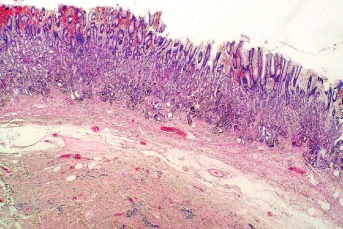
Fig. 3. Characteristic appearance of CFT - hyalinized hypocellular fibrous lesion with variably prominent lymphoplasmacytic infiltration and calcifications (van Giesonʼs stain, 100x). 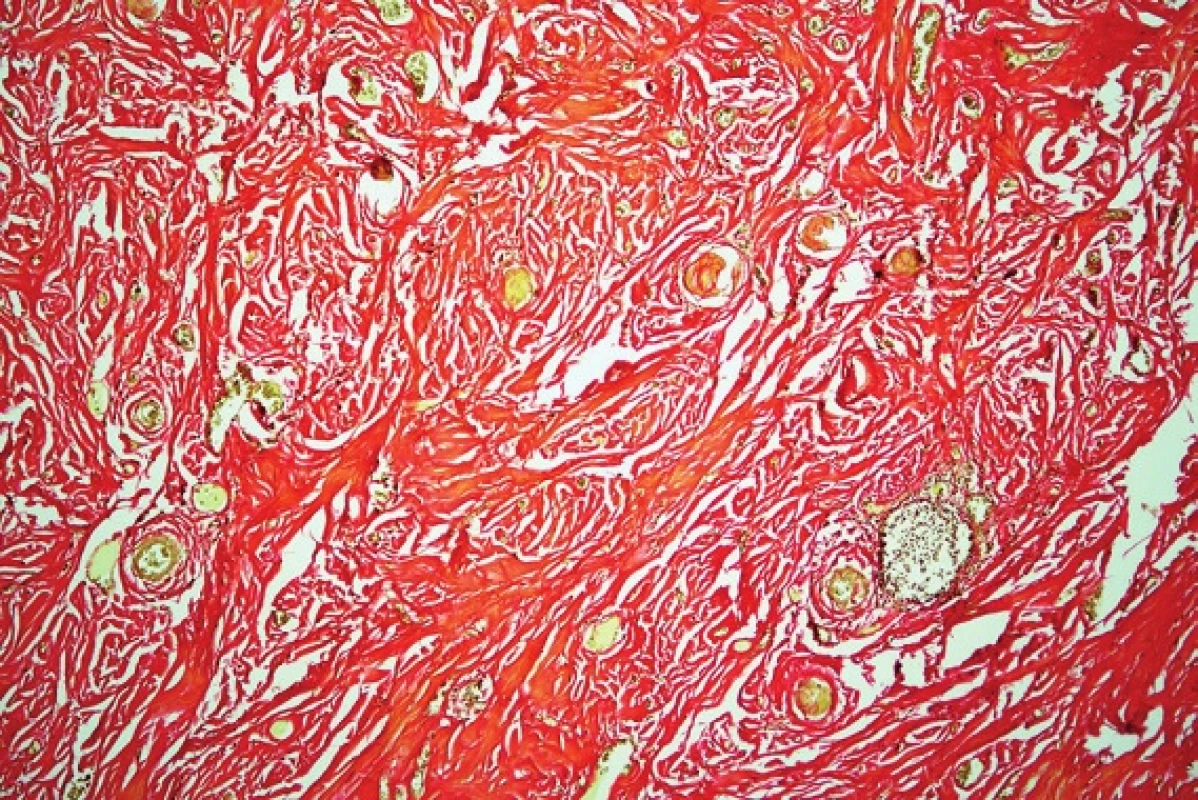
Fig. 4. Mononuclear inflammatory cells are mildly dispersed throughout the lesion and focally form lymphoid follicles (HE, 200x). 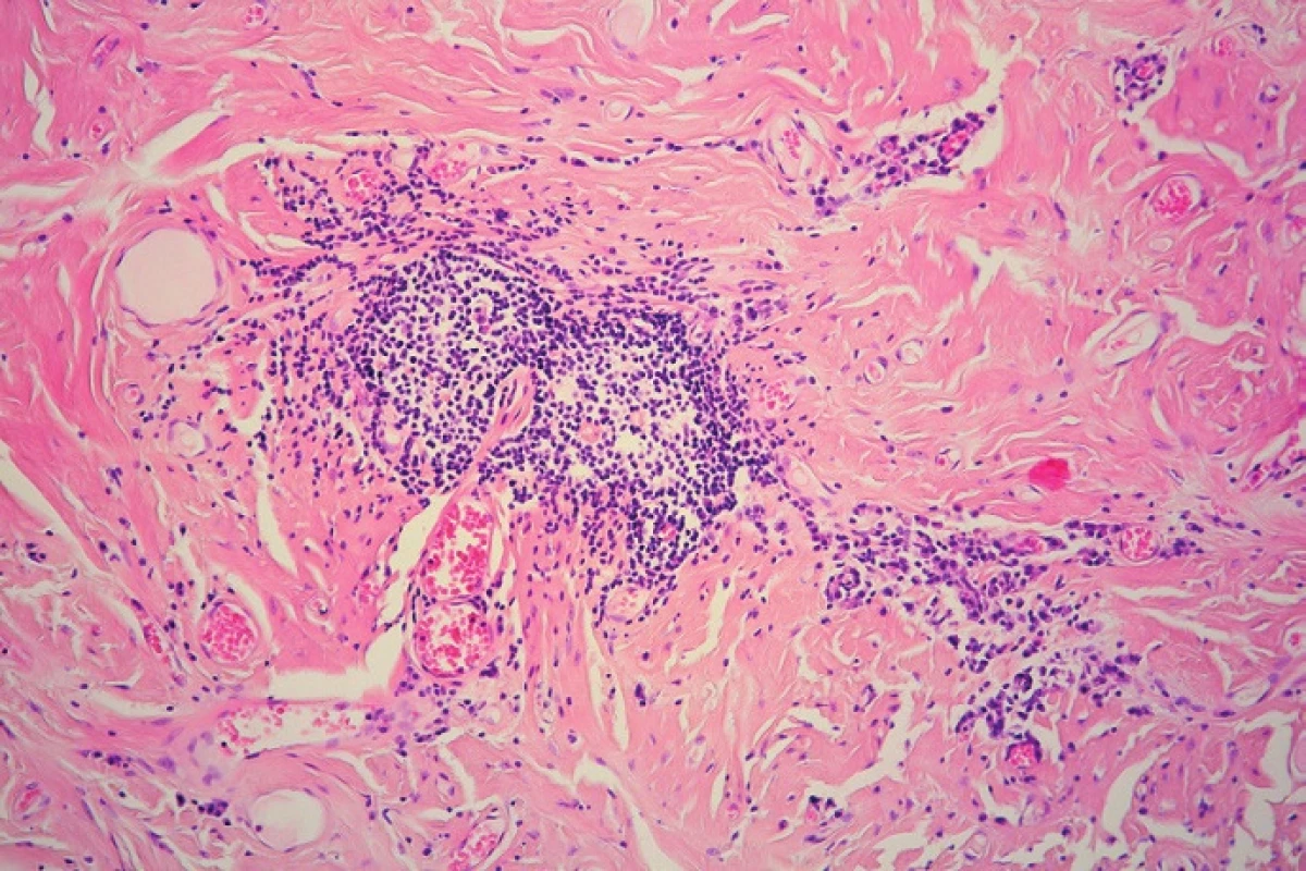
Fig. 5. Calcifications occur mostly in the form of psammoma bodies (HE, 200x). 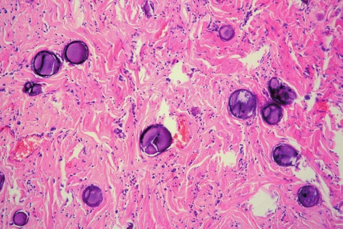
Tab. 1. The list of antibodies used. 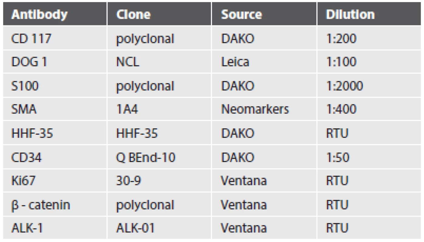
CD – cluster of differentiation, DOG 1 – discovered on GIST-1, SMA – smooth muscle actin, HHF-35 – muscle specific actin, ALK-1- anaplastic lymphoma kinase-1, RTU – ready to use DISCUSSION
CFT was firstly reported by Rosenthal and Abdul-Karim as a “childhood fibrous tumor with psammoma bodies” in 1988 (1). Later, in 1993, Fetsch et al. renamed it as calcifying fibrous pseudotumor and pointed out the probable reactive nature of the lesion (2). Nowadays, CFT is considered to represent a true neoplasm, because of the tendency of local recurrence which is occasionally reported and according to recent WHO classification it is named calcifying fibrous tumor (4,23). At present, ethiopathogenesis has not been yet fully understood. Association with trauma, Castleman disease or IgG4-related sclerosing disease has ben reported (21,24-26). Postulation that CFT represents a late sclerosing stage of inflammatory myofibroblastic tumor has not been confirmed (4,27-28). Authors of the largest study of gastric CFT to date (11) acknowledge that lesions may arise in the process of localized inflammatory fibrosclerosis in response to injury of the muscularis propria.
By careful searching in the English-language literature we found 27 reported cases of CFT affecting the gastric wall (4,6-21). Patient age ranged from 17 to 77 years (mean age 49.9, median 51) and men slightly outnumbered women (16 male to 11 female cases, ratio 1 : 0.7). The tumors reached an average diameter of 21.3 mm and were most often found incidentally. Some of them manifested with abdominal pain or more dramatically with bleeding from ulceration (6,12-13,18,21). CFT was usually endoscopically described as a solitary submucosal nodule with polypoid growth in various parts of the gastric body. Among all, only 4 CFTs were localized on the lesser curvature as presented in our case (7-9,16). Endoscopic ultrasonography can visualize well-demarcated hypoechoic or isoechoic mass within certain layer of gastric wall with internal hyperechoic foci (16,19). CT scans may identify homogeneous, well-circumscribed round lesions with or without contrast enhancement (12,14). Unfortunately, imaging features of CFT are not sufficiently distinctive to rule out a potentially malignant lesion, like gastrointestinal stromal tumor (GIST) or neuroendocrine tumor (10,16). Patients were treated by gastric wedge excision during laparoscopy or laparotomy. Some tumors were removed by newer and less invasive technique of endoscopic submucosal dissection or endoscopic full-thickness resection (10,16,19,21). The complete tumor removal is still regarded as an important aspect of successful treatment to prevent the recurrence and provide the tissue for a definitive histological diagnosis. Consistent with the view that CFT represents a part of the spectrum of IgG4-related sclerosing disease some authors suggest that treatment with steroids should be attempted before surgical resection (21). Gastric CFTs seem to behave in a benign fashion and none of the authors noticed any recurrence. As in other anatomic sites, metastatic spreading has not been reported yet (22).
Interestingly, some clinicopathological differences between soft tissue, peritoneal and gastric CFTs have been described. In comparison to soft tissue CFTs, gastric tumors are smaller, occur in those of an older age and do not recur (11). Peritoneal lesions are commonly multifocal with a predominance in females (11). Gross appearance of gastric CFTs is quite typical, they have a spherical to lobulated shape, a firm consistency and a white to grey cut surface (22).
Immunohistochemical examination typically reveals positivity for vimentin. Among the other investigated markers, CD34 and factor XIIIa are variably positive and markers such as smooth muscle actin, desmin, HHF-35, S100, CD117 and ALK1 are usually negative (3,7,11,28).
Differential diagnosis of CFT in gastric locations comprises other more common spindle cell lesions, including GIST, schwannoma or leiomyoma. Immunohistochemistry can be diagnostically helpful according to their possible morphological resemblance to CFT. GISTs can undergo regression with sclerosis and dystrophic calcification, however they do not contain psammoma bodies or lymphoplasmacytic infiltrates and neoplastic cells coexpress CD117, CD34 and DOG1 (11,29). Gastric schwannomas may be hyalinized and are at least partially surrounded by a lymphoid cuff which sometimes extends into the tumor tissue, but they show diffuse and strong S100 positivity (22). Leiomyomas can be paucicellular with extensive sclerosis and focal calcification, especially in larger tumors, but still should include smooth muscle cells exhibiting muscle marker positivity. Other lesions that could also be taken into account when CFT diagnosis is considered are inflammatory myofibroblastic tumors (IMT), inflammatory fibroid polyps (IFP), solitary fibrous tumors (SFT), reactive nodular fibrous pseudotumors (RNFP) or amyloidoma. IMT arises mainly in young patients, predominantly in the abdomen and may contain stromal calcification or heavily hyalinized stroma. Commonly, it harbors a prominent inflammatory infiltrate composed of plasma cells, lymphocytes and eosinophils dispersed in a proliferation of myofibroblasts, which are often ALK1 and SMA positive (4,28). IFP is a submucosa-based lesion with varying morphological features. Classically, it is composed of predominantly eosinophilic inflammatory infiltration and stromal cells expressing CD34, growing in an onion-shaped pattern around prominent blood vessels. Mutations in the PDGFR-A gene exons 12 and 18 have been implicated as a key actor in molecular pathogenesis of IFP (30). SFT, although extremely rare in the stomach, may mimic CFT depending on the degree of cellularity and hyalinization. In most cases, SFT has a distinctive hemangiopericytic vasculature, shows CD34 and STAT6 expression and calcification or inflammation are not prominent features (22,31). RNFP represents a very rare reactive benign lesion originating probably from multipotential subserosal cells. Typically, it arises in connection with the peritoneal surface in intestinal locations, but a case of gastric RNFP has been documented (32,33). Histologically it consists of paucicellular or moderately cellular proliferation of stellate or spindle cells arranged in poorly formed fascicles in a dense collagenous background, showing positivity for SMA and variable reactivity for cytokeratins AE1/AE3 and CD117 (32,33). Amyloid deposition can rarely form a mass lesion and thus mimic abundant pinkish stroma of CFT, but staining for amyloid is negative in CFT (2).
In conclusion, CFT is a rare, but histologically and immunohistochemically characteristic entity which can affect many different body sites, including the gastric wall. Despite its benign nature it should be considered in the differential diagnosis of wide spectrum of fibrous or spindle cell lesions of the stomach.
CONFLICT OF INTEREST
The authors declare that there is no conflict of interest regarding the publication of this paper.
Correspondence address:
MUDr. Magdaléna Štofíková
Cytopathos, Ltd.
Limbová 5, 83307 Bratislava, Slovakia
tel.: +421 907760058, fax: +421 232660571
e-mail: stofikova@cytopathos.sk
Zdroje
1. Rosenthal NS, Abdul-Karim FW. Childhood fibrous tumor with psammoma bodies. Clinicopathologic features in two cases. Arch Pathol Lab Med 1988; 112(8): 798–800.
2. Fetsch JF, Montgomery EA, Meis JM. Calcifying fibrous pseudotumor. Am J Surg Pathol 1993; 17(5): 502–508.
3. Kocová L, Michal M, Šulc M, Zámečník M. Calcifying fibrous pseudotumour of visceral peritoneum. Histopathology 1997; 31(2): 182–184.
4. Nascimento AF, Ruiz R, Hornick JL, Fletcher CDM. Calcifying fibrous “pseudotumor”: clinicopathologic study of 15 cases and analysis of its relationship to inflammatory myofibroblastic tumor. Int J Surg Pathol 2002; 10(3): 189–196.
5. Lee D, Haam SJ, Choi S, Park CH, Kim TH. Calcifying fibrous tumor of the pleura: a rare case with an unusual presentation on CT and MRI. J Korean Soc Radiol 2015; 72(2): 123-127.
6. Puccio F, Solazzo M, Marciano P, Benzi F. Laparoscopic resection of calcifying fibrous pseudotumor of the gastric wall: a unique case report. Surg Endosc 2001; 15(10): 1227.
7. Delbecque K, Legrand M, Boniver J, Lauwers GY, de Leval L. Calcifying fibrous tumour of the gastric wall. Histopathology 2004; 44(4): 399-400.
8. Attila T, Chen D, Gardiner GW, Ptak TW, Marcon NE. Gastric calcifying fibrous tumor. Can J Gastroenterol 2006; 20(7): 487–489.
9. Elpek GO, Küpesiz GY, Ögüs M. Incidental calcifying fibrous tumor of the stomach presenting as a polyp. Pathol Int 2006; 56(4): 227–231.
10. Sato S, Ooike N, Yamamoto T, et al. Rare gastric calcifying fibrous pseudotumor removed by endoscopic submucosal dissection. Dig Endosc 2008; 20(2): 84–86.
11. Agaimy A, Bihl MP, Tornillo L, Wünsch PH, Hartmann A, Michal M. Calcifying fibrous tumor of the stomach: clinicopathologic and molecular study of seven cases with literature review and reappraisal of histogenesis. Am J Surg Pathol 2010; 34(2): 271–278.
12. Fan SF, Yang H, Li Z, Teng GJ. Gastric calcifying fibrous pseudotumour associated with an ulcer: report of one case with a literature review. Br J Radiol 2010; 83(993): e188–e191.
13. Abbadessa B, Narang R, Mehta R, Martinez J, Leitman IM, Karpeh Jr M. Laparoscopic resection of a gastric calcifying fibrous pseudotumor presenting with ulceration and hematemesis in a teenage patient: a case report and review of the literature. J Surg Radiol 2012; 3(1): 44-47.
14. Jang KY, Park HS, Moon WS, Lee H, Kim CY. Calcifying fibrous tumor of the stomach: a case report. J Korean Surg Soc 2012; 83(1): 56–59.
15. Vasilakaki T, Skafida E, Tsavari A, et al. Gastric calcifying fibrous tumor: a very rare case report. Case Rep Oncol 2012; 5(2): 455–458.
16. Ogasawara N, Izawa S, Mizuno M, et al. Gastric calcifying fibrous tumor removed by endoscopic submucosal dissection. World J Gastrointest Endosc 2013; 5(9): 457-460.
17. Krishnamurthy J, Sanjeeva SM, Mudra KL, Muralidhara ND, Uthaiah S, Kashyap A. Submucosal calcifying fibrous tumor of stomach: a rare case report. World J Pathol 2014; 3 : 53-56.
18. Murdock T, Lee A, Wilcox R. An unusual cause of upper gastrointestinal bleeding. Clin Gastroenterol Hepatol 2014; 12(10): e95–e96.
19. Shi Q, Xu MD, Chen T, et al. Endoscopic diagnosis and treatment of calcifying fibrous tumors. Turk J Gastroenterol 2014; 25(Suppl 1): 153-156.
20. George SA, Abdeen S. Gastric calcifying fibrous tumor resembling gastrointestinal stromal tumor: A Case Report. Iran J Pathol 2015; 10(4): 306-309.
21. Zhang H, Jin Z, Ding S. Gastric calcifying fibrous fumor: a case of suspected immunoglobulin G4-related gastric disease. Saudi J Gastroenterol 2015; 21(6): 423–426.
22. Larson BK, Dhall D. Calcifying Fibrous Tumor of the Gastrointestinal Tract. Arch Pathol Lab Med 2015; 139(7): 943–947.
23. Fletcher CDM, Bridge JA, Hogendoorn PCW, Mertens F (Eds.): WHO Classification of Tumors of Soft Tissue and Bone. Lyon: IARC; 2013 : 69.
24. Zámečník M, Dorociak F, Veselý L. Calcifying fibrous pseudotumor after trauma. Pathol Int 1997; 47(11): 812.
25. Azam M, Husen YA, Pervez S. Calcifying fibrous pseudotumor in association with hyaline vascular type Castleman’s disease. Indian J Pathol Microbiol 2009; 52(4): 527–529.
26. Kuo TT, Chen TC, Lee LY. Sclerosing angiomatoid nodular transformation of the spleen (SANT): clinicopathological study of 10 cases with or without abdominal disseminated calcifying fibrous tumors, and the presence of a significant number of IgG4+ plasma cells. Pathol Int 2009; 59(12): 844–850.
27. Van Dorpe J, Ectors N, Geboes K, D‘Hoore A, Sciot R. Is calcifying fibrous pseudotumor a late sclerosing stage of inflammatory myofibroblastic tumor? Am J Surg Pathol 1999; 23(3): 329–335.
28. Hill KA, Gonzalez-Crussi F, Chou PM. Calcifying fibrous pseudotumor versus inflammatory myofibroblastic tumor: a histological and immunohistochemical comparison. Mod Pathol 2001; 14(8): 784–790.
29. Agaimy A, Wünsch PH, Hofstaedter F, et al. Minute gastric sclerosing stromal tumors (GIST tumorlets) are common in adults and frequently show c-Kit mutations. Am J Surg Pathol 2007; 31(1): 113–120.
30. Liu TC, Lin MT, Montgomery EA, Singhi AD. Inflammatory fibroid polyps of the gastrointestinal tract: spectrum of clinical, morphologic, and immunohistochemistry features. Am J Surg Pathol 2013; 37(4): 586–592.
31. Doyle LA, Vivero M, Fletcher CDM, Mertens F, Hornick JL. Nuclear expression of STAT6 distinguishes solitary fibrous tumor from histologic mimics. Mod Pathol 2014; 27(3): 390-395.
32. Yantiss RK, Nielsen GP, Lauwers GY, Rosenberg AE. Reactive nodular fibrous pseudotumor of the gastrointestinal tract and mesentery: a clinicopathologic study of five cases. Am J Surg Pathol 2003; 27(4): 532–540.
33. Gauchotte G, Bressenot A, Serradori T, Boissel P, Plénat F, Montagne K. Reactive nodular fibrous pseudotumor: a first report of gastric localization and clinicopathologic review. Gastroenterol Clin Biol 2009; 33(12): 1076-1081.
Štítky
Patologie Soudní lékařství Toxikologie
Článek vyšel v časopiseČesko-slovenská patologie
2016 Číslo 3-
Všechny články tohoto čísla
- Novinky v patológii hlavy a krku
- S laureátkou Hlavovej ceny za rok 2016
- MONITOR aneb nemělo by vám uniknout, že...
- HPV-associated head and neck cancer: update and recommendations for practice
- New developments in molecular diagnostics of carcinomas of the salivary glands: “translocation carcinomas”
- Poorly differentiated sinonasal tract malignancies: A review focusing on recently described entities
- Observations of different patterns of dysplasia in barrett’s esophagus - a first step to harmonize grading
- Submucosal calcifying fibrous tumor of the stomach: A case report
- Case report: Diagnosis under the microscope - disseminated echninococcosis, the multilocular form with protoscoleces
- K životnému jubileu prof. MUDr. Štefana Kopeckého, PhD.
- Jaká je Vaše diagnóza?
- Lungs „Hassalloid´s-like“ bodies in children with epidermolysis bullosa junctionalis and bart´s syndrome
- Jaká je Vaše diagnóza? Odpověď
- Dr. h. c. prof. MUDr. Štefan Galbavý, DrSc. 70-ročný
- Česko-slovenská patologie
- Archiv čísel
- Aktuální číslo
- Informace o časopisu
Nejčtenější v tomto čísle- Dr. h. c. prof. MUDr. Štefan Galbavý, DrSc. 70-ročný
- HPV-associated head and neck cancer: update and recommendations for practice
- Poorly differentiated sinonasal tract malignancies: A review focusing on recently described entities
- Case report: Diagnosis under the microscope - disseminated echninococcosis, the multilocular form with protoscoleces
Kurzy
Zvyšte si kvalifikaci online z pohodlí domova
Autoři: prof. MUDr. Vladimír Palička, CSc., Dr.h.c., doc. MUDr. Václav Vyskočil, Ph.D., MUDr. Petr Kasalický, CSc., MUDr. Jan Rosa, Ing. Pavel Havlík, Ing. Jan Adam, Hana Hejnová, DiS., Jana Křenková
Autoři: MUDr. Irena Krčmová, CSc.
Autoři: MDDr. Eleonóra Ivančová, PhD., MHA
Autoři: prof. MUDr. Eva Kubala Havrdová, DrSc.
Všechny kurzyPřihlášení#ADS_BOTTOM_SCRIPTS#Zapomenuté hesloZadejte e-mailovou adresu, se kterou jste vytvářel(a) účet, budou Vám na ni zaslány informace k nastavení nového hesla.
- Vzdělávání


