-
Články
- Vzdělávání
- Časopisy
Top články
Nové číslo
- Témata
- Kongresy
- Videa
- Podcasty
Nové podcasty
Reklama- Kariéra
Doporučené pozice
Reklama- Praxe
Oncology in pictures
Authors: Yosra Yahyaoui 1; Olfa Adouni 2; Imen Harhira 1; Azza Gabsi 1; Fahmi Mghirbi 1; Maha Driss 2; Amel Mezlini 1
Authors place of work: Department of Medical Oncology, Institut Salah-Azaïz de Cancerologie, Tunis, Tunisia 1; Department of Anatomopathology, Institut Salah-Azaïz University of Tunis El Manar, Tunis, Tunisia 2
Published in the journal: Klin Onkol 2020; 33(3): 236-237
Complete Response of Classic Kaposi’s Sarcoma of the Larynx after Radiotherapy
Kaposi’s sarcoma (KS) is a neoplastic vascular disorder. Most frequently, KS arise in mucocutaneous sites, typically the skin of the lower extremities. Laryngeal involvement is not unusual. Approximately 25 cases of KS of the larynx have been recorded in the literature. Most patients have had AIDS-related KS, although HIV-negative persons with laryngeal KS have also been noted. Classic Kaposi’s sarcoma (CKS) is one of the four distinct clinical presentations of KS not related to immunodeficiency. We report a case of CKS of the larynx.
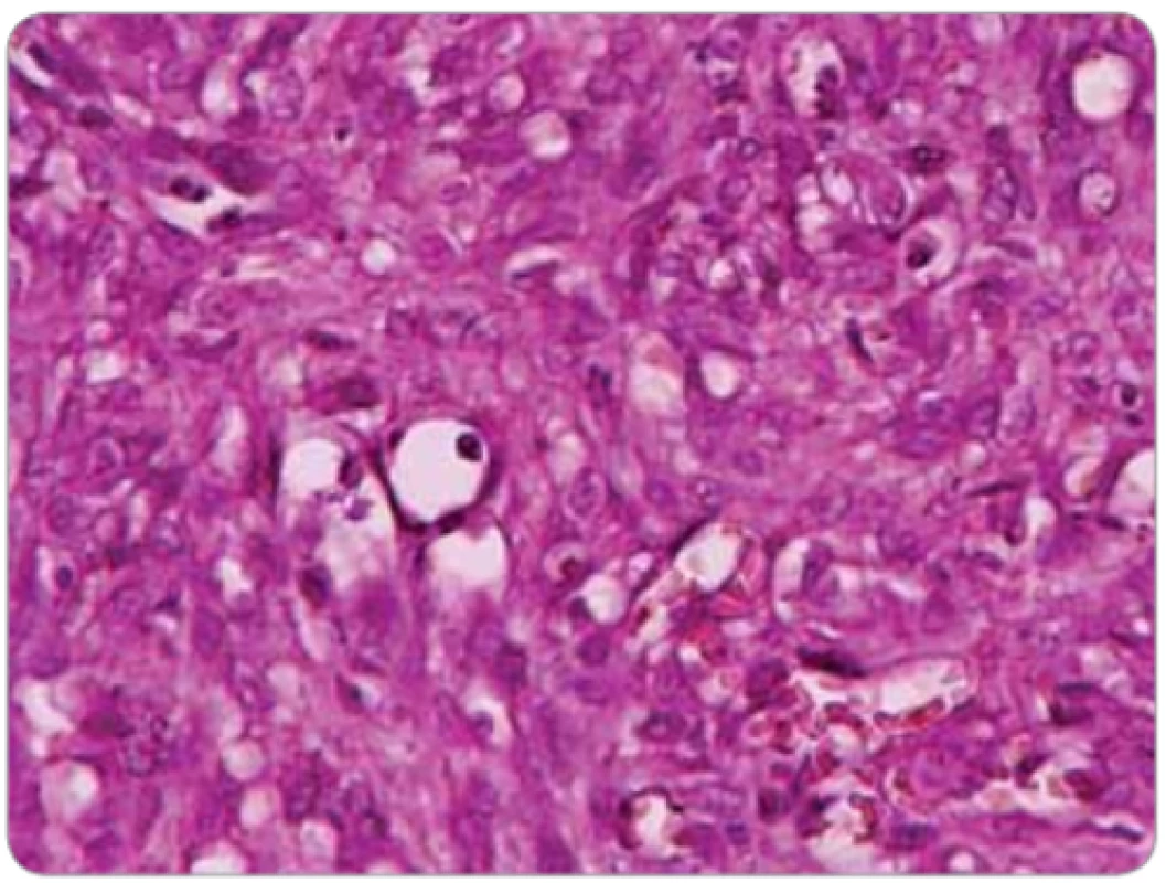
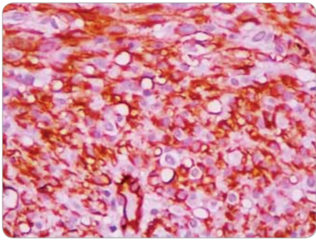
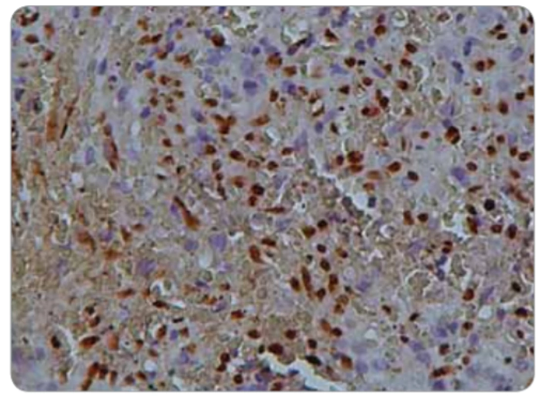
A 77-year-old man consulted with a 4-month history of dyspnea and dysphagia to solids. Nasofibroscopy revealed a globular laryngeal mass. Direct laryngoscopy showed a large globular tumefaction raising from the glottis region and fixing the left vocal cord. A biopsy of the mass was complicated by acute respiratory failure leading to emergency tracheotomy. Histological examination revealed a double mesenchymal and vascular proliferation pushing the intact lingual surface epithelium. The mesenchymal component was made of fascicular spindle cells and the vascular component, was made of dilated vessels with a thickened and hyalinized wall. Spindle cells have swollen nuclei with some cytonuclear atypia and few mitotic figures (Fig. 1). Immunohistochemical examination showed positive immunoreactivity for CD34 (Fig. 2) and human herpes virus 8 (HHV8) (Fig. 3). CD31 and factor VIII related-antigen were negative. No cutaneous manifestation was depicted and serology showed a positive HHV8 status not associated with HIV infection. Body CT didn’t show any secondary lesion. The patient was diagnosed with primary classic Kaposi’s sarcoma. He was treated with radiotherapy. A cobalt-60 external beam was employed. A daily dose of 1.8 or 2 Gy at the midplane was administered in 5 fr/week. The total radiation dose administered was 45 Gy over 5 weeks. At the time of finalization of this report, and 36 months post-treatment, the patient was still in complete response.
Fig. 1. Spindle cells with swollen nuclei with some cytonuclear atypia and few mitotic fi gures. 
Fig. 2. Positive immunoreactivity for CD34. 
Fig. 3. Positive immunoreactivity for human herpes virus 8. 
KS is a vascular lesion of a low grade of malignancy caused by HHV8 infection. It occurs in males in their 6th or 7th decades [1,2]. Four epidemiologic subtypes were identified: Classical type, African endemic KS, iatrogenic KS and AIDS related KS [1]. In 1872, Moritz Kaposi, a physician and dermatologist, described the CKS as a pigmented sarcoma of the skin in elderly European patients with HIV negative status [1]. Before the AIDS epidemic, KS was rarely seen in the head and neck region, especially in the larynx [3,4]. Abramson and Simonsin 1970 reported the largest review in the literature of CKS involving the head and neck. The incidence of laryngeal KS, in this review, was estimated to 17%. All patients with laryngeal KS had extremity skin lesions [5]. The first case report of CKS of larynx was documented in 1983 [6]. The management of KS depends on the extent of lesions and their evolution. Treatment of laryngeal CKS consists on systemic and local therapies. The local therapies as local excision, cryotherapy, radiotherapy and intralesional injections of vinblastine are usually used to reduce symptoms [7,8]. Radiotherapy is frequently used for ear, nose and throat KS with complete remission in about 85% of cases with a fractionated regimen to a total dose of 15–45 Gy. Systemic therapy is used for patients with rapidly progressive and/or wide spread disease [7,9]. The cytotoxic agents that have been used are vincristine, vinblastine, bleomycin, doxorubicin, etoposide, liposomal doxorubicin, liposomal daunorubicin, ABV regimen, as much as immunomodulation therapy with interferon-alpha [9]. Median survival for classic Kaposi sarcoma was 9.35 years [10]. Although CKS is not an aggressive malignancy, follow up is essential for the prevention of new KS lesions or locoregional recurrence.
Classic KS rarely involves larynx. It must be suspected in elderly patients. Early treatment has to be conducted as soon as possible to avoid sudden and or massive airway obstruction. CKS does not impair the quality of life and survival in the short term but aggressive forms may be life-threatening and should be treated urgently.
Dr. Yosra Yahyaoui
Department of Medical Oncology Institut Salah Azaïz Place Bab Saâdoune 1006 Jabberi Tunis Tunisia
e-mail: yosyahyaoui@gmail.com
Submitted/Obdrženo: 9. 2. 2020
Zdroje
1. Cesarman E, Damania B, Krown SE et al. Kaposi sarcoma. Nat Rev Dis Primers 2019; 5 (1): 9. doi: 10.1038/s41572-019-0060-9.
2. Shiels RA. A history of Kaposi‘s sarcoma. J R Soc Med 1986; 79 (9): 532–534. doi: 10.1177/014107688607900910.
3. Schiff NF, Annino DJ, Woo P et al. Kaposi‘s sarcoma of the larynx. Ann Otol Rhinol Laryngol 1997; 106 (7): 563–567. doi: 10.1177/000348949710600706.
4. Singh B, Harel G, Lucente FE. Kaposi‘s sarcoma of the head and neck in patients with acquired immunodeficiency syndrome. Otolaryngol Head Neck Surg 1994; 111 (5): 618–624. doi: 10.1177/019459989411100513.
5. Abramson AL, Simons RL. Kaposi‘s sarcoma of the head and neck. Arch Otolaryngol 1970; 92 (5): 505–507. doi: 10.1001/archotol.1970.04310050087014.
6. Coyas A, Eliadellis E, Anastassiades O. Kaposi‘s sarcoma of the larynx. J Laryngol Otol 1983; 97 (7): 647–649. doi: 10.1017/s0022215100094755.
7. Mohd Tahir J, Gopalan KN, Marina MB et al. A rare case of laryngeal Kaposi‘s sarcoma. Bangladesh Journal of Medical Science 2010; 9 (2): 107–109. doi: 10.3329/bjms.v9i2.5660.
8. Pantanowitz L, Dezube BJ. Kaposi sarcoma in unusual locations. BMC Cancer 2008; 8 : 190. doi: 10.1186/1471-2407-8-190.
9. Angouridakis N, Constantinidis J, Karkavelas G et al. Classic (Mediterranean) Kaposi‘s sarcoma of the true vocal cord: a case report and review of the literature. J Eur Arch Otorhinolaryngol 2006; 263 (6) : 537–40. doi: 10.1007/ s00405-006-0007-0.
10. Franceschi S, Arniani S, Balzi D et al. Survival of classic Kaposi‘s sarcoma and risk of second cancer. Br J Cancer 1996; 74 (11) : 1812–1814. doi: 10.1038/ bjc.1996.635.
Štítky
Dětská onkologie Chirurgie všeobecná Onkologie
Článek vyšel v časopiseKlinická onkologie
Nejčtenější tento týden
2020 Číslo 3- Metamizol jako analgetikum první volby: kdy, pro koho, jak a proč?
- Nejlepší kůže je zdravá kůže: 3 úrovně ochrany v moderní péči o stomii
- Metamizol v léčbě různých bolestivých stavů – kazuistiky
-
Všechny články tohoto čísla
- Editorial
- Levels of NT-proBNP and Troponin T in Cancer Patients – Mini-Review
- Importance of Aberrantly Activated Hedgehog/Gli Pathway in Tumour Progression
- Association of XPG rs17655G>C and XPF rs1799801T>C Polymorphisms with Susceptibility to Cutaneous Malignant Melanoma: Evidence from a Case-Control Study, Systematic Review and Meta-Analysis
- Assessment of Quality of Life in Patients with Head and Neck Cancer
- The Relationship of FOXR2 Gene Expression Profile with Epithelial-Mesenchymal Transition Related Markers in Epithelial Ovarian Cancer
- Current Possibilities of Early Detection of Cardiotoxicity of Cytostatic Treatment
- Prognostic Survival Factors of Hepatocellular Carcinoma Treated with Transarterial Chemoembolization
- Complete Response to Chemotherapy in Metastatic Pancreatic Carcinoma Associated with Double Heterozygous Germline Mutation in BRCA2 and CHEK2 Genes – a Case Report
- Long-Term Effect of Erlotinib Therapy in the Third Line of Anticancer Treatment in a Patient with Non-Small-Cell Lung Cancer – a Case Report
- Aktuality z odborného tisku
- Predstavujeme nové knihy
- Therapy with Trifluridine/Tipiracil and Regorafenib in Patientswith Pre-treated Metastatic Colorectal Cancer – Experience from the Czech Republic
- Oncology in pictures
- Klinická onkologie
- Archiv čísel
- Aktuální číslo
- Informace o časopisu
Nejčtenější v tomto čísle- Levels of NT-proBNP and Troponin T in Cancer Patients – Mini-Review
- Complete Response to Chemotherapy in Metastatic Pancreatic Carcinoma Associated with Double Heterozygous Germline Mutation in BRCA2 and CHEK2 Genes – a Case Report
- Assessment of Quality of Life in Patients with Head and Neck Cancer
- Importance of Aberrantly Activated Hedgehog/Gli Pathway in Tumour Progression
Kurzy
Zvyšte si kvalifikaci online z pohodlí domova
Autoři: prof. MUDr. Vladimír Palička, CSc., Dr.h.c., doc. MUDr. Václav Vyskočil, Ph.D., MUDr. Petr Kasalický, CSc., MUDr. Jan Rosa, Ing. Pavel Havlík, Ing. Jan Adam, Hana Hejnová, DiS., Jana Křenková
Autoři: MUDr. Irena Krčmová, CSc.
Autoři: MDDr. Eleonóra Ivančová, PhD., MHA
Autoři: prof. MUDr. Eva Kubala Havrdová, DrSc.
Všechny kurzyPřihlášení#ADS_BOTTOM_SCRIPTS#Zapomenuté hesloZadejte e-mailovou adresu, se kterou jste vytvářel(a) účet, budou Vám na ni zaslány informace k nastavení nového hesla.
- Vzdělávání


