-
Články
Top novinky
Reklama- Vzdělávání
- Časopisy
Top články
Nové číslo
- Témata
Top novinky
Reklama- Kongresy
- Videa
- Podcasty
Nové podcasty
Reklama- Kariéra
Doporučené pozice
Reklama- Praxe
Top novinky
ReklamaP21-Associated ncRNA DNA Damage-Activated Expression in Bladder Cancer
Exprese ncRNA spojené s P21 aktivovaná poškozením DNA u karcinomu močového měchýře
Úvod: Dlouhé nekódující ribonukleové kyseliny (long non-coding ribonucleic acids – lncRNA) jsou v poslední době vzhledem ke své úloze v procesu karcinogeneze předmětem zkoumání vědců zabývajících se nádory. Tyto transkripty regulují kritické kroky v normálních buněčných procesech, takže dysregulace jejich exprese se účastní patogeneze karcinomů. Z důvodu své blízkosti k lokusu CDKN1A má ncRNA spojená s P21 aktivovaná poškozením DNA (P21-associated ncRNA DNA damage activated – PANDA) v tomto ohledu zvláštní pozici. Podílí se na regulaci reakce na poškození DNA, stárnutí buněk a proliferace.
Materiály a metody: V této studii jsme metodou kvantitativní polymerázové řetězové reakce hodnotili expresi této lncRNA ve tkáních karcinomu močového měchýře, sousedních nerakovinných tkání (adjacent non-cancerous tissues – ANCT) a v normálních vzorcích močového měchýře.
Výsledky: Nebyl detekován žádný významný rozdíl v expresi PANDA, a to ani mezi nádorovými tkáněmi a ANCT (poměr exprese = 1,75; p = 0,11) nebo mezi nádorovými tkáněmi a normálními tkáněmi (poměr exprese = 2,72; p = 0,57). Úroveň exprese této lncRNA nebyla spojena s žádnými demografickými ani klinickými údaji o pacientech, jako je grade nádoru nebo recidiva, ani s rizikovými faktory souvisejícími s rakovinou, mezi něž patří např. kouření cigaret nebo závislost na opiu.
Závěr: Tato studie tedy naznačuje, že PANDA není zapojena do patogeneze karcinomu močového měchýře. Hodnocení exprese jiných lncRNA by mohlo pomoci při identifikaci biomarkerů pro tyto karcinomy.
Tato studie byla podpořena grantem univerzity Shahid Beheshti University of Medical Sciences.
Autoři deklarují, že v souvislosti s předmětem studie nemají žádné komerční zájmy.
Redakční rada potvrzuje, že rukopis práce splnil ICMJE kritéria pro publikace zasílané do biomedicínských časopisů.
Obdrženo: 27. 5. 2019
Přijato: 28. 7. 2019
Klíčová slova:
ncRNA spojená s P21 – aktivace poškozením DNA – PANDA – dlouhé nekódující – karcinomy močového měchýře
Authors: Feraydoon Abdolmaleki 1; Soudeh Ghafouri-Fard 1; Mohammad Taheri 2; Davood Mir Omrani 3
Authors place of work: Department of Medical Genetics, Shahid Beheshti University of Medical Sciences, Tehran, Iran 1; Urogenital Stem Cell Research Center, Shahid Beheshti University of Medical Sciences, Tehran, Iran 2; Urology and Nephrology Research Center, Shahid Beheshti University of Medical Sciences, Tehran, Iran 3
Published in the journal: Klin Onkol 2019; 32(4): 277-280
Category: Původní práce
doi: https://doi.org/10.14735/amko2019277Summary
Úvod: Dlouhé nekódující ribonukleové kyseliny (long non-coding ribonucleic acids – lncRNA) jsou v poslední době vzhledem ke své úloze v procesu karcinogeneze předmětem zkoumání vědců zabývajících se nádory. Tyto transkripty regulují kritické kroky v normálních buněčných procesech, takže dysregulace jejich exprese se účastní patogeneze karcinomů. Z důvodu své blízkosti k lokusu CDKN1A má ncRNA spojená s P21 aktivovaná poškozením DNA (P21-associated ncRNA DNA damage activated – PANDA) v tomto ohledu zvláštní pozici. Podílí se na regulaci reakce na poškození DNA, stárnutí buněk a proliferace.
Materiály a metody: V této studii jsme metodou kvantitativní polymerázové řetězové reakce hodnotili expresi této lncRNA ve tkáních karcinomu močového měchýře, sousedních nerakovinných tkání (adjacent non-cancerous tissues – ANCT) a v normálních vzorcích močového měchýře.
Výsledky: Nebyl detekován žádný významný rozdíl v expresi PANDA, a to ani mezi nádorovými tkáněmi a ANCT (poměr exprese = 1,75; p = 0,11) nebo mezi nádorovými tkáněmi a normálními tkáněmi (poměr exprese = 2,72; p = 0,57). Úroveň exprese této lncRNA nebyla spojena s žádnými demografickými ani klinickými údaji o pacientech, jako je grade nádoru nebo recidiva, ani s rizikovými faktory souvisejícími s rakovinou, mezi něž patří např. kouření cigaret nebo závislost na opiu.
Závěr: Tato studie tedy naznačuje, že PANDA není zapojena do patogeneze karcinomu močového měchýře. Hodnocení exprese jiných lncRNA by mohlo pomoci při identifikaci biomarkerů pro tyto karcinomy.
Keywords:
P21-associated ncRNA – DNA damage-activated – PANDA – long non-coding – urinary bladder neoplasms
Introduction
Long non-coding ribonucleic acids (lncRNAs) with sizes of more than 200 nucleotides constitute the main part of the human transcriptome. Although they do not encode proteins, they participate in multiple biological activities [1,2]. Dysregulation of their expression contributes to conferring malignant phenotypes in diverse tissues [3]. The lncRNA P21 associated ncRNA deoxyribonucleic acid damage activated (PANDA) gene location is near the cyclin-dependent kinase inhibitor 1A (CDKN1A) gene and is transcribed antisense to CDKN1A [4]. The expression of this lncRNA is induced in a p53-dependent fashion following deoxyribonucleic acid (DNA) damage. Its interaction with the transcription factor NF-YA leads to suppression of the expression of pro-apoptotic genes [4]. Furthermore, this lncRNA interacts with scaffold attachment factor A to recruit polycomb repressive complexes and inhibit the expression of senescence-enhancing genes [5]. Peng et al. have reported a lower expression of PANDA in hepatocellular carcinoma samples compared with peri-tumour tissues [6]. However, forced overexpression of this lncRNA has enhanced cell proliferation and tumourigenesis potential both in vitro and in vivo [6]. In the osteosarcoma cell line, PANDA stimulates G1-S transition and increases cell proliferation by suppressing p18 transcription [7]. Zhan et al. have demonstrated up-regulation of this lncRNA in bladder cancer tissue compared with the corresponding adjacent non-cancerous tissues (ANCTs) [8]. In addition, they reported positive correlations between PANDA over-expression and higher histological and advanced tumour, node, metastasis (TNM) stage [8]. Based on the inconsistency of data regarding the expression pattern of PANDA in diverse cancer types, we designed the current study to evaluate its expression in bladder cancer tissues, ANCTs and normal bladder tissues.
Materials and Methods
Study participants
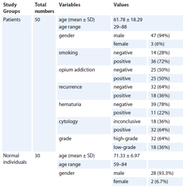
In the current study, we recruited 50 patients with histopathologically-defined bladder cancer. Both tumour tissue and ANCTs were excised during bladder surgery. The patients received no prior chemo/radiotherapy. Furthermore, 30 samples were excised from the bladder tissue of corpses to be used as controls. The individuals recruited as controls had no history of urogenital disease or cancer. Permission to use these tissues was obtained from the guardians of the deceased. The study protocol was approved by the ethical committee of the Shahid Beheshti University of Medical Sciences. All the patients signed written informed consent forms.
Assessment of PANDA expression
Total RNA was extracted from the tissue samples using TRIzol™ Reagent (Invitrogen, Carlsbad, California, USA). The quality of the RNA was assessed using a Thermo Scientific NanoDrop Spectrophotometer. The RNA purity was assessed by measuring the ratio of absorbance at 260 and 280 nm. Samples with ratios around 1.9 were regarded as acceptable. About 500 ng of RNA was converted to complementary DNA (cDNA) using a High-Capacity cDNA Reverse Transcription Kit (Applied Biosystems, USA) according to the manufacturer’s instructions. The expression levels of PANDA were compared between tumour tissues, ANCTs and control samples in a Rotor Gene 6000 Real-Time PCR Machine (Corbett, Australia) using a TaqMan® Universal PCR Master Mix (Applied Biosystems, USA). The HPRT1 gene was used as the endogenous control. The PCR programme included a preliminary step at 94 °C for 10 min, forty cycles of 94 °C for 20 sec and 60 °C for 40 sec and a final extension step at 72 °C for 5 min. The sequences of the primers and probes are shown in Tab. 1.
Tab. 1. The nucleotide sequence of primers and probes. 
Statistical analysis
The transcript levels of PANDA in tumour tissues were compared with ANCTs/controls using REST 2009 software. The significance of the difference in the expression of PANDA between the tumour tissues and the ANCTs/controls was evaluated using a t-test. The association between the clinical data and relative expression of PANDA was evaluated using a Chi-square test. P values of less than 0.05 were considered as significant.
Results
General information on the recruited persons
General information on the study participants has been summarised in Tab. 2.
Tab. 2. General information of study participants. 
SD – standard deviation Relative expression of PANDA in bladder cancer tissues, ANCTs and normal tissues
No significant difference has been detected in PANDA expression either between tumour tissues and ANCTs (expression ratio 1.75, P = 0.11) or between tumour tissues and normal tissues (expression ratio 2.72, P = 0.57) (Graph 1).
Graph 1. Relative expression of PANDA in bladder cancer tissues, ANCTs compared with normal tissues.
ANCT – adjacent non-cancerous tissues
Association between relative expression of PANDA in bladder cancer tissues and tumour features
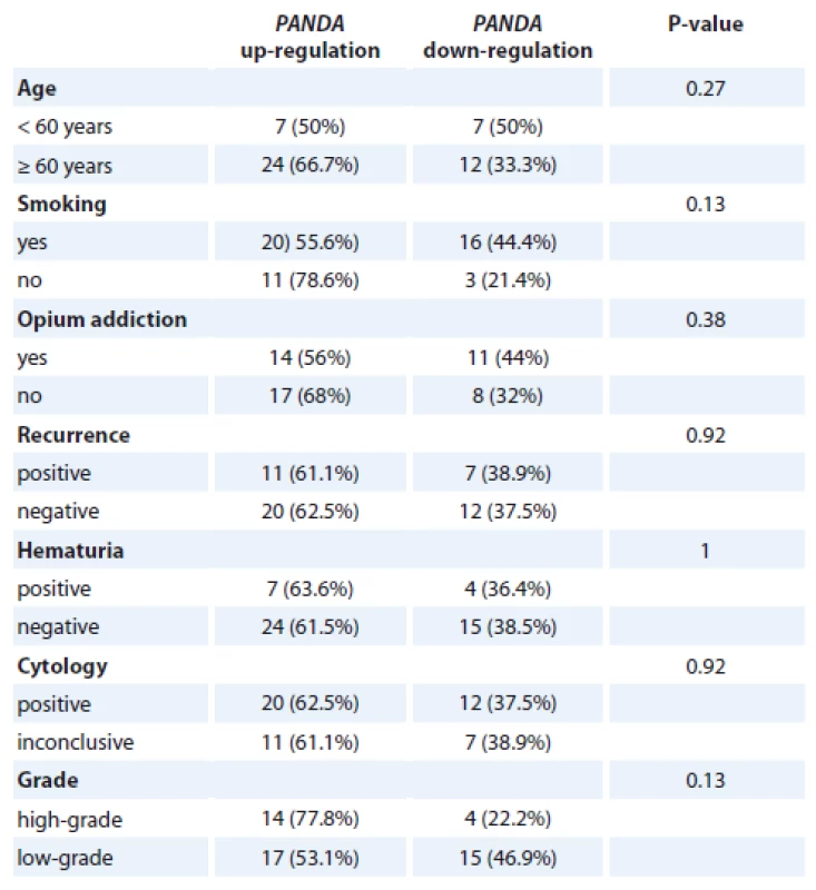
Based on the relative expression of PANDA in each tumour tissue compared with the corresponding ANCT, the patients were categorised into two groups (up-regulated vs. down-regulated). Subsequently, the associations between PANDA expression and clinicopathological features were assessed. The expression level of this lncRNA was not associated with any of the demographic or clinical data of patients or cancer-associated risk factors such as cigarette smoking or opium addiction (Tab. 3).
Tab. 3. Association between relative expression of PANDA in bladder cancer tissues and tumor features. 
Discussion
Bladder cancer is one of the most frequently occurring cancers worldwide [9]. Based on the lack of specific symptoms in the initial phases of bladder cancer evolution, diagnosis of this malignancy is delayed and subsequently the therapeutic options are less effective [10]. The need for identification of diagnostic biomarkers has prompted researchers to evaluate the expression of several genes in the tissues or body fluids of the patients [11,12]. LncRNAs are among the putative biomarkers and therapeutic targets for this kind of human malignancy [1].
In the present study, we assessed the expression levels of PANDA in three types of bladder tissues including normal, ANCT and tumour tissues and found no significant difference in its expression between these three sets of samples. This lncRNA has been suggested to be involved in a variety of human disorders including neuroinflammatory and malignant conditions [6,13]. Moreover, it participates in several cancer-related processes such as DNA damage response, cell proliferation and cell senescence [4,5]. Previous studies in bladder cancer cells have revealed that PANDA knock-down suppresses proliferation/migration and stimulates cell apoptosis. Based on these observations, the authors proposed PANDA as a potent tumour biomarker and a therapeutic target in bladder cancer [8]. However, we could not find any difference in the expression of this lncRNA between normal, peri-tumoural and tumour tissues. Moreover, we could not detect any association between the expression levels of this gene and any of the clinical data of the patients. This lack of association further disproves the theory that lncRNA is a tumour biomarker. The inconsistency between our results and those of Zhan et al. [8] might be attributed to ethnic-based factors or differences in environmental risk factors.
We also detected a high prevalence of opium addiction in the patients. Based on the small sample size of the study, we cannot suggest opium addiction as a risk factor for bladder cancer. Previous studies have shown similar roles for both cigarette smoking and opium addiction in the development of bladder cancer [14]. However, we could not find any association between the expression of PANDA and these two risk factors.
The main advantage of the current study was the incorporation of two sets of control samples including normal tissue and ANCT. The former tissue was used to control the intervening effects of tumour cells or tumour microenvironment on the expression of genes in the peri-tumour tissues, while the latter control was applied to adjust the effects of personal risk factors or environmental hazards. However, our study had some limitations regarding sample size and lack of mechanistical studies. Consequently, we propose large-scale studies in different populations to assess the diagnostic power of this lncRNA as a putative biomarker for cancer.
The current study was supported by a grant from the Shahid Beheshti University of Medical Sciences.
The authors declare they have no potential conflicts of interest concerning drugs, products, or services used in the study.
Redakční rada potvrzuje, že rukopis práce splnil ICMJE kritéria pro publikace zasílané do biomedicínských časopisů.
Mir Davood Omrani, M.D.
Urology and Nephrology Research Center
Shahid Beheshti University of Medical Sciences
9th Boostan St, No. 103
Tehran, Iran
e-mail: davood_omrani@yahoo.co.uk
Submitted: 27. 5. 2019
Accepted: 28. 7. 2019
Zdroje
1. Taheri M, Omrani MD, Ghafouri-Fard S. Long non-coding RNA expression in bladder cancer. Biophys Rev Aug 2018; 10 (4): 1205–1213. doi: 10.1007/s12551-017-0379-y.
2. Dianatpour A, Ghafouri-Fard S. The role of long non-coding RNAs in the repair of DNA double strand breaks. Int J Mol Cell Med 2017; 6 (1): 1–12.
3. Taheri M, Omrani MD, Ghafouri-Fard S. Long non-coding RNAs expression in renal cell carcinoma J Biol Today‘s World 2017; 6 (12): 240–247. doi: 10.15412/J.JBTW.01061201.
4. Hung T, Wang Y, Lin MF et al. Extensive and coordinated transcription of noncoding RNAs within cell-cycle promoters. Nat genet 2011; 43 (7): 621–629. doi: 10.1038/ng.848.
5. Puvvula PK, Desetty RD, Pineau P et al. Long non-coding RNA PANDA and scaffold-attachment-factor SAFA control senescence entry and exit. Nat Commun 2014; 5 : 5323. doi: 10.1038/ncomms6323.
6. Peng C, Hu W, Weng X et al. Over expression of long non-coding RNA PANDA promotes hepatocellular carcinoma by inhibiting senescence associated inflammatory factor IL8. Sci Rep 2017; 7 (1): 4186. doi: 10.1038/s41598-017-04045-5.
7. Kotake Y, Goto T, Naemura M et al. Long non-coding RNA PANDA positively regulates proliferation of osteosarcoma cells. Anticancer Res 2017; 37 (1): 81–85. doi: 10.21873/anticanres.11292.
8. Zhan YH, Lin JH, Liu YC et al. Up-regulation of long non-coding RNA PANDAR is associated with poor prognosis and promotes tumorigenesis in bladder cancer. J Exp Clin Canc Res 2016; 35 (1): 83. doi: 10.1186/s13046-016-03 54-7.
9. Siegel R, Naishadham D, Jemal A. Cancer statistics, 2013. CA Cancer J Clin 2013; 63 (1): 11–30. doi: 10.3322/caac.21166.
10. Stenzl A, Cowan NC, De Santis M et al. Treatment of muscle-invasive and metastatic bladder cancer: update of the EAU guidelines. Eur Urol 2011; 59 (6): 1009–1018. doi: 10.1016/j.eururo.2011.03.023.
11. Nekoohesh L, Modarressi MH, Mowla SJ et al. Expression profile of miRNAs in urine samples of bladder cancer patients. Biomark Med 2018; 12 (12): 1311–1321. doi: 10.2217/bmm-2018-0190.
12. Yazarlou F, Mowla SJ, Oskooei VK et al. Urine exosome gene expression of cancer-testis antigens for prediction of bladder carcinoma. Cancer Manag Res 2018; 10 : 5373–5381. doi: 10.2147/CMAR.S180389.
13. Dastmalchi R, Ghafouri-Fard S, Omrani MD et al. Dysregulation of long non-coding RNA profile in peripheral blood of multiple sclerosis patients. Mult Scler Relat Disord 2018; 25 : 219–226. doi: 10.1016/j.msard.2018.07. 044.
14. Afshari M, Janbabaei G, Bahrami MA et al. Opium and bladder cancer: a systematic review and meta-analysis of the odds ratios for opium use and the risk of bladder cancer. PloS One 2017; 12 (6): e0178527. doi: 10.1371/journal.pone.0178527.
Štítky
Dětská onkologie Chirurgie všeobecná Onkologie
Článek vyšel v časopiseKlinická onkologie
Nejčtenější tento týden
2019 Číslo 4- Metamizol jako analgetikum první volby: kdy, pro koho, jak a proč?
- Nejasný stín na plicích – kazuistika
- Nejlepší kůže je zdravá kůže: 3 úrovně ochrany v moderní péči o stomii
- Hojení análních fisur urychlí čípky a gel
-
Všechny články tohoto čísla
- Farmakoekonomické studie a procesy HTA při hodnocení nákladů a benefitů nákladné inovativní léčby u nás i ve světě
- Current Perspective on HPV-Associated Oropharyngeal Carcinomas and the Role of p16 as a Surrogate Marker of High-Risk HPV
- Role of the Microbiome in the Formation and Development of Colorectal Cancer
- Methods of Classical and Molecular Cytogenetics Suitable for Biodosimetry of Persons with Professional Exposure to Carcinogens
- P21-Associated ncRNA DNA Damage-Activated Expression in Bladder Cancer
- BMI and Odds of Endometrial Adenocarcinoma in Czech Women – a Case Control Study
- The Pharmacoeconomic Analysis of Cetuximab and Panitumumab in the 1st Line Treatment of mCRC in Real Clinical Practice in the Czech Republic
- Incidence and Risk Factors of Distant Metastases of Head and Neck Carcinoma
- The Role of Radiotherapy in Skull Metastasis of Thyroid Follicular Carcinoma
- Oncology Case Report – When Is the Appropriate Time to Integrate Palliative Care?
- Aplikovat jeden cyklus nebo jednu sérii chemoterapie?
- Urgent Surgical Treatment of GIST of Esophago-Gastric Junction in a Patient with Giant Hiatal Hernia
- Zemřel prof. MUDr. Josef Koutecký, DrSc., zakladatel dětské onkologie
- Proposed Strategies for Improving Adherence to Tyrosine Kinase Inhibitors in Patients with Chronic Myeloid Leukaemia
- Onkologie v obrazech Kožní toxicita při cílené léčbě generalizovaného melanomu
- Klinická onkologie
- Archiv čísel
- Aktuální číslo
- Informace o časopisu
Nejčtenější v tomto čísle- Role of the Microbiome in the Formation and Development of Colorectal Cancer
- Current Perspective on HPV-Associated Oropharyngeal Carcinomas and the Role of p16 as a Surrogate Marker of High-Risk HPV
- Oncology Case Report – When Is the Appropriate Time to Integrate Palliative Care?
- Incidence and Risk Factors of Distant Metastases of Head and Neck Carcinoma
Kurzy
Zvyšte si kvalifikaci online z pohodlí domova
Autoři: prof. MUDr. Vladimír Palička, CSc., Dr.h.c., doc. MUDr. Václav Vyskočil, Ph.D., MUDr. Petr Kasalický, CSc., MUDr. Jan Rosa, Ing. Pavel Havlík, Ing. Jan Adam, Hana Hejnová, DiS., Jana Křenková
Autoři: MUDr. Irena Krčmová, CSc.
Autoři: MDDr. Eleonóra Ivančová, PhD., MHA
Autoři: prof. MUDr. Eva Kubala Havrdová, DrSc.
Všechny kurzyPřihlášení#ADS_BOTTOM_SCRIPTS#Zapomenuté hesloZadejte e-mailovou adresu, se kterou jste vytvářel(a) účet, budou Vám na ni zaslány informace k nastavení nového hesla.
- Vzdělávání


