-
Články
- Vzdělávání
- Časopisy
Top články
Nové číslo
- Témata
- Kongresy
- Videa
- Podcasty
Nové podcasty
Reklama- Kariéra
Doporučené pozice
Reklama- Praxe
A Rare Neoplastic Growth on the Ear Lobe
Neobvyklá nádorová infiltrace ušního lalůčku
Prezentujeme případ 83leté ženy, dosud bez závažných onemocnění, u které byla zjištěna rychle rostoucí, zarudlá nafialovělá infiltrace levého ušního lalůčku, s hyperkeratotickým povrchem cibulovitého vzhledu a ojedinělou ulcerací. Po odstranění byla léze diagnostikována jako atypický fibroxanthom – zcela ojedinělý tumor představující kožní variantu maligního fibrózního histiocytomu. Článek v krátkosti popisuje diagnostiku, terapii a prognózu onemocnění.
Klíčová slova:
kůže – světobuněčný atypický fibroxanthom – imunohistochemie – diferenciální diagnóza – nádory hlavy a krku – novotvary – prognóza
Autoři deklarují, že v souvislosti s předmětem studie nemají žádné komerční zájmy.
Redakční rada potvrzuje, že rukopis práce splnil ICMJE kritéria pro publikace zasílané do biomedicínských časopisů.Obdrženo:
31. 3. 2014Přijato:
1. 4. 2014
Authors: R. K. Rovere 1; S. F. Hilgert 2; Da Costa Camara P. 3; De Lima A. S. 4
Authors place of work: Department of Medical Oncology, Santo Antonio Hospital, Blumenau, Santa Catarina, Brazil 1; Santo Antonio Hospital, Blumenau, Santa Catarina, Brazil 2; Department of Surgical Oncology, Santo Antonio Hospital, Blumenau, Santa Catarina, Brazil 3; Private Practice of Dermatology, Brusque, Santa Catarina, Brazil 4
Published in the journal: Klin Onkol 2014; 27(5): 367-368
Category: Kazuistiky
doi: https://doi.org/10.14735/amko2014367Summary
We report a case of an 83 year old previously healthy female patient presenting with a swiftly evolving erythematous violaceous, infiltrative, ulcerated onion like mass with hyperkeratotic surface on the left ear lobe. The lesion was excised and resulted as an atypical fibroxanthoma, an extremely rare neoplastic growth, being a superficial variant of pleomorphic malignant fibrous histiocytoma. A brief review of diagnosis, treatment and prognosis is discussed.
Key words:
skin – clear cell atypical fibroxanthoma – immunohistochemistry – differential diagnosis – head and neck neoplasms – neoplasms – prognosisCase report
An 83 year old previously healthy female patient, agriculturist, presents with a history of an erythematous violaceous infiltrative, ulcerated onion like mass with hyperkeratotic surface on the left ear lobe (Fig. 1 – 4). As the patient had a long history of chronic sun exposure and lived in one of the highest melanoma rates areas in the world [1], it was initially thought to be a metastatic melanoma by the surgeon.
The lesion was then completelly excised and sent for pathological analysis, with the result coming as a malignant ulcerated fusocellular neoplasia with negative margins. Further, an immunohistochemical analysis was performed and was negative for all markers, including protein S 100, all the cytokeratins, Melan A/ MART 1, protein p53, CD 23and desmin, compatible with an atypical fibroxanthoma, a very rare form of skin cancer. The atypical fibroxanthoma is a superficial variant of pleomorphic malignant fibrous histiocytoma [2]. Our case has followed the classic presentation as a head and neck tumor in an elderly individual, and to the best of our knowledge just one case in medical literature has been reported in a different topography on the dorsum of the hand, described almost three decades ago [3].
In a retrospective analysis of Mohs surgery, only 0.2% of the malignant findings were fibroxantomas out of 42,279 patients [4].
Fig. 1. A solitary erythematous nodule with hyperkeratotic and ulcerated surface on the left ear lobe (front view). 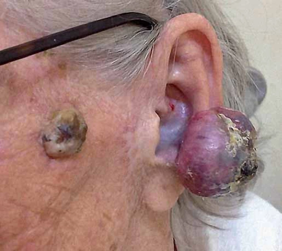
Fig. 2. A solitary erythematous nodule with prominent vessels, hyperkeratotic and ulcerated surface on the left ear lobe (side view). 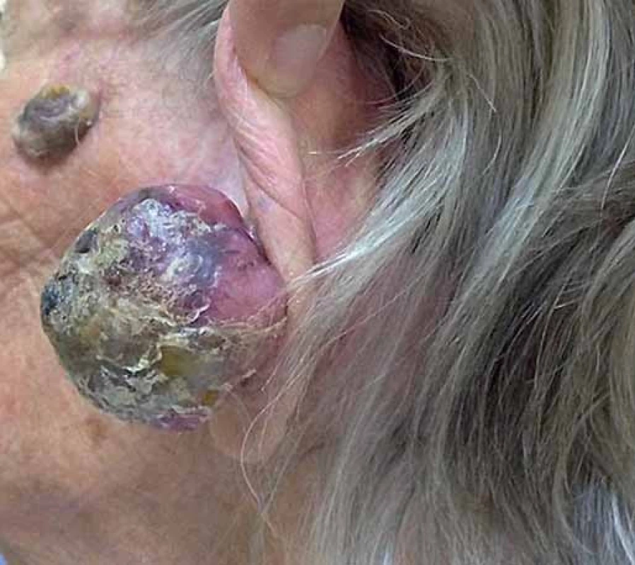
Fig. 3. Fusocelular proliferation in multi-directional bundles (100×). 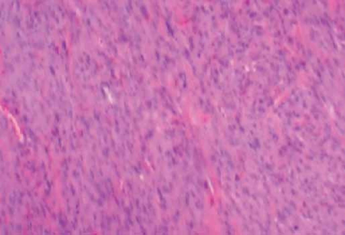
Fig. 4. Proliferation of elongated cells with poorly defined limits, dense eosinophilic cytoplasm, vesiculous or dense nuclei, irregular nuclear membrane and small to moderate diameter variation (400×). 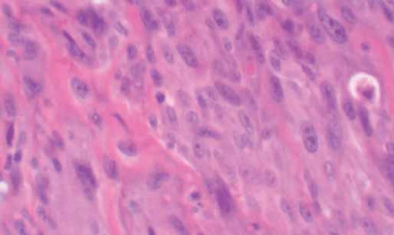
Conclusion
Atypical fibroxanthoma normally appears as a swiftly growing nodular or nodulo ulcerative lesion. It may be composed predominantly of either pleomorphic, spindle, epithelioid cells, or a mixture of these cells. The differential diagnosis includes pleomorphic dermal sarcoma, squamous cell carcinoma, malignant melanoma and leiomyosarcoma [5].
It occurs mostly in older adults and in sun exposed areas [6], with male predominance [7] and is a diagnosis of exclusion. The treatment is surgical and the preferred method is the aforementioned Mohs surgery [8]. Even though fibroxanthoma may be locally aggressive [9], the prognosis is usually very good if margins are adequate and these tumors rarely metastasise [7,10].
The authors declare they have no potential conflicts of interest concerning drugs, products, or services used in the study.
The Editorial Board declares that the manuscript met the ICMJE “uniform requirements” for biomedical papers.
Rodrigo Kraft Rovere, MD
Oncology Unit
Santo Antonio Hospital
Rua Itajai 545
Blumenau, Santa Catarina
CEP 89050100 Brazil
e-mail: rodrigorovere@hotmail.com
Submitted: 31. 3. 2014
Accepted: 1. 4. 2014
Zdroje
1. Naser N. Cutaneous melanoma: a 30‑year ‑ long epidemiological study conducted in a city in southern Brazil, from 1980 – 2009. An Bras Dermatol 2011; 86(5): 932 – 941.
2. Bedir R, Agirbas S, Sehitoglu I et al. Clear cell atypical fibroxantoma: a rare variant of atypical fibroxanthoma and review of the literature. J Clin Diagn Res 2014; 8(6): FD09 – FD11. doi: 10.7860/ JCDR/ 2014/ 8798.4466.
3. Patterson JW, Konerding H, Kramer WM. „Clear cell“ atypical fibroxanthoma. J Dermatol Surg Oncol 1987; 13(10): 1109 – 1114.
4. Anderson HL, Joseph AK. A pilot feasibility study of a rare skin tumor database. Dermatol Surg 2007; 33(6): 693 – 696.
5. Hussein MR. Atypical fibroxanthoma: new insights. Expert Rev Anticancer Ther 2014; 14(9): 1075 – 1088.
6. Hammerschmidt M, Azevedo LM, Ruaro A et al. Case for diagnosis. Atypical fibroxanthoma. An Bras Dermatol 2012; 87(4): 647 – 648.
7. Wollina U, Schonlebe J, Ziemer M et al. Atypical fibroxanthoma: a series of 56 tumors and an unexplained uneven distribution of cases in southeast Germany. Head Neck. In press 2014. doi: 10.1002/ hed.23673.
8. McCoppin HH, Christiansen D, Stasko T et al. Clinical spectrum of atypical fibroxanthoma and undifferentiated pleomorphic sarcoma in solid organ transplant recipients: a collective experience. Dermatol Surg 2012; 38(2): 230 – 239. doi: 10.1111/ j.1524 ‑ 4725.2011.02180.x
9. Inskip M, Magee J, Weedon D et al. Atypical fibroxanthoma of the cheek ‑ case report with dermatoscopy and dermatopathology. Dermatol Pract Concept 2014; 4(2): 77 – 80. doi: 10.5826/ dpc.0402a16.
10. Cooper JZ, Newman SR, Scott GA et al. Metastasizing atypical fibroxanthoma (cutaneous malignant histiocytoma): report of five cases. Dermatol Surg 2005; 31(2): 221 – 225.
Štítky
Dětská onkologie Chirurgie všeobecná Onkologie
Článek vyšel v časopiseKlinická onkologie
Nejčtenější tento týden
2014 Číslo 5- Metamizol jako analgetikum první volby: kdy, pro koho, jak a proč?
- Nejasný stín na plicích – kazuistika
- Nejlepší kůže je zdravá kůže: 3 úrovně ochrany v moderní péči o stomii
- Jak souvisí postcovidový syndrom s poškozením mozku?
-
Všechny články tohoto čísla
- Editorial
- Soutěž o nejlepší práci
- Pomalidomide in the Treatment of Relapsed and Refractory Multiple Myeloma
- Cereblon – a New Target of Therapy in the Treatment of Multiple Myeloma
- The Role of MicroRNAs in the Pathophysiology of Neuroblastoma and Their Possible Use in Diagnosis, Prognosis and Therapy
- Function of CDK12 in Tumor Initiation and Progression and Its Clinical Consequences
- Informace z České onkologické společnosti
- Prognostic Markers of Advanced Non-small Cell Lung Carcinoma – Assessing the Significance of Oncomarkers Using Data-mining Techiques RPA
- Breast Cancer Patient Satisfaction with Immediate Two-stage Implant-based Breast Reconstruction
- Soutěž na podporu autorských týmů publikujících v zahraničních odborných titulech
- Influence of Preoperative Chemoradiotherapy on Changes of Epidermal Growth Factor Receptor Expression in Patients Treated by Preoperative Chemoradiotherapy for Local Advanced Rectal Carcinoma
- A Rare Neoplastic Growth on the Ear Lobe
- To Whom it May Concern – Photodiagnosis and Photodynamic Therapy
- Aktuality z odborného tisku
- Prof. Žaloudík šedesátiletý
- Karcinomová lymfangiopatie při pokročilém karcinomu prsu
- Klinická onkologie
- Archiv čísel
- Aktuální číslo
- Informace o časopisu
Nejčtenější v tomto čísle- Karcinomová lymfangiopatie při pokročilém karcinomu prsu
- Prognostic Markers of Advanced Non-small Cell Lung Carcinoma – Assessing the Significance of Oncomarkers Using Data-mining Techiques RPA
- Breast Cancer Patient Satisfaction with Immediate Two-stage Implant-based Breast Reconstruction
- Cereblon – a New Target of Therapy in the Treatment of Multiple Myeloma
Kurzy
Zvyšte si kvalifikaci online z pohodlí domova
Autoři: prof. MUDr. Vladimír Palička, CSc., Dr.h.c., doc. MUDr. Václav Vyskočil, Ph.D., MUDr. Petr Kasalický, CSc., MUDr. Jan Rosa, Ing. Pavel Havlík, Ing. Jan Adam, Hana Hejnová, DiS., Jana Křenková
Autoři: MUDr. Irena Krčmová, CSc.
Autoři: MDDr. Eleonóra Ivančová, PhD., MHA
Autoři: prof. MUDr. Eva Kubala Havrdová, DrSc.
Všechny kurzyPřihlášení#ADS_BOTTOM_SCRIPTS#Zapomenuté hesloZadejte e-mailovou adresu, se kterou jste vytvářel(a) účet, budou Vám na ni zaslány informace k nastavení nového hesla.
- Vzdělávání


