-
Medical journals
- Career
Effect of earlier load on healing of calcaneus fractures after stable osteosynthesis
Authors: Roman Litner 1; Leopold Pleva 1; Jiří Kohut 3; Pavel Koscielnik 2; Jan Kubíček 3; Jiří Szeliga 1
Authors‘ workplace: Klinika úrazové chirurgie FN Ostrava 1; Radiodiagnostický ústav FN Ostrava 2; Vysoká škola báňská TU Ostrava 3
Published in: Úraz chir. 26., 2018, č.3
Overview
The authors present the results of a workgroup of patients with earlier burden of injured heel bone treated with osteosynthesis by LCP plate and C-nail compared to the control group with standard procedure of injured extremity loading after osteosynthesis.
Goal: To assess the suitability of earlier loading of extremity with calcaneous bone fracture treated with stable osteosynthesis with recommendations of postoperative care for these patients.
Material and methodology: The working and the control group of patients were monitored clinically and by optoelectronic stereophotogrammetry and evaluated by ultrasound examination and spectral analysis. We managed to include 18 patients in the working group and 6 patients in the control 6 patients treated 11 times by means of a plate and 13 times with a nail.
Results: After evaluation of ultrasound examination and a part of patients in terms of spectral analysis, the trends in graphs show a steeper growth pattern for the working group with earlier loading than for control group with later loading of the injured extremity.
Conclusion: In a smaller group of patients, we have demonstrated that earlier loading of an injured extremity with full body weight is more appropriate for the process of healing of calcaneus fractures after stable osteosynthesis.
Keywords:
Dislocated calcaneus fractures – LCP plate – C-nail – early postoperative loading of affected extremity
Introduction
Calcaneus fractures do not belong to frequent ones. It is reported that they account for about 2 % of all fractures [12]. Plate osteosynthesis whose origins date back to 1921 [11] was considered a stable osteosynthesis of calcaneus fractures for a long time. Its use spread in the early 1980s in connection with the development of CT scans and the creation of Sanders and later Zwipp classification.
A problem in the treatment of calcaneus fractures by plate osteosynthesis is the healing of a wide extensive hind limb wounds, particularly in patients at risk [21], with up to 21 % of superficial infectious complications reported [6, 7].
To reduce the incidence of these complications, a new implant has been developed for miniinvasive osteosynthesis of calcaneus fractures - C-nail, whose use dates back to 2011 [10].
These two types of osteosynthesis of calcaneus fractures are considered to be stable and, with the use of stereophotogrammetry and pedopedic pads, we performed an earlier load on the affected limb to assess this earlier loading on repair-induced bone changes, the correct gait stereotype with the technique of proper foot detachment from support.
1. Extended lateral approach used to apply plate and titanium LCP plate 
2. Postoperative CT images after osteosynthesis of calcaneus fracture by plate 
3. Postoperative VRT images after osteosynthesis of calcaneus fracture by plate osteosynthesis 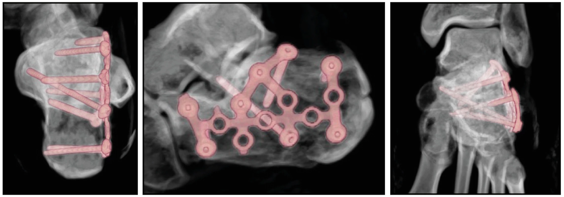
4. C – nail and perioperative representation in osteosynthesis by this nail 
5. Postoperative CT images after osteosynthesis of calcaneus fracture with C-nail 
6. Postoperative VRT images after osteosynthesis of calcaneus fracture with C-nail 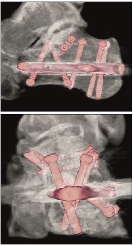
Methodology and material
In the selected group of patients, in addition to earlier loading of the injured limb, we also monitored biomechanical parameters after stable osteosynthesis of calcaneus fractures. In addition to clinical investigation, we used ultrasound to measure parameters in patients (measurement of callus size in mm), examination by optoelectronic stereophotogrammetry, evaluation of repair-induced changes in X-ray images by spectral analysis and the use to monitor patient load using pedopedic insoles.
We divided the group of patients into two groups: working and control. We were deciding for early loading of the limb in case of this fracture in the working group by the magnitude of the injury violence, type of fracture of the calcaneus, type of surgery, by the stability of osteosynthesis, healing of soft tissues in the post-operative period and by the patient‘s cooperation. We were selecting patients with a single bone fracture (monotrauma) in cooperating patients whose surgical wounds healed primarily, patients were able to progressively cope with the dosed load and no other structure on the given limb was injured in addition to calcaneus.
After the healing of wounds, we carried out X-ray examination of the injured heel bone in the working group in addition to clinical examination and rehabilitation in the 3rd, 6th, 9th and 12th postoperative week, ultrasound and optoelectronic stereophotogrammetric examinations were performed in week 3, 4, 5, 6, 9 and 12, and a part of the patients were monitored in terms of loading also using pedopedic insoles. We started to load the operated limb in week 3, 4, 5 and 6 after surgery to 1/3, 1/2, 2/3 and ultimately to the full weight of the body, as far as it was possible for the patients with the exclusion of painful stimuli. Subsequently, X-ray documentation was evaluated by spectral analysis with a focus on the dynamics of bone callus growth in predilection sites. In the control group, the above-mentioned accompanying examinations were performed in the same manner, only the injured limb load was 1/3 of the body weight ranging from 6 to 12 weeks postoperatively.
In the years 2012–2015, we treated 206 patients with calcaneous fractures at the Clinic of Traumatic Surgery of the University Hospital in Ostrava. 98 % of these were intra-articular fractures and 54 fractures were treated with plate osteosynthesis, and 102 calcaneus fractures were treated with nail.
So far, we have managed to include to the working group only eighteen patients who were able to agreed to research and six patients in the control group. ORIF surgical method was applied to 7 patients from the working group and 4 patients from the control group and surgery was applied to 11 patients from the working group and 2 patients from the control group.
Ultrasound
In patients after postoperative CT examination, the available fracture line in the calcaneus was selected, which was then traceable by the ultrasound device. The medial and lateral sides of the calcaneus were monitored in patients with nails, and the medial side of the calcaneus was followed by ultrasound in patients after PLATE osteosynthesis. The ultrasound examination of the patients of both groups was performed on a GE LogiQ 7 ultrasound device, with a linear probe with a frequency range of 8–12 MHz. An internally created setting has been selected as the instrument‘s preset, which is routinely used for soft tissue examination. The TCG setting was in the middle position.
In the course of examination, the aim was to display the soft tissues in the periostal region of the calcaneus in the best possible manner - i.e. at a maximum possible frequency of - 12 MHz. However, this showed that the highest quality settings could not be applied to all patients. Limiting was the amount of soft tissue and changes that were not controllable by patients - especially post-traumatic/postoperative soft tissue edema. In some patients, it was necessary to reduce the frequency of the probe and thus to display deeper structures even at the cost of reduced image quality. If we proceeded to this step during the initial examination, we used a lower frequency also for further checks of the respective patient.
Even with the same instrumentation, there were noticeable differences in the quality of the image, mainly due to the different degree of edema in the patients during the day and the examination.
Periostal healing changes were shown on the ultrasound as a differently thick hypoechogenic region around the fracture line. In the initial phase in the ultrasound image, these changes correspond to the periosteal hematoma. Over time, there was a noticeable increase in small hyperechogenities, i.e. ossification. Of course, the presence of intraoseal corrective changes cannot be evaluated from the principle of the method.
Ultrasound monitoring of calcaneus callus is possible, but it is technically demanding in terms of performance and is loaded with factors that we are unable to influence. Changes in the area of the callus were more apparent when comparing the examination at a longer time interval. Within the monitoring, it was not possible to draw on documentation that would accurately define the appearance of the callus. Tie studies dealing with examination of callus are rare, with focus rather on long bones [17, 21].
Stereofotogrammetry
7. Possibilities of examination on orthopedic footpath 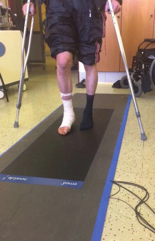
It is one of the diagnostic methods where we use the orthopedic footpath to investigate the physiological gait stereotype and the patient‘s load on the limbs. The orthopedic footpath that we use in rehabilitation is a measuring electronic system for recording and evaluating foot pressure distribution in static and dynamic conditions. This method provides us with accurate and reliable information for foot function analysis and diagnostics of possible pathophysiological abnormalities. Deformities and gait disturbances can be detected by data analysis.
Possibilities of examination on orthopedic footpath
- Plantar foot pressure measurement in dynamic and static mode
- Providing accurate information for gait diagnostics
- Detection of deformities and physiological deviations of gait
- 3D display of limb pressure values under load
- Time progress of the load
Methodology of load examination
During the clinical examination, a suitable cooperating patient was selected, and a measuring method for sensing the limb load while walking was specified. The sensing methods under assessment are based on the principle of load measurement, where the force the patient loads the limb and the contact pressures in the defined areas are evaluated. Furthermore, the dynamic pressure distribution in the individual phase of the foot detachment is evaluated. The examination took place from the third week after osteosynthesis of the fracture to the sixth week with a weekly frequency and further every third week until the 12th week.
The figure shows the load distribution on the plantar side of the foot and a graphical representation of the load distribution. The patient loads the limb with all his weight (71kg), the crutches serve as a stabilizing aid. The load is always compared to the standard of a healthy limb and physiological deviations from the correct gait stereotype can be evaluated with a precise distribution of force acting on the selected foot areas and the patient receives instructions for further rehabilitation based on this data.
8. Loading of the patient‘s limb after the third week - load distribution on the plantar side of the foot and a graphical representation of the load distribution. The patient starts with one third of the load by his own weight. In this particular case, it is 27 kg 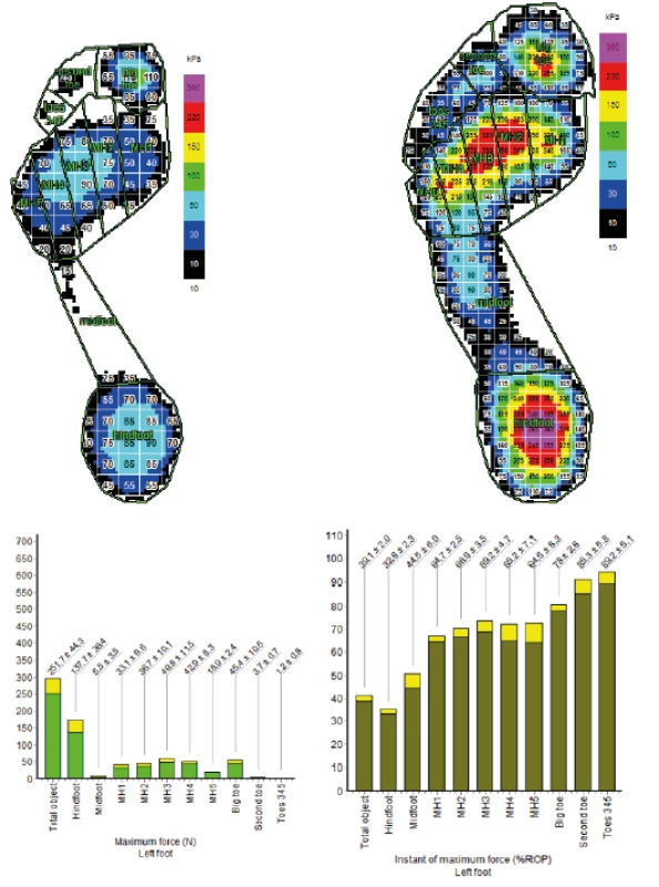
9. Loading of the patient‘s limb after the twelfth week 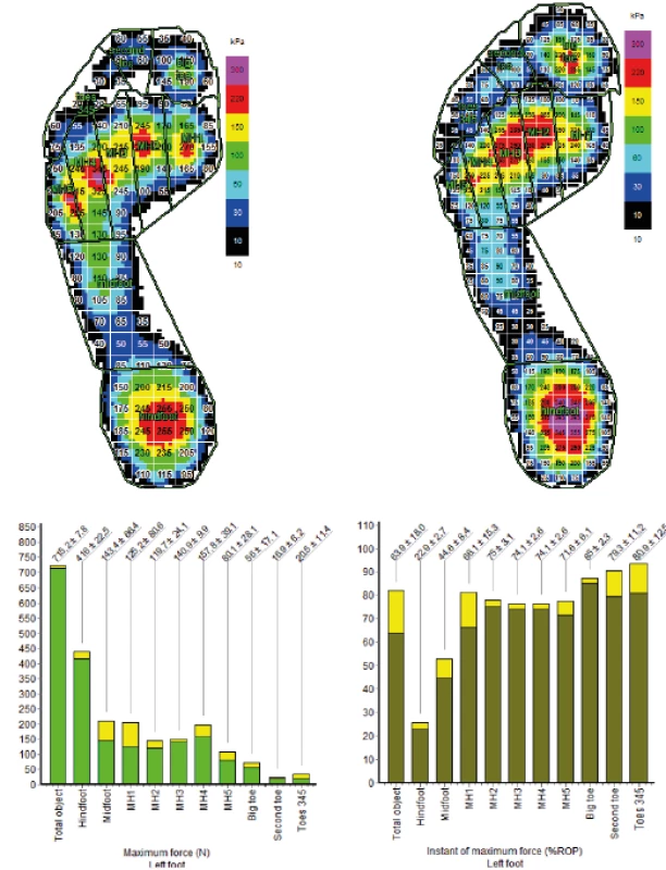
Pedopedic insoles
Rehabilitation also took place in home care, where pedopedic insoles were used to monitor the load; these insoles can be used to continuously measure the limb load even at home. The patient has pedopedic insoles paired with the phone, where he has preset gait parameters according to the examination on the load footpath. If the patient rehabilitates at home and overloads the injured limb, the patient will be alerted by a phone beep or vibration. Before using pedopedic insoles, the patient was instructed on how to use, calibrate, and operate the mobile pedoped App so that the patient could fully use it in home care. Patients had to be chosen to be at least technically literate and able to operate a smart mobile device, computer or laptop.
Load changes during treatment, when the load gradually increased, were always checked and consulted with the patient during hospital checks. Each check was also accompanied by a test on the load footpath, where the correct step detachment, the load distribution on the foot and the total load of the injured limb were determined by a test.
10. Pedoped insoles that meet the required parameters 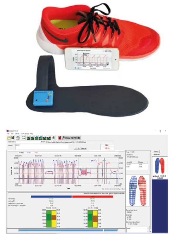
Spectral analysis
In terms of the development of fracture healing, we evaluated the development of the periosteal callus. Since the callus does not have constant geometric parameters over time, it develops dynamically depending on the fracture healing. While these changes may be observed in X-ray images, they are subjectively evaluated depending on the investigator‘s experience. Based on recent trends allowing the extraction of clinical information from image information, there is an effort to introduce a backward computer support link for automated and objective assessment of clinical X-ray images. As part of the analysis, we designed a segmentation method that allows for the autonomous extraction of the periosteal callus at a particular time of examination. This method allows objective dynamic monitoring of changes in callus development over time and thus provides effective feedback for clinical diagnosis.
From the point of view of the periosteal callus manifestation, it is crucial to determine the bone-callus interface, indicating boundaries and defining the callus for analysis. For this reason, we had to exclude some patients from the analysis because the contrast was unusable in terms of segmentation technique. In the presented spectral analysis, two groups of patients were tested: control group (3 patients) and working group (7 patients) whose heel bone was differently loaded during treatment. The obtained values are shown in the table 1.
1. Results of ultrasound measurement of callus (in mm) 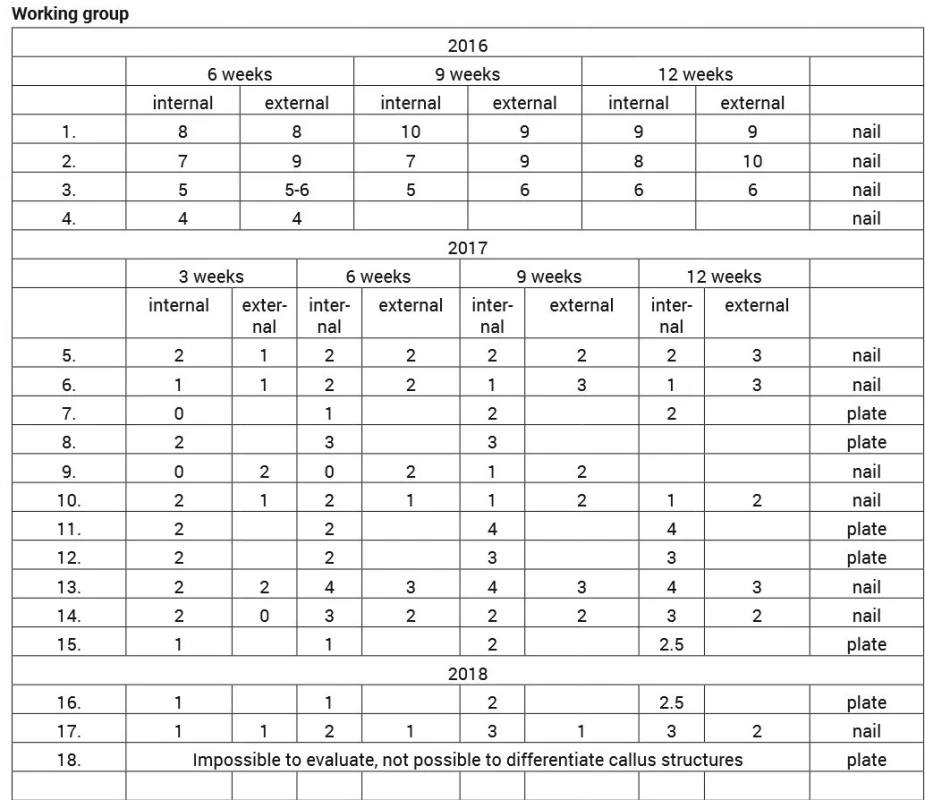
The dynamic model of the periosteal callus allows us to monitor the development of the callus on the basis of X-ray image data obtained at a time interval. This Dynamic Periosteal Callus Model uses the principle of the so-called regional segmentation of X-ray image data. In general, regional segmentation is a mathematical procedure that allows the entire X-ray image domain to be separated into non-overlapping parts that characterize individual identifiable objects, such as bones, muscles, and others.
The specific type of regional segmentation used in the periosteal callus modeling is the so-called active contour method (Fig. 11). This method falls within the category of the so-called evolutionary segmentation methods. The method can be imagined as an arbitrary regular curve (a regular geometric shape) that is set within the periosteal callus. This geometric curve has the ability to expand in all directions towards the periosteal callus boundary (interface) during the preset number of the so-called interaction steps. The iteration step represents a gradual change of the active contour. E.g. when 100 iteration steps are set, the active contour 100 changes its shape. During segmentation, this active contour is deformed to the periosteal callus boundaries and is stopped at these boundaries. In this way, a curve is created that surrounds the periosteal callus boundary (Fig. 11).
11. Application of the active contour method (green curve) that detect the periosteal callus area in clinical X-ray image data 
12. Binary model of the periosteal callus, classifying the area of the callus in white 
13. The final model of the periosteal callus reflecting the area of the callus based on white colour and the surrounding structures are filtered black 
Results
The obtained results of ultrasound measurement and spectral analysis were organized into tables with subsequent graphs of spectral analysis of repair X-ray changes.
Spectral analysis
The periosteal callus region is determined, in the X-ray image, based on the segmentation curve. In the next step, the genesis of the final periosteal callus model is applied (Fig. 2). The so-called image binarization is used for this purpose. This procedure unambiguously classifies the inner area of the callus, based on the white fill and the image background, where other structures are visible (Fig. 2). Binarization is finalized based on suppressing surrounding image structures into the background. This operation finalizes the periosteal callus model where the surrounding structures are filtered off in black (Fig. 3). A separate model of callus would be only little informative. It is essential that this proposed model allows for the extraction of determining clinical signs that characterize and objectify the periosteal callus. As a gradual expansion of the callus is anticipated over time, the callus model area is calculated, which, according to clinical assumptions, should increase depending on the mode of loading the the same. Technically, the area of this callus is calculated as the frequency of the white pixels that define the area of the callus model.
In this section we present experimental numerical results of the proposed model on real clinical data. For the purpose of model analysis, 7 patients in the working group (Table 2) and 3 patients in the control group (Table 2) were followed. Patients received x-ray images of the calcaneus 3, 6, 9 and 12 weeks after surgery. For each of the images, a periosteal callus model was created based on the procedure described. Based on the model, the area of the model was always calculated (Table 2). The development of these areas was plotted in the graphs and interlaced with a linear function (line) to identify trends in individual patient groups (Graph 1, 2).
2. The results of the periosteal callus model calculated for the area of the callus in pixels for each week of study for the working and control groups 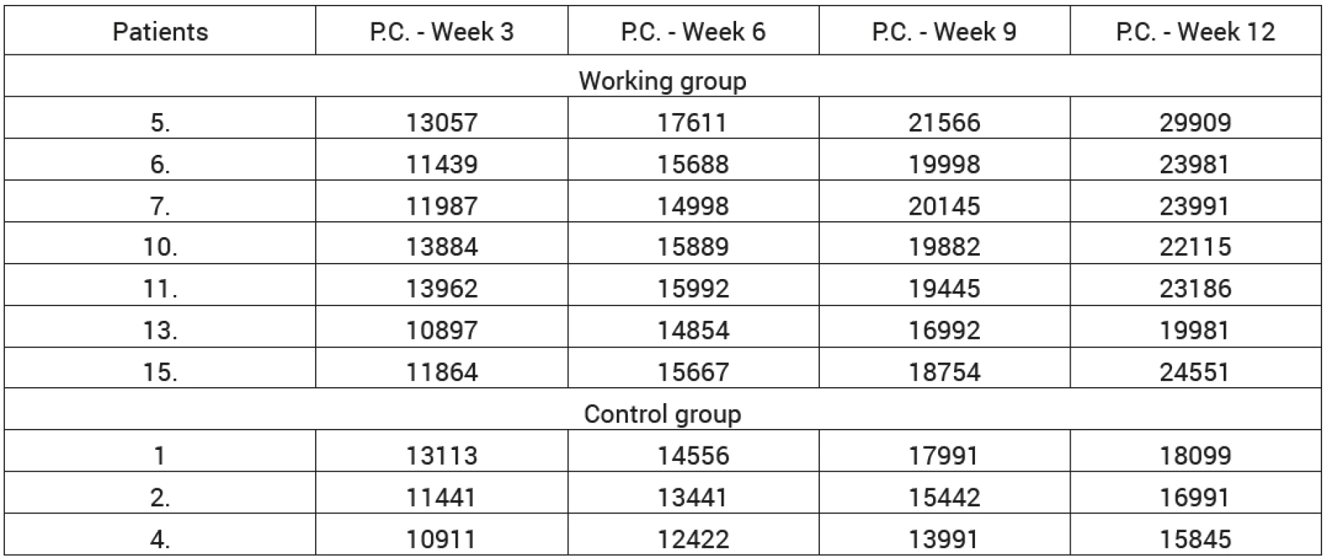
1. , 2.: The dynamic trend of evolution of periosteal callus, calculated on the basis of results in Tab. 2 for the working group group (left) and the control group (right) 
Graf 3: Comparison of trend functions of Fig. 4 for both monitored patient groups. The trends clearly show a steeper increase of pattern for the working group, as clinically expected 
Discussion
Most authors report the initiation of loading of the affected limb with a fracture of the calcaneus after stable osteosynthesis in the range of 6-12 weeks, depending on the severity of the fracture of the calcaneus [11, 12, 24] and the authors mainly deal with the types of osteosynthesis and their complications [10, 14, 20].
Our aim was to prove the benefit of earlier loading of the affected limb with fracture of the calcaneus after stable osteosynthesis for the growth of the periosteal callus, i.e. healing of this bone. Stable osteosynthesis, including plate osteosynthesis or osteosynthesis by the nail, timely loading of the affected limb and early rehabilitation lead to faster induction of physiological gait conditions and, according to our results, to faster bone callus formation. The above results show greater repair bone changes, a more rapid return to symmetric gait with correct feet detachment, with the correct positioning of the joints placed on the affected limb in the working group ather than the control group, although we recognize that the number of subjects in the groups is not high.
During the follow-up of patients one year after osteosynthesis, only two patients experienced complications - one patient was affected by phlebotrombosis and one patient´s condition was complicated by another injury in the spine area followed by later surgery. Overall, our complications corresponded to those reported in worldwide literature [10, 12, 14, 24].
In a smaller number of patients from both groups, we were able to perform post-traumatic magnetic resonance imaging with assessment of damage to the cartilage of the posterior subtalar calcaneus. We would like to compare the result of this examination with the result of the same examination one year after surgery in the given patient.
We see the future in the monitoring of the earlier dosed loading of the injured limbs of cooperating patients by means of pedopedic insoles for footwear at a larger scale, enlargement of the monitored groups and monitoring of the consequences of earlier loading of the subtalar joint.
Conclusion
The correct indication of patients for timely loading of the injured limb, careful management and monitoring of the patient‘s injured limb load in the postoperative period will lead to fewer complications and faster physiological gait training.
Faster training of correct gait with the return of foot proprioception along with early rehabilitation care leads to higher patient satisfaction without any more pronounced negative observed phenomena compared to patients with later load of the injured limb.
MUDr. Roman Litner
Sources
- Birkfellner, W. Applied medical image processing: a basic course. 2nd ed. Boca Raton : CRC Prress, c2014. ISBN 978-1-4665-5557-0
- Berry, GK, Stevens, DG, Kreder, HJ et al. Open fractures of the calcaneus: a review of treatement and outcome. J Orthop Trauma. 2004, 18, 202–206. ISSN 1531-2291
- Coste, C., Asloum, Y., Marcheix, PS et al. Percutaneous iliosacral screw fixation in unstable pelvic ring lesions: the interest of O-ARM CT-guided navigation. Orthop Traumatol Surg Res. 2013, 99, S273–278. ISSN 1877-0568
- Goldzak, M., Mittlmeier, T., Simon, P. Locked nailing for treatement of displaced articular fractures of the calcaneus with calcanail. Eur J Orthop Surg Traumatol. 2012, 22, 345–349. ISSN 1633-8065
- Hofer, M. Kurz sonografie. Praha : Grada, 2005. 240 s. ISBN 80-247-0956-2
- Howard, JL., Buckley, R., Cormack, RM et al. Complications following management of displaced intraarticular calcaneal fractures, J Orthop Trauma. 2003, 17, 241–249. ISSN 1531-2291
- Charles, M. , Court, B., Matthias, S. et al. Factors affecting infection after calcaneal fracture fixation. Injury. 2011, 40, 1313–1315. ISSN 0020-1383
- Jan, J. Medical image processing, reconstruction and restoration: concepts and methods. Boca Raton : Taylor & Francis, 2006. ISBN 0-8247-5849-8.
- Oberst, M., Hauschild, O., Konstantinidis, L. et al. Effects of three-dimensional navigation on intraoperative management and early postoperative outcome after open reduction and internal fixation of displaced acetabular fractures. J Trauma Acute Care Surg. 2012, 73, 950–956. ISSN 2163-0763
- Pompach, M., Carda, M., Žilka, L. et al. Nailing of the calcaneal bone C-nail. Úraz chir. 2015, 2, 31–38. ISSN 1211-7080
- Popelka, V., Zamborský, R., Schmidt, F. et al. Otvorená repozícia a interná fixácia u dislokovaných intraartikulárnych zlomenín. Úraz chir. 2011, 1, 17–23. ISSN 1211-7080
- Rak, V., Buček, P., Ira, D. et al. Operační metoda léčby nitrokloubních zlomenin patní kosti. Rozhl chir. 2006, 6, 311–317. ISSN 0035-9351
- Ruan, Z., Luo, CF, Zeng, BF et al. Percutaneous screw fixation for the acetabular fracture with quadrilateral plate involved by three-dimensional fluoroscopy navigation: surgical technique. Injury. 2012, 43, 517–521. ISSN 0020-1383
- Sanders, R., Fortin, P., DiPasqualem, T. et al. Operative treatement in 120 displaced intraarticular calcaneal fractures. Clinical orthopaedics and related research. 1993, 290, 87–95. ISSN 1528-1132
- Seligson, D., MAUFFREY, C., ROBERTS, C. et al. External fixation in orthopedic traumatology. London : Springer, 2012. ISBN 978-1-4471-2199-2.
- Schepers, T. The sinus tarsi approach in displaced intra-articular calcaneal fractures. Int Orthopaedics. 2011, 35, 697–703. ISSN 2313-1462
- Semmlow, J., GRIFFEL, B. Biosignal and medical image processing. 3rd ed. Boca Raton : CRC Press, 2014. ISBN 978-1-4665-6736-8.
- Sushil, G., Kachewar, D., Kulkarni, S. Utility of Diagnostic Ultrasound in Evaluating Fracture Healing. J Clin Diagn Res. 2014, 8, 179–180. doi: 10.7860/JCDR/2014/4474.4159 ISSN 0973-709X
- Suri, JS, WILSON, DL, LAXMINARAYAN, S. et al. Handbook of biomedical image analysis. Segmentation Models. Vol. I. New York : Kluwer Academic/Plenum Publishers, c2005. ISBN 0-306-48550-8.
- Svatoš, F., Bartoška, R., Skála-Rosenbaum J. et al. Zlomeniny patní kosti léčené otevřenou repozicí a dlahovou osteosyntézou - prospektivní studie. Část I: Základní analýza souboru pacientů. Acta Chir Ortop Traum Čech. 2011, 78, 136–140. ISSN 0001-5415
- Vaněček, L., Malkus, T., Dungl, P. Léčba zlomenin patní kosti otevřenou repozicií z mediálního přístupu. Acta Chir Ortop Traum Čech. 2003, 70, 100–107. ISSN 0001-5415
- Wawrzyk, M., Sokal, J., Andrzejewska, E. et al. The Role of Ultrasound Imaging of Callus Formation in the Treatment of Long Bone Fractures in Children. Pol J Radiol. 2015, 80, 473–478. doi: 10.12659/PJR.894548. eCollection 2015. ISSN 1899-0967
- Zwipp, H., Tscherne, H., Thermann, H. et al. Osteosynthesis of displaced intraarticular fractures of the calcaneus. Clinical orthopaedics and related research. 1993, 290, 76–86. ISSN 1528-1132
- Zwipp, H., Rammelt, S., Bartel, S. Calcaneal fractures-open reduction and internal fixation (ORIF). Injury. 2004, 35, 46–54. ISSN 0020-1383
- Žvák, I. Traumatologie ve schématech a RTG obrazech. Praha : Grada Publishing, 2006. ISBN 80-247-1347-0. 207 s.
Labels
Surgery Traumatology Trauma surgery
Article was published inTrauma Surgery
2018 Issue 3-
All articles in this issue
- Current approach to the management of forearm and elbow dislocation in children
- Medial malleolar fractures
- Examples of use of outpatient negative pressure therapy in chronic wound healing – a set of case reports
- Forefoot Navicular Bone Fractures - Short Communication and Case Report
- Assessment of the injuring effect of loose objects in a vehicle in a traffic accident
- Effect of earlier load on healing of calcaneus fractures after stable osteosynthesis
- Trauma Surgery
- Journal archive
- Current issue
- Online only
- About the journal
Most read in this issue- Current approach to the management of forearm and elbow dislocation in children
- Forefoot Navicular Bone Fractures - Short Communication and Case Report
- Effect of earlier load on healing of calcaneus fractures after stable osteosynthesis
- Examples of use of outpatient negative pressure therapy in chronic wound healing – a set of case reports
Login#ADS_BOTTOM_SCRIPTS#Forgotten passwordEnter the email address that you registered with. We will send you instructions on how to set a new password.
- Career


