-
Články
- Vzdělávání
- Časopisy
Top články
Nové číslo
- Témata
- Kongresy
- Videa
- Podcasty
Nové podcasty
Reklama- Kariéra
Doporučené pozice
Reklama- Praxe
Eyelid edema as a first sign of lymphoma
Authors: J. Halička 1; P. Žiak 1; B. Jakubovičová 1; K. Janurová 1; T. Balhárek 2; L. Plank 2; Ľ. Váleková 3; D. Žiak 4,5
Authors place of work: Očná klinika, Jesseniova Lekárska fakulta v Martine, Univerzita Komenského v Bratislave, SR, Prednosta MUDr. Peter Žiak, Ph. D 1; Ústav patologickej anatómie, Jesseniova Lekárska fakulta v Martine, Univerzita Komenského v Bratislave, SR, Prednosta: prof. MUDr. Lukáš Plank, CSc. 2; Klinika hematológie a transfúziológie, Jesseniova lekárska fakulta v Martine, Univerzita Komenského v Bratislave, SR 3; CGB laboratory a. s., Ostrava, ČR 4; Ústav patologie, Fakultní nemocnice Ostrava, Ostrava, ČR 5
Published in the journal: Čes. a slov. Oftal., 75, 2019, No. 6, p. 323-328
Category: Kazuistika
doi: https://doi.org/10.31348/2019/6/5Summary
Chronic eyelid edema may be a symptom of different disease. The most common are autoimmune diseases such as orbital pseudotumor, vasculitis, sarcoidosis, or impaired vascular or lymphatic drainage. Rarely has it been reported as the sole manifestation of the lymphoma. Eyelid lymphoma is a special clinical entity in the spectrum of hematological malignancies. Here we present our clinical experience with eyelids lymphomas. First case is a 76-year-old female patient with bilateral edema of upper eyelid non-responding to anti-inflammatory therapy. Histological examination diagnosed mantle cells lymphoma. In the second case, 58-year-old patient was diagnosed with solitary unilateral tumor of the lower eyelid, where primary biopsy was ordered and diagnosis of MALT lymphoma was established after histological examination. In both cases, it was not solitary eyelid tumor, but systemic disease with multiple lymphadenopathy and bone marrow infiltration were found in follow-up examinations. Subsequently, patients care was given to the hemato-oncologist.
Keywords:
B-lymphoma – orbit – eyelid edema
INTRODUCTION
Lymphomas are malignant tumours of a heterogeneous group with more than 40 subtypes [1]. They may occur in lymph nodes or extranodally, and originate from monocular proliferation of B-cells, T-cells or in rarer cases NK-cells. Lymphomas of ocular adnexa represent 2% of all extranodal lymphomas [2], and 5% of these occur on the eyelids [3].
Eyelid lymphoma is defined as a lymphoma incorporating preseptal tissues: skin, subcutaneous bonding material and the musculus orbicularis oculi [4]. It is a rare pathology, with 199 published cases to date. Clinical trials are limited, according to our knowledge only one study on a large cohort has been published to date [5]. Independent case reports exist on eyelid edema as the first symptom of an oncological pathology [6]. In the published works, lymphomas of B-cell origin are represented in 56% of cases, while 44% are of T-cell origin together with NK cells. B-cell lymphomas of a low degree of malignancy include extranodal marginal zone lymphoma (EMZL MALT associated), follicular lymphoma (FL) and extramedullary plasmacytoma. B-cell lymphomas of a high degree of malignancy include diffuse large B-cell lymphoma (DCBCL) and mantle cell lymphoma (MCL) [7]. The most commonly occurring T-cell lymphoma of the eyelids is mycosis fungoides (MF) [5].
Pathogenically lymphomas may originate in lymphoid tissue as the result of chronic inflammation or autoimmune disease. The latest molecular and cytogenetic observations indicate that lymphomas may be linked with the infection Chlamydia psittaci [8].
The majority of subtypes occur in older patients aged between 50 and 70 years, with the exception of lymphoblastic lymphoma of B-cell and T-cell origin, and Burkitt lymphoma, which occur in children and adolescents.
Tumour and swelling of the eyelid are a manifestation of lymphomas of B-lymphocytes and T-cells. Ulceration and erythema occur more frequently in patients with T-cell lymphoma. An important pathognomonic sign is palpation rigidity of such lesions.
The prognosis of the pathology depends on the subtype according to the histological finding [7]. Overall the prognosis of eyelid lymphomas is considered to be worse than for lymphomas in other ocular adnexa [9], and for this reason eyelid lymphomas have staging of T3 or higher, irrespective of other factors [4]. The majority of subtypes, especially of a low degree, have a good prognosis, with a small proportion of recurrences, the five-year rate of survival for EMZL and FL is more than 85%. Other subtypes, including mycosis fungoides, have a worse prognosis [7].
Radiotherapy with or without a surgical procedure is the treatment of choice for a low degree of solitary lymphomas, whereas chemotherapy with or without adjuvant therapy is the choice for lymphomas of a higher stage, or diffuse lymphomas [10].
CASE REPORT 1
A 76 year old female patient was sent to the centre from the local outpatient unit with tumescence of the upper eyelids persisting for approximately six months. At the first examination natural visual acuity of 5/15 bilaterally was determined, which was not improved by correction due to the presence of a cataract. The patient had no noteworthy features in her personal and family medical history with respect to the diagnosis in question.
The patient had objectively clearly palpable rigid, smooth, painless formations beneath the skin of the upper eyelids, passing into the orbit in the temporal region, where the edges of the orbit were unclear, otherwise well palpable (Fig. 1). Ptosis of the upper eyelids was similarly present. Eyeballs without protrusion, pacific. Finely delineated chemosis of conjunctiva. Motility of eyeballs slightly obstructed in the upward direction, in other directions free. Intrabulbar presence of cataract with pseudoexfolative syndrome, pupils isocoric, otherwise without pathological finding. Symmetrical finding on fundus, without signs of congestion on the disc of the optic nerve, without increased perfusion of capillaries, finding commensurate to age.
Fig. 1. Patient 1 – bilateral, hyperemic tumours of the upper eyelids 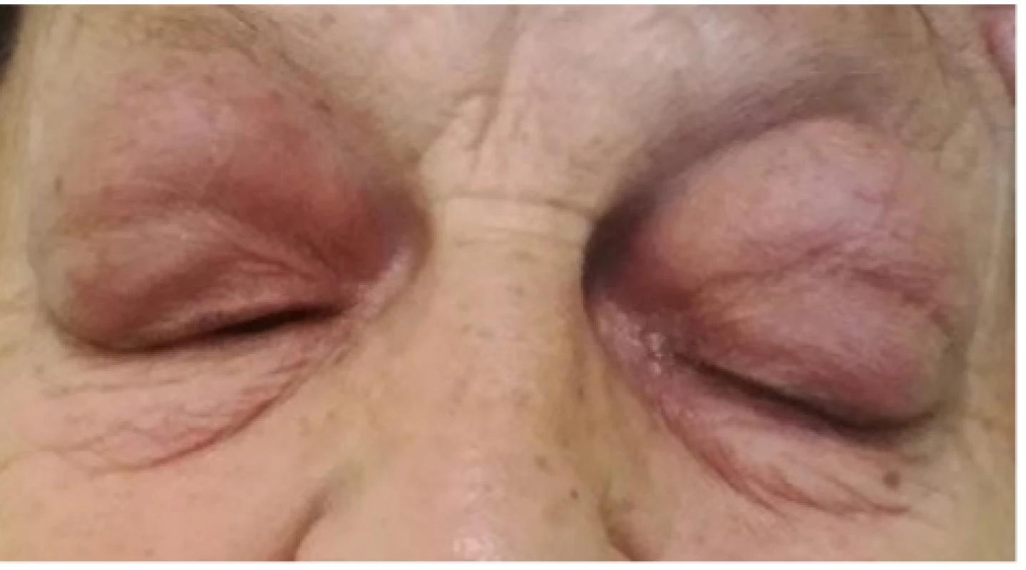
According to the description of B-scan ultrasonography and magnetic resonance imaging of the orbits, bilateral expansive lesions were recorded, which were well bordered, pressing craniolaterally on the eyeball, forcing out the adjacent muscle structures medially (Fig. 2A), with post-contrast occurrence of homogeneous saturation of both expansions.
Fig. 2. Fig. 2. Patient 1 – magnetic resonance of head: axial cross-section, T2 weighting, bilateral lymphoma of upper eyelids
A) at time of diagnosis 02/2018
B) following chemotherapy and immunological treatment 01/2019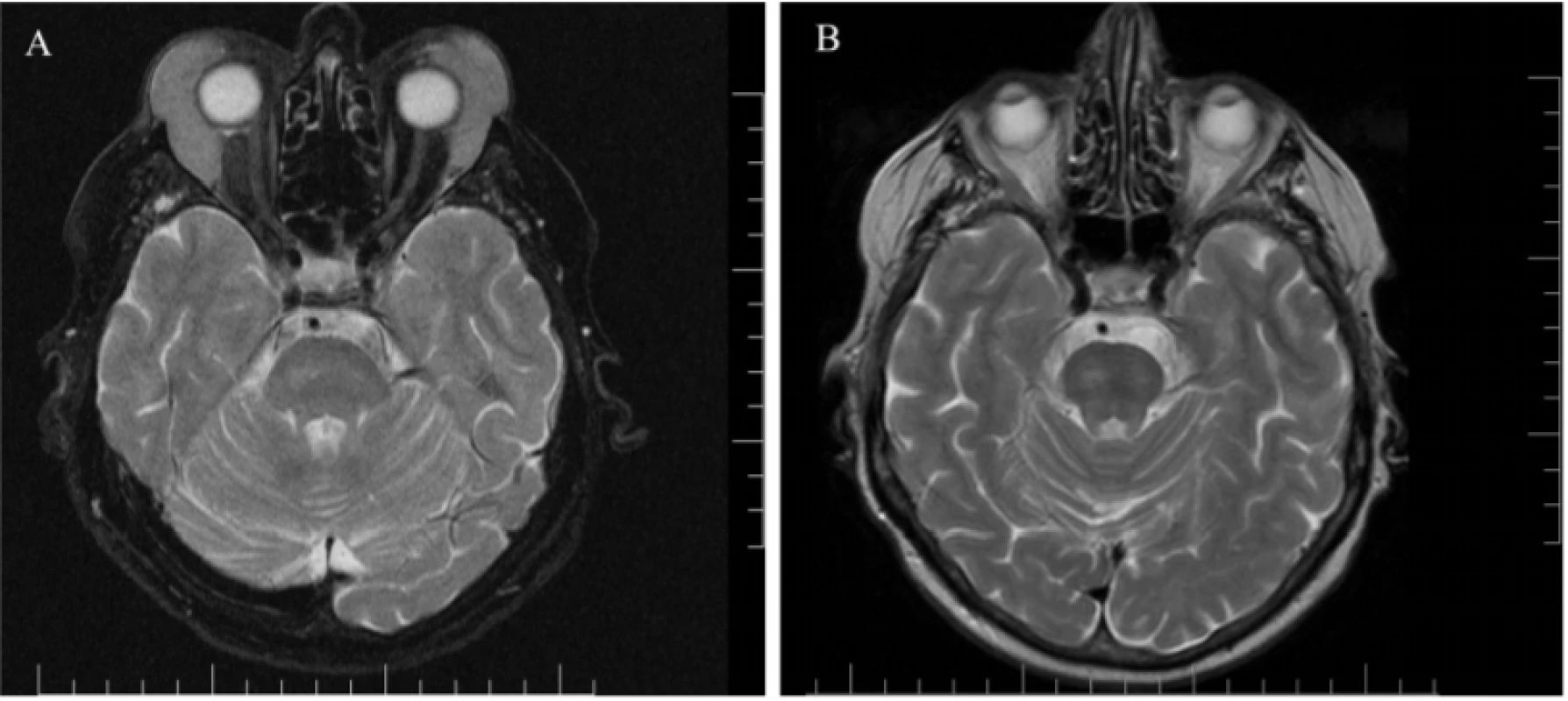
At the outset the condition was diagnosed as bilateral enlargement of the lachrymal glands, which did not respond to treatment by local antibiotics or to corticoids. Subjectively the patient’s condition fluctuated. Within the framework of differential diagnostics (inflammatory pseudotumour, sarcoidosis, amyloidosis, lymphoma, tumour of lachrymal gland, metastasis of unknown origin), the patient underwent a stomatosurgical, dermatological and haematological consultation with a negative result.
Subsequently a probatory excision was indicated, in which a whitish, rigid fatty substance was found and a sample taken from it. It was histologically diagnosed as a small-cell CD20+ B-NHL type mantle cell lymphoma (Fig. 3A and 3B).
Fig. 3. Fig. 3. Patient 1 – B-NHL type MCL, biopsy finding:
A) Infiltration of soft tissues by dense, pseudonodular growing tumorous lymphoproliferation from
small to medium-sized lymphoid cells (HE, enlargement 100x)
B) Immunohistochemical positivity of CD20+ marker in tumour cells (enlargement 400x)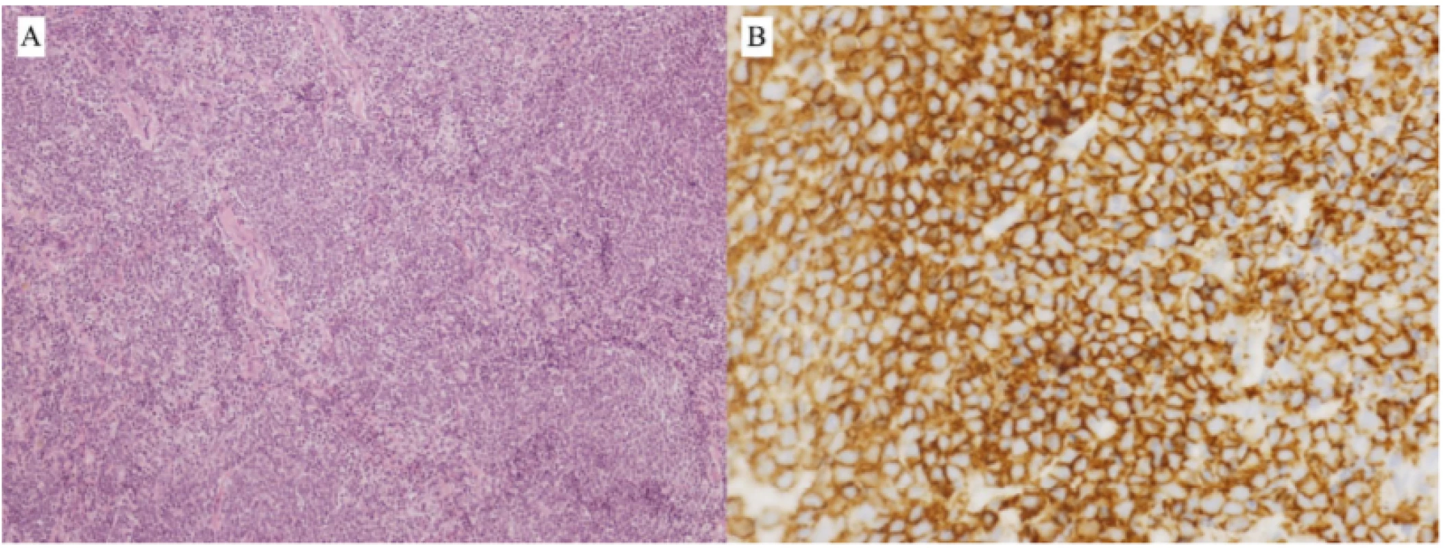
With these results the patient was sent to the department of haematology, where supplementary examinations (biopsy of bone marrow, CT of head, neck, chest and abdomen) classified the finding as small-cell CD20+ B-NHL type of mantle cell lymphoma (MCL), morphologically conventional variant, SOX 11+, IgH(14q32) negative, CCND1 negative, MLPA P377 negative. The clinical stage was determined as IV-B due to infiltration of the lachrymal and parotid gland bilaterally, bilateral enlargement of jugular and abdominal lymph nodes, lymphadenopathy in region of uterus with minimal infiltration of bone marrow less than 1%.
At present the patient is under the supervision of a haemato-oncologist. The selected treatment was immunotherapy and chemotherapy by rituximab-CHOP (cyclophosphamide, doxorubicin, vincristine and prednisone). After 4 cycles a significant regression of the finding (Fig. 2B) was observed on magnetic resonance (MR) from January 2019. Due to the persistence of the pathology, in July 2019 DHAP chemotherapy was commenced (dexamethasone, cytarabine, cisplatin), at the time of writing (December 2019) 5 doses have been administered.
CASE REPORT 2
A 58 year old male patient was sent for an ocular examination due to an asymmetrical pouch beneath the left eye. He stated no subjective visual complaints. He had no general pathology, increased temperature, inflammation of the eyelids or paranasal sinuses, and no symptoms of toothache. A biopsy was taken from the region of the lower eyelid. Objectively on the lower eyelid of the left eye two bordered, freely motile rigid formations were described with a size of approximately 2x1, 5 cm, slight tumescence (Fig. 4), the eyeball was freely motile, without diplopia, the other anterior segment was within the norm. In the left axilla there was a palpable lymph node with a size of approximately 5 cm, and in the inguinal regions only small lymph nodes up to a size of 1 cm.
Fig. 4. Patient 2 – tumour of left lower eyelid 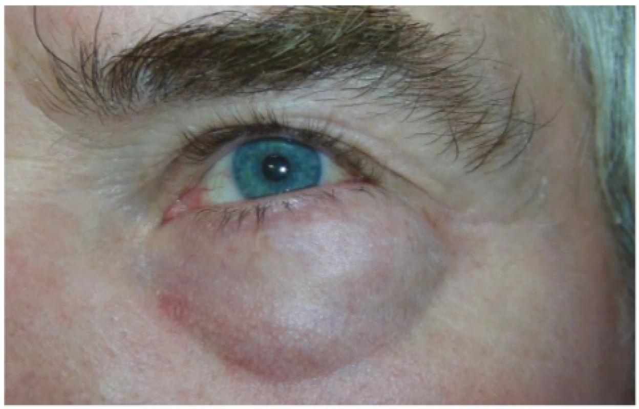
On magnetic resonance imaging there was an enlarged lachrymal gland latero-caudally, in T1 isointensity in comparison with muscle structures, in T2 slight non-homogeneous signal activity of higher intensity in comparison with muscles, lower in comparison with fat tissue, which pressed closely against the musculus rectus lateralis in its ventral third, and also pressed closely on the eyeball latero-ventrally, and in a short section ventrally also on the musculus rectus superior and inferior. This expansion has partial retrobulbar localisation, in total on a surface of 2.5 x 3.5 x 3.3 cm (Fig. 5A). There is post-contrast slight diffuse saturation of the lesion – in differential diagnostics described as a tumour of the lachrymal gland, metastasis, rhabdomyosarcoma, idiopathic inflammatory process or other.
Fig. 5. Fig. 5. Patient 2 – magnetic resonance of head: axial cross-section, T2 weighting, lymphoma of lower left eyelid
A) at time of diagnosis 01/2019
B) following chemotherapy and immunological treatment 11/2019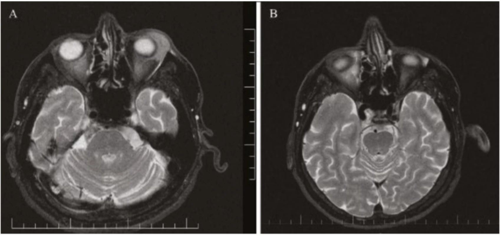
Fig. 6. Patient 2 – B-NHL from spectrum of MZBL type malt, biopsy finding:
A) Infiltration of fibrolipomatous conjunctiva with dense, diffuse tumorous lymphoproliferation from
small to medium-sized lymphoid cells (HE, enlargement 100x)
B) Immunohistochemical positivity of CD20+ marker in tumour cells (enlargement 630x)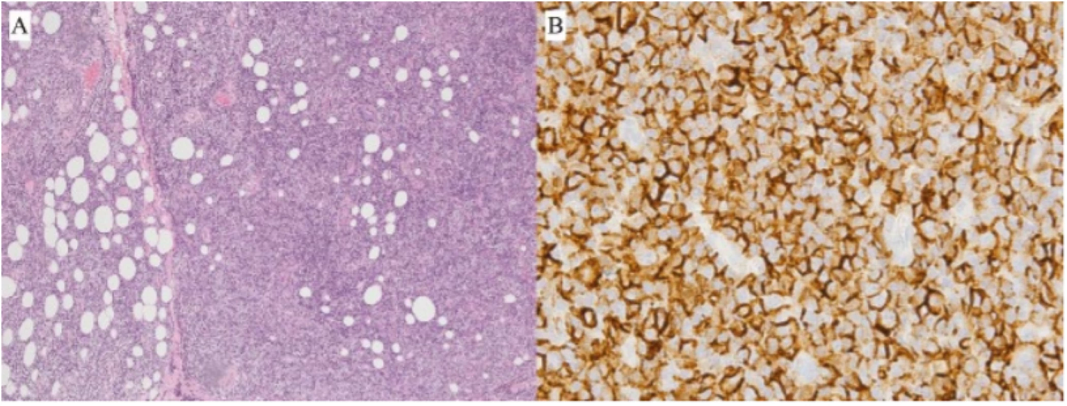
A histological analysis of the extirpated tumour diagnosed small-cell B-NHL from a spectrum of lymphomas from marginal zone B-cells (MZBL), of the type of MALT-lymphoma (CD20+, CD5-, cyclin-D1/isolated+, bcl-2+) with plasmacytoid and plasmacytic differentiation (monotypical c-lg type lambda+) and reproduction of blastocytes, but without signs of blastic transformation. Clinical stage IV.A – tumour of lachrymal gland, positive supraclavicular lymph nodes bilaterally, and axial lymph nodes on left side with 15% infiltration of bone marrow.
With regard to the type and extent of the pathology, the haemato-oncologist indicated 8 doses of immuno-chemotherapy in a combination of rituximab and bendamustine. The treatment was terminated in October 2019, at which time the patient was in remission. The finding on MR is regression of the tumour (Fig. 5B).
DISCUSSION
Chronic edema of the eyelids may be a symptom of either local or general pathologies. The most frequent of these include autoimmune diseases such as pseudotumour or the orbit, amyloidosis, vasculitis, sarcoidosis or deteriorated vascular or lymphatic drainage due to a primary tumour of the orbit or metastasis of a remote tumour. In rare cases it is stated as a manifestation of lymphoma of the eyelids. In the literature separate case reports are described with edema of the eyelids as the first symptom of an oncological pathology [6].
Lymphomas of the eyelids constitute 5% of all lymphomas of the ocular adnexas. According to published cases of eyelid lymphomas, 56% are of B-cell origin and 44% of T-cell origin. The most common B-cell lymphomas are extranodal marginal zone lymphoma and diffuse large-cell lymphoma. The majority of subtypes occur in older patients, with the exception of lymphoblastic lymphoma of B-cell and T-cell origin and Burkitt lymphoma, which occur in children and adolescents.
In a cohort studied by the Czech authors Krásný et al., the most frequently diagnosed orbital tumour was pseudotumour of the orbit in 40% of cases, in addition to which sarcoidosis was verified in four out of 87 cases (4.5%), lymphoma in 19.5% of cases (MALT-lymphoma verified in 59% of these), primary carcinomas of the orbito-palpebral region in 14% of cases and secondary melanoma of the orbit originating from the conjunctiva in 5.5% of cases [11]. In a cohort of patients with lymphoma of the orbit studied by the Slovak authors Furdová et al. [12], LPL (lymphoplasmacytic lymphoma) occurred in 3%, Burkitt lymphoma 6%, FK 6%, MCL 8%, DLBCL 17%, while MALT lymphoma had the largest representation of 60%.
Extranodal lymphoid tumours may occur in the eyelids, orbit or conjunctiva, either as isolated lesions or as part of a systemic pathology. Lymphoid tumours in the conjunctiva have a tendency to be localised and have a better prognosis, because this location is normally inhabited by lymphocytes [13]. In our female patient an infiltration of lymph nodes occurred throughout the body, but infiltration of bone marrow was only minimal.
In general, clinical MCL manifests extensive lymphadenopathy and splenomegaly. It may manifest pancytopenia or a leukaemic image with leukocytosis [14]. MCL usually represents clinical stage IV, with affliction of bone marrow. The patient may but need not necessarily have proptosis, diplopia, tumescence of the conjunctivas (salmon-pink), reddening of the edges and ptosis caused by affliction of the skin or musculus orbicularis of the upper eyelids [15].
Almost all lymphomas of the ocular adnexas are of B-cell type. Lymphomas of T-cell origin are rare, and are usually linked with systemic conditions such as mycosis fungoides [16]. Even though unilateral lymphoid tumours are more common than bilateral [5], the overall prognosis is approximately equal [17]. Approximately 17% of patients with lymphatic lesions of the ocular adnexas have bilateral affliction.
In the case of solitary tumours of a low degree, the therapeutic procedures used are radiotherapy [17] and surgical resection [10]. In the case of a high degree or diffuse lymphomas, chemotherapy is used, with or without the use of adjuvant therapy [19]. In clinical stages III and IV of extranodal MALT lymphomas, as well as MCL lymphomas, therapy is based on the treatment of other indolent non-Hodgkins lymphomas [20]. In the case of MCL, R-CHOP is not considered curative, but may induce long-term remission. More intensive chemotherapeutic regimens are not recommended for patients aged over 60 years due to their high toxicity [21]. Immunotherapy in the form of monoclonal antibodies against CD-20, rituximab, is added as standard to treatment on the basis of improvement of the overall survival rate of patients in retrospective studies [22].
Both cases of lymphoma of the eyelids demonstrate that even in the case of an edema of the eyelids of benign appearance it is necessary, within the framework of differential diagnostics, to reckon with the manifestation of a general pathology – lymphoma, especially in cases with a longer-term course, which do not respond to immunosuppressant and antibiotic therapy. A beneficial and unequivocally indicated procedure is treatment in co-operation with a haemato-oncologist.
The authors of the study declare that no conflict of interest exists in the compilation, theme and subsequent publication of this professional communication, and that it is not supported by any pharmaceuticals company.
MUDr. Juraj Halička
Očná klinika,
Jesseniova Lekárska fakulta v Martine,
Univerzita Komenského v Bratislave,
SR
Korespondující autor:
MUDr. Peter Žiak, Ph.D.
Očná klinika JLF UK a UNM
Kollárova 2,
036 59 Martin,
SR
Received: 14. 10. 2019
Accepted: 15. 12. 2019
Available on-line: 20. 5. 2020
Zdroje
1. Swerdlow, S.H., International Agency for Research on Cancer.: World Health Organization. WHO classification of tumours of haematopoietic and lymphoid tissues. 4th ed. Lyon, France: International Agency for Research on Cancer, 2008 : 1-439.
2. Freeman, C., Berg, J.W., Cutler, S.J.: Occurrence and prognosis of extranodal lymphomas. Cancer, 29; 1972 : 252-260.
3. Sjo, L.D.: Ophthalmic lymphoma: epidemiology and pathogenesis. Acta ophthalmologica. 2009;87 Thesis 1 : 1-20.
4. Amin, M.B., Edge, S., Greene, F., et al.: AJCC cancer staging manual. 8th ed. Springer, New York, USA, 2017 : 1-1024.
5. Svendsen, F.H., Rasmussen, P.K., Coupland, S.E. et al.: Lymphoma of the Eyelid - An International Multicenter Retrospective Study. Am J Ophthalmol, 177; 2017 : 58-68.
6. Smith, L.B., Pynnonen, M.A., Flint, A., et al.: Progressive eyelid and facial swelling due to follicular lymphoma. Arch Ophthalmol., 127; 2009 : 1068-70.
7. Svendsen, F.H., Heegaard, S.: Lymphoma of the eyelid. Surv Ophthalmol, 62; 2017 : 312-331.
8. McKelvie, P.A.: Ocular adnexal lymphomas: a review. Adv Anat Pathol, 17; 2010 : 251-61.
9. Jakobiec, F.A.: Ocular adnexal lymphoid tumors: progress in need of clarification. American journal of ophthalmology, 145; 2008 : 941-950.
10. Furdová, A., Oláh, Z.: Nádory oka a okolitých štruktúr. Brno, CERM, 2010, 152 s.
11. Krásný, J., Šach, J., Brunnerová, R., et al.: Orbitální tumory u dospělých – desetiletá studie. Čes. a slov. Oftal., 64; 2008 : 219-227.
12. Furdová, A., Marková, A., Kapitánová, K., et al.: Výsledky liečby pacientov s lymfómovým ochorením v oblasti očnice. Cesk Slov Oftalmol, 73; 2017 : 211–217.
13. Jakobiec, F.A., Iwamoto, T., Patell, M., et al.: Ocular adnexal monoclonal lymphoid tumors with a favorable prognosis. Ophthalmology, 93; 1986 : 1547-1557.
14. Vose, J.M.: Mantle cell lymphoma: 2012 update on diagnosis, risk-stratification, and clinical management. American Journal of Hematology, 87; 2012 : 604–609.
15. Seregard, S., Milton, B.E.: Ocular Adnexal Lymphomas Five Case Presentations. Survey of ophthalmology, 47; 2002 : 9-10.
16. Kirsch, L.S., Brownstein, S., Codere, F.: Immunoblastic T-cell lymphoma pre - senting as an eyelid tumor. Ophthalmology, 97; 1990 : 1352-1357.
17. McNally, L., Jakobiec, F.A., Knowles, D.M.II.: Clinical, morphologic, immuno - phenotypic, and molecular genetic analysis of bilateral ocular adnexal lymphoid neoplasms in 17 patients. Am J Ophthalmol, 103; 1987 : 555-568.
18. Yahalom. J., Illidge. T., Specht. L., et al.: International Lymphoma Radiation Oncology Group. Modern radiation therapy for extranodal lymphomas: field and dose guidelines from the International Lymphoma Radiation Oncology Group. Int J Radiat Oncol Biol Phys. 92; 2015 : 11-31.
19. Houghton, O., Gordon, K.: Abeloff‘s Clinical Oncology (Sixth Edition): Ocular Tumors, Elsevier, 2020: p 968-998.e9.
20. Zucca, E., Conconi, A., Martinelli., G., et al.: Final Results of the IELSG-19 Randomized Trial of Mucosa-Associated Lymphoid Tissue Lymphoma: Improved Event-Free and Progression-Free Survival With Rituximab Plus Chlorambucil Versus Either Chlorambucil or Rituximab Monotherapy. J Clin Oncol., 35; 2017 : 1905-1912.
21. Ganti, A.K., Bierman, P.J., Lynch, J.C., et al.: Hematopoietic stem cell transplantation in mantle cell lymphoma. Ann Oncol., 16; 2005 : 618-24.
22. Ghielmini, M., Schmitz, S.F., Cogliatti, S., et al.: Swiss Group for Clinical Cancer Research. Effect of single-agent rituximab given at the standard schedule or as prolonged treatment in patients with mantle cell lymphoma: a study of the Swiss Group for Clinical Cancer Research (SAKK). J Clin Oncol., 23; 2005 : 705-11.
Štítky
Oftalmologie
Článek vyšel v časopiseČeská a slovenská oftalmologie
Nejčtenější tento týden
2019 Číslo 6- Stillova choroba: vzácné a závažné systémové onemocnění
- Familiární středomořská horečka
- První schválený léčivý přípravek pro terapii Leberovy hereditární optické neuropatie dostupný rovněž v ČR
- Diagnostický algoritmus při podezření na syndrom periodické horečky
- Možnosti využití přípravku Desodrop v terapii a prevenci oftalmologických onemocnění
-
Všechny články tohoto čísla
- Innovative strategies for treating retinal diseases
- Assessment of the efficacy of photodynamic therapy in patients with chronic central serous chorioretinopathy
- Sensitivity and specificity in methods for examination of the eye astigmatism
- Evaluation of retinal light scattering, visual acuity, refraction and subjective satisfaction in patients after Acrysof IQ PanOptix intraocular lens implantation
- Eyelid edema as a first sign of lymphoma
- Ocular Symptoms of Rosacea
- 100 let od narození prof. MUDr. Heleny Lomíčkové, DrSc. 40 let oční kliniky dětí a dospělých v Motole
- OCENĚNÍ ČLS JEP
- CENA PREZIDENTA ČLK
- Česká a slovenská oftalmologie
- Archiv čísel
- Aktuální číslo
- Informace o časopisu
Nejčtenější v tomto čísle- Ocular Symptoms of Rosacea
- Eyelid edema as a first sign of lymphoma
- Assessment of the efficacy of photodynamic therapy in patients with chronic central serous chorioretinopathy
- Innovative strategies for treating retinal diseases
Kurzy
Zvyšte si kvalifikaci online z pohodlí domova
Autoři: prof. MUDr. Vladimír Palička, CSc., Dr.h.c., doc. MUDr. Václav Vyskočil, Ph.D., MUDr. Petr Kasalický, CSc., MUDr. Jan Rosa, Ing. Pavel Havlík, Ing. Jan Adam, Hana Hejnová, DiS., Jana Křenková
Autoři: MUDr. Irena Krčmová, CSc.
Autoři: MDDr. Eleonóra Ivančová, PhD., MHA
Autoři: prof. MUDr. Eva Kubala Havrdová, DrSc.
Všechny kurzyPřihlášení#ADS_BOTTOM_SCRIPTS#Zapomenuté hesloZadejte e-mailovou adresu, se kterou jste vytvářel(a) účet, budou Vám na ni zaslány informace k nastavení nového hesla.
- Vzdělávání


